RESUMO.- [Cultura de tecido profundo e hemocultura em cães com feridas e sepse.] As feridas contaminadas e infectadas em cães ocorrem com grande frequência na me-dicina veterinária e podem causar síndrome da resposta
inflamatória sistêmica, sepse e morte. Os objetivos do pre
-sente trabalho foram verificar a viabilidade da técnica de coleta de material da ferida por biópsia para realização de cultura microbiana, determinar a frequência das bactérias nas culturas das feridas e hemoculturas e a susceptibilida
-de -destes agentes aos antimicrobianos, bem como avaliar
parâmetros clínicos que pudessem ser relacionados ao prognóstico em 30 cães com feridas e sinais de SIRS/sepse. Foram isoladas bactérias de todas as feridas e a técnica de coleta de material para cultura por biópsia permitiu a ob
-tenção de 41 agentes microbianos, sendo isoladas 53,66% bactérias Gram negativas e 46,34% Gram positivas, princi
-palmente Pseudomonas aeruginosa, Klebsiella pneumoniae e Enterococcus spp. As bactérias gram positivas isoladas fo -ram Streptococcus spp., Enterococcus spp. e Staphylococcus spp. A taxa de sobrevivência foi 66,67%. Na hemocultura constatou-se bacteremia em sete pacientes, com predomi
-nância de bactérias Gram negativas, o que influenciou ne
-gativamente na sobrevivência dos pacientes, pois seis cães vieram a óbito. A hipoglicemia (≤60mg/dL) ou hiperglice
-mia severa (≥180mg/dL), também influenciaram negativa
-mente a sobrevivência, pois 23,33% dos pacientes hipo/ hiperglicêmicos vieram a óbito. Já fatores como nível sérico de lactato na admissão do paciente, pressão arterial média (PAM) e hematócrito não apresentaram correlação estatís
-tica com o óbito ou sobrevivência destes pacientes.
Deep tissue culture and hemoculture in dogs with wounds
and sepsis
1Mônica V. Bahr Arias2*, Flávia N. Padilha3 and Marcia R.E. Perugini4
ABSTRACT.- Bahr Arias M.V., Padilha F.N. & Perugini M.R.E. 2017. Deep tissue culture and hemoculture in dogs with wounds and sepsis.Pesquisa Veterinária Brasileira 37(12):1483-1490. Departamento de Clínicas Veterinárias, Universidade Estadual de Londrina, Rodovia Celso Garcia Cid Km 380, Londrina, PR 86057-970, Brazil. E-mail: [email protected]
Contaminated and infectedwounds occur very frequently in veterinary medicine and can cause systemic inflammatory response syndrome, sepsis, and death. This study aimed to test the feasibility of collecting wound material by deep-tissue or punch biopsy for mi
-crobial culture, determine the frequency of bacteria in the wound(s) and blood cultures and the susceptibility of these microbes to antimicrobials, and evaluate clinical parameters that could be related to prognosis. Thirty dogs with wounds and signs of SIRS/sepsis were included in this study. Bacteria were isolated from all wounds and 41 bacterial isolates could be identified based on culture of the materials collected by punch biopsy; 53.66% of the isolates were gram-negative, mainly involving Pseudomonas aeruginosa, Klebsiella pneumoniae, and Enterococcus spp., and 46.34% were gram-positive bacteria such as Strep-tococcus spp., Enterococcus spp., and Staphylococcus spp. The survival rate was 66.67%. Based on blood culture analysis, we identified bacteremia in seven patients, predominantly of gram-negative bacteria, which negatively affected patient survival, as six dogs died. Hy
-poglycemia (≤60mg/dL) and severe hyperglycemia (≥180mg/dL) also negatively affected survival as 23.33% of the hypo/hyperglycemic dogs died. Factors such as blood lactate le
-vel at admission and hematocrit le-vels, and mean arterial pressure were not significantly correlated with death or survival of the dogs.
INDEX TERMS: Tissue culture, hemoculture, dogs, sepsis, bacteria, bacteremia, blood glucose.
1 Received on January 18, 2016.
Accepted for publication on April 6, 2017.
2 Departamento de Clínicas Veterinárias, Universidade Estadual de Lon -drina (UEL), Rodovia Celso Garcia Cid Km 380, Lon-drina, PR 86057-970, Brazil. *Autor para correspondência: [email protected]
3 Universidade Norte do Paraná (Unopar), Unidade Arapongas, PR 218 Km 1, Jd. Universitário, PR 86702-067, Brazil.
TERMOS DE INDEXAÇÃO: Cultura de tecido, hemocultura, cães, sepse, bactérias, bacteriemia, glicemia.
INTRODUCTION
Contaminated and infected wounds occur very frequently in veterinary medicine (Arias et al. 2008). Factors such as type, quantity, and virulence of microorganisms and pa
-tient-linked aspects such as age, immunosuppression, and concomitant diseases influence the wound healing. Charac
-teristics of bacteria that colonize and infect wounds, as pa
-thogenicity and pattern of susceptibility to antimicrobials, can be affected by the wound care and use of topical and systemic antimicrobials. There are wounds which are not infected initially but have the potential to become so. Pro
-per care of patient and antibiotic therapy may have good results, however the opposite also occurs, as overuse or mi
-suse of antibiotics can result in development of antibiotic
--resistant bacteria (Hedlund 2007).
Systemic inflammatory response syndrome (SIRS) is the clinical manifestation of the inflammatory response that occurs in response to an infectious or non-infectious insult to the animal as burns, trauma, heatstroke, pancre
-atitis ot immune-mediated disease (Gebhardt et al. 2009). Patients with wounds have a significant risk of developing secondary complications, because severe tissue trauma, with or without the presence of infections can initiate SIRS. The term sepsis is used in SIRS patients with concomitant infection (Gebhardt et al. 2009). Bacteriological confirma
-tion of systemic infec-tion by blood culture can be difficult, although negative results of the cultures do not rule out the presence of infection (Calvert & Thomason 2011).
According to Amalsadvala & Swaim (2006), different microorganisms can coexist in a wound; thus, it is impor
-tant to differentiate the agents found on the wound surface from those actually responsible for infectious complica
-tions. In human medicine, identification of bacteria pre
-sent in the wounds based on collecting wound fragments by biopsy is considered the gold standard (Bowler et al. 2001), as it provides precise information about the bacte
-ria responsible for infections, especially in wounds covered by necrotic tissue (Steer 1996).
The aims of this study were to verify in dogs with con
-taminated and infected wounds the feasibility of collecting wound fragments by skin punch biopsy, the bacteria pre
-sent in wounds and in positive blood cultures and antimi
-crobial susceptibility of these bacteria. Further, the epi
-demiologic data and hematocrit levels, blood glucose and lactate concentration were evaluated.
MATERIALS AND METHODS
The study was performed at the inpatient section of the Compa -nion Animal Surgical Service at the Veterinary Hospital from Uni -versidade Estadual de Londrina (HV/UEL), between April 2012 and January 2013. Thirty dogs with contaminated and infected wounds and clinical and laboratory signs of SIRS/sepsis were mo -nitored. The study was approved by the Ethics Committee on Ani -mal Use of the Universidade Estadual de Londrina (CEUA-UEL), recorded under process no.22863.2011.51.
Clinical cases. The presence of wounds and two or more of
the following clinical signs was used as a criterion for SIRS diagno -sis: heart rate higher than 150 beats per min, respiratory rate hi -gher than 40 breaths per min, temperature above 39.4°C or below 37.2°C, and leukocyte counts above 19,000 or below 5,000mm3
(Otto 2007). Lesion duration and macroscopic appearance were used as a criterion to classify the degree of wound contamination; a wound was considered contaminated when presented 6-12 hours after occurrence of injury, and infected when presented for longer than 12 hours of duration with the presence of devitalized tissue and purulent discharge (Dernell 2006).
Epidemiology. Information regarding breed, age, sex, weight, cause of the wound, presence of secondary insult(s), concomitant factor(s), type of injury, quantity of injuries, time elapsed betwe -en the occurr-ence of injury and hospital care, initial treatm-ent(s), and clinical evolution were obtained.
Wound sample collection. Small fragments of the wound(s) were collected for microbiological analysis when the dogs were under general anesthesia for surgical debridement of the wound, after prior antisepsis for 10min with chlorhexidine gluconate, followed by washing with sterile physiological solution. In woun -ds smaller than 5cm in diameter, a 6mm wide fragment was exci -sed from the center of the wound using a punch biopsy instrument (Kolplast Ltda®), in wounds between 5 and 10cm in diameter, two
fragments were collected, and in wounds larger than 10cm in dia -meter, three fragments were collected. The tissues were placed in sterile flasks within isothermic boxes and transported to the Clinical Microbiology Sector of the Clinical Analysis Laboratory of the University Hospital at the State University of Londrina (LAC --HU/UEL) for processing on the same day.
Microbiological analysis. The wound fragments were incu -bated overnight at 37oC in soybean tryptone (SBT) solution and
replated on non-selective media including chromogenic, chocola -te, and blood agar, and on selective medium such as MacConkey agar, and incubated at 35-37°C for 24h. Then, the Petri dishes were inspected macroscopically and microscopically for bacterial growth, and those that did not exhibit growth were reincubated for a further 24h. In the positive cultures, the isolated bacteria were manually identified on the basis of colony development on the different culture media, Gram staining, and biochemical/en -zymatic tests for microbiological identification according to the method standardized by Versalovic et al. (2011).
For blood cultures, blood was collected from the jugular vein with a syringe and needle, after trichotomy and surgical antisepsis of the ventral cervical region. From small dogs, 5mL was collected, while from large ones, 10mL was collected. The blood was seeded into commercial flasks for automated culture containing culture medium (BD-Bactec Peds PlusTM and BD Bactec Plus+Anaerobic)
that were placed into a BD BactecTM 9000 automated blood culture
system (BD Diagnostics, Spars, MD, USA), which detects bacterial growth in a period of up to five days. For the flasks that exhibited positive growth, one aliquot of the material seeded onto agar was collected for later identification of the microbial agent and for an -tibiogram analysis.
In the cases in which correct identification of the microor -ganism was uncertain, automated identification was performed using VITEK® (bioMérieux–Grenoble, Isère, France) and/or BD
PhoenixTM systems (BD Diagnostics, Spars, MD, USA), which also
determined the sensitivity pattern by microdilution.
-dations of the Clinical and Laboratory Standards Institute (CLSI),
and added maximally 15min after seeding of the test organisms. A maximum of five disks were used per 100mm Petri dish, and the plates were incubated at 35-37°C. The test was read between 18-24h after incubation. The susceptibility profile was determi -ned by measuring the growth inhibition zone and the results were reported as sensitive, intermediately resistant, or resistant, based on the interpretation of the CLSI breakpoints.
Clinical parameters. Blood was collected from all dogs to ob -tain a complete blood count. Glucose and lactate blood levels were obtained using Optium Xceed® (Abbott®) and Accutrend Plus®
(Roche®) portable analyzers, respectively. Lactate levels were
measured at admission time only and were considered normal when <2.0mmol/L, moderately increased when 2.0-4.0mmol/L, and severely increased when >4.0mmol/L. The glycemic index was classified into four categories: hypoglycemic (<60mg/dL), normoglycemic (60-130mg/dL), moderately hyperglycemic (130-180mg/dL), and severely hyperglycemic (>180mg/dL).
Mean arterial pressure (MAP) was calculated from three consecutive measurements in one of the forelimbs using the os -cillometric method (petMAPTM graphic). Dogs with a MAP of
65-100mmHg were considered normotensive dogs, while those with a MAP <65mmHg are in a state of hypotension, and those with a MAP >100mmHg are hypertensive at the time of evaluation.
Statistical analysis. The statistical package R (R Core Team 2013) was used. The results were analyzed using descriptive sta -tistics (frequencies and percentages, including measures of posi -tion and dispersion). Quantitative data were compared by ANOVA. In the cases where the assumptions for ANOVA were not met, the data were transformed (Box & Cox 1964). Due to the sample size and distribution, the variables with categorical data were evalu -ated using Fisher’s exact test. P<0.05 was considered significant in all cases.
RESULTS AND DISCUSSION Epidemiology
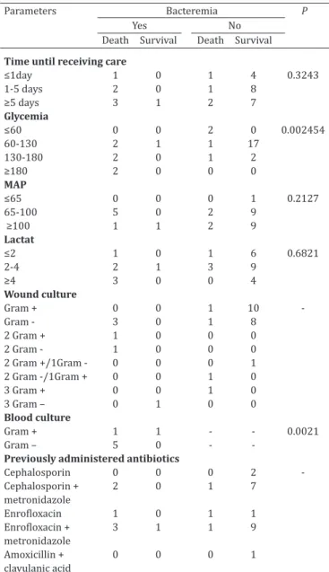
The clinical and laboratory parameters related to the presence or absence of bacteremia evaluated in the 30 dogs with wounds and signs of SIRS/sepsis, and the correlation of these parameters with survival are shown in Table 1. The overall survival rate was 66.67%. The age of the 30 patients ranged from six months to 16 years (mean age of five years and eight months) and the weight ranged between three and 45 kg (mean of 14.6kg), and these parameters exerted no effect on patient survival. Twenty (66.66%) dogs were intact males and 10 were females, of which eight (26.67%) were non-spayed and two (6.67%) were spayed. With re
-gard to breeds, 21 (70%) were mongrel dogs, two (6.67%) were poodles, and one (3.33%) was of each of the follo
-wing: German Shepherd, Belgian Shepherd, Dachshund, Pit-bull Terrier, Rottweiler, and Dalmatian. Regarding ani
-mal care, 18 (60%) dogs were domiciled, nine (30%) were semi-domiciled, and three (10%) were strays dogs. Similar results have been obtained in other studies performed in dogs with traumatic lesions in soft tissues, except that the majority of the individuals had a defined breed (Griffin & Holt 2001, Buriko et al. 2008, Meyers et al. 2008, Simpson et al. 2009). This may be related to regional factors, as the
-se studies were conducted in areas with different socioe
-conomic characteristics than that of the present study.In a study of 94 dogs with severe wounds caused by bites from other dogs, Ateca et al. (2015) found an equal proportion of
males and females, predominantly mongrel dogs, and 68% weighed less than 10 kg, because the small dogs suffered more attacks from large dogs.
In the present study, wounds were of unknown etiology in 10 (33.33%) patients, due to biting in nine (30%), due to being run over in four (13.33%), and due to each of the following causes in one (3.33%) dog: burning, cat scratch, reaction to drug application, ulcerated skin nodules, dehis
-cence of stitches after ovariohysterectomy due to pyometra, mastitis, and decubitus sores. The wounds of unknown etio
-logy were most likely due to bites or being run over, as 40% of the dogs had free access to the street. In small animals, the largest portion of traumatic wounds is caused by being run over (Dernell 2006, Arias et al. 2008) or by bites (Ateca et al. 2015). According to Pavletic & Trout (2006), wounds due to biting correspond to 10-15% of the trauma cases in small animal medicine; however, in the present study, the
Table 1. Clinical and laboratory observations in 30 dogs with wounds and signs of sepsis regarding the presence or absence of bacteremia, correlation of these parameters with
survival and mortality (p)
Parameters Bacteremia P
Yes No
Death Survival Death Survival
Time until receiving care
≤1day 1 0 1 4 0.3243
1-5 days 2 0 1 8
≥5 days 3 1 2 7
Glycemia
≤60 0 0 2 0 0.002454
60-130 2 1 1 17
130-180 2 0 1 2
≥180 2 0 0 0
MAP
≤65 0 0 0 1 0.2127
65-100 5 0 2 9
≥100 1 1 2 9
Lactat
≤2 1 0 1 6 0.6821
2-4 2 1 3 9
≥4 3 0 0 4
Wound culture
Gram + 0 0 1 10
Gram - 3 0 1 8
2 Gram + 1 0 0 0
2 Gram - 1 0 0 0
2 Gram +/1Gram - 0 0 0 1
2 Gram -/1Gram + 0 0 1 0
3 Gram + 0 0 1 0
3 Gram – 0 1 0 0
Blood culture
Gram + 1 1 - - 0.0021
Gram – 5 0 -
-Previously administered antibiotics
Cephalosporin 0 0 0 2
Cephalosporin + 2 0 1 7
metronidazole
Enrofloxacin 1 0 1 1
Enrofloxacin + 3 1 1 9
metronidazole
Amoxicillin + 0 0 0 1
percentage was doubled (30%), which can explain the pre
-sence of signs of SIRS/sepsis in these animals. Ateca et al. (2015) found that SIRS occurred in 54% of the patients in dogs with wounds caused by bites. This type of wound has a higher risk of developing clinical complications, as all bite wounds should be considered contaminated because they contain a polymicrobial flora that reflects the aerobic and anaerobic florae of the oral cavity of the bitter, skin of the victim, and environment (Pavletic & Trout 2006).
The time elapsed between observing the primary insult and receiving care varied between <1 h and 11 days, with a mean of 4.88 days after occurrence of the lesion. As a result, both by criteria time as well the appearance of the lesion, wounds were classified as infected in 80% (24/30) of the patients and as contaminated in 20%. Although this factor did not negatively affect patient survival, it is known that the presence of tissue infection may promote sepsis deve
-lopment, as the skin’s protective barrier is more vulnerable in case of endothelial disruption, which may promote he
-matogenous migration of pathogens. Most of authors des
-cribes early treatment for dogs with wounds, with no com
-plications in most of patients (Griffin & Holt 2001, Meyers et al. 2008, Mouro et al. 2010). Ateca et al. (2015) found that the mean duration between wound occurrence and hospitalization was 3 h, and that this factor did not affect mortality and treatment duration, although it did affect the occurrence of positive bacterial cultures, as seen in the pre
-sent study.
Signs of infection in the wounds and deep-tissue invol
-vement, such as subcutaneous tissue and/or muscle fas
-cia, were observed in all of the patients; lacerated injury was found in 14 (46.67%) cases, abscess was found in six (20%), penetrating and contused wounds most likely due to bite was found in six (20%), phlegmon was found in three (10%), and second and third-degree burns were found in one (3.33%) patient. In multiple animals, we observed se
-condary insults and/or concomitant systemic alterations, such as dehydration in 19 animals (63.33%); bone fracture and exposure in six (20%); gastroenteritis induced by dru
-gs in five (16.67%); major hemorrhage at the wound site in two (6.67%), and each one of the following affections in five (16.67%) dogs: urolithiasis, hepatopathy, pneumonia, pneumothorax, and pyothorax. Of the 30 treated dogs, 20 (66.67%) survived, and 10 (33.33%) died, and death oc
-curred between 1-20 days after the initial care, with a mean of four days. The degree of deeper tissue involvement and presence of SIRS/sepsis are related to higher mortality rate (Buriko et al. 2008). In dogs with sepsis due to several etio
-logies, Gebhardt et al. (2009) reported a survival rate of 58%, a value similar to that of the present study. Ateca et al. (2015) found that in dogs with wounds due to bites, the
presence of SIRS was directly related to higher mortality, with a rate of 24%, which was lower than that found in the present study.
Microbiological analysis
Bacteria were isolated from all of the samples collected from the wounds. In total, it was obtained 41 bacterial isola
-tes (Table 2). The frequencies of bacteria in the wounds are shown in Tables 2 and 3. Pseudomonas aeruginosa, Strepto-coccus spp., and Enterococcus spp.were the most frequently observed. Dryden (2010) stated that Staphylococcus aureus, followed by P. aeruginosa, Escherichia coli, and Enterococcus spp. were the most frequently identified species in skin and soft tissue infections in dogs. Except for the low presence of Staphylococcus aureus in the current study, these findings are in agreement with ours. Meyers et al. (2008) and Mouro et al. (2010) identified Staphylococcus intermedius, Pasteu-rella multocida, and pyogenic streptococcias the most com
-mon agents in wounds due to biting.
Arias et al. (2008, 2011), studying traumatic wounds in dogs, found Pseudomonas spp., Proteus spp. and Sta-phylococcus spp. as the most frequently isolated bacteria. However, in these two studies, the wounds were sampled using swabs, which frequently identify the contaminant mi
-croorganisms on the wound surface, such as Staphylococ-cus spp. (Pallua et al. 1999). Similarly, Mouro et al. (2010) used swabs to collect sample from wounds during initial presentation, resulting in the isolation of mainly bacteria belonging to the cutaneous flora. The culture from the ma
-terial collected by biopsy in the present study allowed for identifying bacteria not belonging to the normal skin mi
-crobiota, emphasizing the importance of differentiating be
-tween pathogens responsible for tissue infection and the skin-colonizing agents, which usually do not require treat
-ment with antimicrobials and do not interfere with the hea
-ling duration (Buchanan et al. 1986, Amalsadvala & Swaim 2006, Meyers et al. 2008, Dryden 2010).
Table 2. Bacterial isolates from wounds of 30 dogs with signs of sepsis
Gram-positive bacteria Number of isolates (%) Gram-negative bacteria Number of isolates (%) Total Streptococcus spp. 07 (36.84) Pseudomonas aeruginosa 11 (50.00)
Enterococcus spp. 07 (36.84) Klebsiella pneumoniae 06 (27.27) Staphylococcus spp. 05 (26.32). Escherichia coli 03 (13.64)
Proteus spp. 02 (9.09)
Total 19 (46.34) 22 (53.66) 41 (100)
Table 3. Frequencies of bacterial species isolated from wounds of 30 dogs with signs of sepsis
Bacteria Number of Isolates (%)
Pseudomonas aeruginosa 11 (26.83) Enterococcus spp. 7 (17.07) Klebsiella pneumoniae 6 (14.63) Streptococcus pyogenes 6 (14.63) Escherichia coli 3 (7.32) Staphylococcus pseudointermedius 3 (7.32) Staphylococcus aureus 2 (4.88) Proteus mirabilis 1 (2.44) Proteus vulgaris 1 (2.44) Streptococcus milleri 1 (2.44)
Only few veterinary studies on collecting samples from wounds by biopsy have been reported. This techni
-que is considered the gold standard in human medicine for diagnosing infection in skin wounds, because it allows obtaining the bacteria that infect the deeper tissues (Steer 1996, Pallua et al. 1999, Bowler et al. 2001, Amalsadvala & Swaim 2006, Laitano et al. 2008). In veterinary practice, there is still resistance to applying this technique because it is considered invasive and prone to complications such as hemorrhage (Pallua et al. 1999, Laitano et al. 2008); ho
-wever, no complications occurred in any of the patients in the present study.
The number of bacterial species in relation to patient is in Table 1. Of the ten dogs that died, three showed polymi
-crobial infection of the wounds: S. aureus, Proteus vulgaris, and E. coli were isolated from one animal; Streptococcus pyogenes, Enterococcus spp., and S. aureus were isolated from another; and E. coli and Enterococcus spp. were iso
-lated from the third dog.Various types of microorganisms can coexist in the same wound, especially Pseudomonas spp., Streptococcus spp., and Staphylococcus spp. (Amal
-sadvala & Swaim 2006). Polymicrobial type of infection is often associated with complications, i.e., bacteremia and/ or endotoxemia (Cowell & Penwick 1989, Amalsadvala & Swaim 2006, Dryden 2010), as found in the present stu
-dy. Any type of wound has the potential to be colonized and/or infected by a wide variety of aerobic and anaero
-bic microorganisms originating from the environment, the endogenous medium, or skin, and environmental sources frequently predominate in polymicrobial traumatic woun
-ds (Krahwinkel & Boothe 2006). Enterococcus spp. are pre
-sent in the soil, water, and endogenous flora of the animals, and can cause infections of the surgical site and wounds in dogs. Although we found Enterococcus spp. at high fre
-quency in the present study, we observed no resistance to vancomycin, which would be a major concern for public health because of possible animal-to-human transmission (Abbot et al. 2008).
Presence of bacteremia (23.33% of cases) and clinical evolution are in Table 4. These patients showed a negati
-ve clinical evolution, considering that of the se-ven animals in which bacteremia was confirmed, six died, representing 20% of the total mortality rate, indicating that bacteremia has a significant effect on mortality. Blood culture is indica
-ted in any seriously ill patient exhibiting fever, leukocytosis with deviation to the left or neutropenia, hypoglycemia, cir
-culatory collapse, tachycardia, tachypnea, dyspnea, anuria or oliguria, jaundice, thrombocytopenia, or signs of wides
-pread intravascular clotting (Calvert & Thomason 2011). Because the dogs with wounds in the present study were intensively monitored to identify signs of SIRS/sepsis, we succeeded in obtaining a number of positive blood cultu
-res. Bacteremia represents a serious clinical complication, with an important role in prognosing sepsis; however, blood cultures are positive in only one third of the cases in human patients with severe sepsis (Hodgin & Moss 2008). In veterinary medicine, information regarding prevalence of bacteremia in dogs and cats is still quite scarce (Dow et al. 1989, Greiner et al. 2008), but is has been estimated to yield positive results in 30–50% of blood cultures when performed appropriately (Calvert & Thomason 2011). Ide
-ally, samples should be collected at three time points (Cal
-vert & Thomason 2011), which was not performed in the present study, but could increase the percentage of positive blood cultures.
The microbial agents isolated from the blood cultures are in Table 4. The wound and blood cultures contained the same species in two dogs (Table 4). In human patients, before 1987, blood stream infections by gram-negative bacteria were predominant; however, after this period, gram-positive bacteria became the most common agent responsible for the development of severe sepsis (Davies & Hagen 1997, Otto 2007, Hodgin & Moss 2008). In veterina
-ry medicine, the predominance of gram-positive or gram
--negative bacteria in sepsis remains poorly described (Otto 2007). In blood cultures from dogs with infectious diseases of different origins, S. aureus and Streptococcus spp. are considered the most frequently isolated agents (Greiner et al. 2008), followed by E. coli, although gram-negative mi
-croorganisms can be more common in critically ill patients (Calvert & Thomason 2011), as observed in the present study. E. coli and Pseudomonas spp. are gram-negative mi
-croorganisms most commonly cultured in blood cultures of human patients with severe sepsis, and polymicrobial in
-fections are responsible for 5% of sepsis cases; however, they account for 30% of cases in patients with severe sep
-sis (Hodgin & Moss 2008).
Antibiotics were empirically administered in the initial treatment. In most cases, a combination of drugs, involving broad-spectrum antibiotics such as enrofloxacin and first
--generation cephalosporins, such as cephalexin or cephalo
-thin, was used. Enrofloxacin combined with metronidazole Table 4. Bacterial isolates from the wounds and blood cultures and evolution
of the condition in the seven patients with positive blood culture
Patient Bacterial growth Wound Bacterial growth Blood Evolution
02 Proteus mirabilis Staphylococcus aureus death
05 Staphylococcus aureus Staphylococcus aureus survival 16 Klebsiella pneumoniae/ Klebsiella pneumoniae/ death Pseudomonas aeruginosa Escherichia coli/Enterococcus spp.
17 Pseudomonas aeruginosa Pseudomonas aeruginosa death 19 Staphylococcus pseudointermedius/ Brucella melitensis death Streptococcus pyogenes
24 Pseudomonas aeruginosa/ Acinetobacter baumannii death Klebsiella pneumoniae
was used in 43.33% of cases, followed by first-generation cephalosporins combined with metronidazole (33.33%), and amoxicillin/clavulanic acid was administered to one (3.33%) dog. These antibiotics were also used alone, al
-though at a lower frequency (Table 1).
It was not observed the growth of anaerobic bacteria in blood cultures, and this may be occurred as a consequence of metronidazole administration in 24/30 (80%) patients. However anaerobic culture of the wound material was not performed. Anaerobic microbial growth also did not occur in a low number of cases in which the drug was not used, and although few dogs had not received this drug, it raises the issue whether the use of this antibiotic is really neces
-sary. As metronidazole is metabolized in the liver, critical patients show a trend of developing multiple organ dys
-function, which can contribute to the worsening of clinical status and sepsis (Calvert & Thomason 2011). Another im
-portant point is that anaerobes make up a significant por
-tion of the normal bacterial flora, on most mucosal surfa
-ces, and they play an important role in protection (Greene & Jang 2011), so it can occur imbalances as a consequence of antibiotic misuse.
AST
We did not detect any microorganisms exhibiting ab
-solute multiresistence to the antimicrobials tested in vitro. However, among gram-negative bacilli, a high rate of resis
-tance (61.18-95.45%) to first and third-generation cepha
-losporins was observed. The bacteria also exhibited high resistance to enrofloxacin (59.1%), amoxicillin/clavulanic acid (59.1%), and sulfa (72.72%). Gram-negative bacte
-ria exhibited lower resistance to aminoglycosides such as amikacin (13.64%) and gentamicin (27.27%). Mouro et al. (2010) recommend empirical use of sulfas in dogs with wounds, and in the case of infected wounds, they recom
-mend ampicillin combined with fluoroquinolones, amino
-glycosides, or amoxicillin/clavulanic acid. However, these recommendations cannot be simply extrapolated to the location in which the present study was performed, as it is highly advisable to use culturing results and antibiograms to guide the choice of antibiotics (Guardabassi et al. 2010). There was 100% sensitivity to imipenem and ticarcillin/ clavulanic acid. Nevertheless, its use as well as that of other carbapenems and vancomycin should be discouraged in ve
-terinary medicine because of the possible development of multiresistant bacteria, which pose a risk to humans (Guar
-dabassi et al. 2010). However, these antimicrobials must be tested to obtain information relevant to public health (Mouro et al. 2010).
In the AST of the gram-positive bacteria isolated from the wounds, among the five Staphylococcus spp. isolates, there was higher resistance (60%) to clindamycin, genta
-micin, sulfamethoxazole/trimethoprim, and tetracycline and 100% sensitivity to cephalosporins and oxacillin. Strep-tococcus spp. exhibited the lowest resistance (0-28.6%) to the antimicrobials tested and were 100% sensitive to amo
-xicillin/clavulanic acid and enrofloxacin. Among the seven Enterococcus spp. isolates, there was 100% sensitivity to amoxicillin/clavulanic acid, ampicillin, and vancomycin,
71.4% resistance to ceftiofur and gentamicin, 42.9% resis
-tance to erythromycin and tetracycline, and 28.6% resis
-tance to cephalexin and cefazolin. Because of the high va
-riation in the sensitivity patterns of the agents found, it is impossible to recommend a single antimicrobial or a com
-bination of agents effective against all of the species found in the wounds (Mouro et al. 2010).
The AST results of the microorganisms isolated from the blood cultures were similar to those of the wound cul
-tures, both for gram-positive and gram-negative microor
-ganisms. Among the five gram-negative isolates, 60% sho
-wed resistance to amoxicillin/clavulanic acid, ampicillin, sulfamethoxazole/trimethoprim, and tetracycline, 40% to cephalexin, cephalothin, cefazolin, and cefoxitin, and 20% to enrofloxacin and gentamicin, while 100% displayed sen
-sitivity to amikacin, imipenem, and ticarcillin/clavulanic acid. The two S. aureus samples exhibited 100% sensitivity to amoxicillin/clavulanic acid, ampicillin, cephalosporins, enrofloxacin, gentamycin, oxacillin, sulfamethoxazole/tri
-methoprim, tetracycline, and vancomycin, and 50% resis
-tance to clindamycin, penicillin, and erythromycin. Entero-coccus spp. were resistant to erythromycin and gentamycin, and sensitive to amoxicillin/clavulanic acid, ampicillin, ce
-phalosporins, enrofloxacin, tetracycline, and vancomycin.
Clinical and laboratory parameters
The mean cell volume (MCV) was 21.82%, with hemato
-crit levels between 8.9% and 41.6%, but these values were not correlated with mortality. Positive blood culture was not correlated with MCV too. Although anemia is not consi
-dered a specific marker of sepsis, these abnormalities may complicate the clinical status of severely ill patients (John
-son et al. 2004).
The admission lactate level ranged from 1.20 to 4.80mmol/L with a mean of 3.03mmol/L, whereas the lac
-tate blood level was normal in eight dogs (26.37%), and hyperlactatemia was observed in 22 dogs (73.33%) (Table 1). The lactate level was higher than 3mmol/L in 50% of the dogs that died. However, this parameter was not corre
-lated with higher mortality or occurrence of positive blood culture. The limitation of the present study was that serial measurements of lactate concentration was not performed. The current Surviving Sepsis Campaign guidelines recom
-mend that blood lactate concentration be serially moni
-tored during resuscitation of patients with severe sepsis (Ateca el al. 2015). In human and veterinary medicine, the blood lactate concentration has been used as indirect pa
-rameter for evaluating systemic tissue perfusion in critical patients, especially in those exhibiting clinical parameters compatible with SIRS/sepsis, and higher clinical attention should be given to patients with a concentration above 2.0mmol/L (Pachtinger & Drobatz 2008).In some studies, a positive correlation has been found between high lacta
-te values and poor prognosis, mainly in patients with SIRS, severe tissue infections, polytrauma, babesiosis, gastric di
-latation-volvulus, and peritonitis (Ateca et al. 2015, Buriko et al. 2008, Butler 2011).
dL, where 21 (70%) dogs exhibited normoglycemia, five (16.66%) were moderately hyperglycemic, two (6.67%) were severely hyperglycemic, and two (6.67%) exhibited hypoglycemia. It is estimated that 16% of dogs exhibit hyperglycemia when severely ill (Torre et al. 2007, Surman & Fleeman 2013). It was found that glucose level was po
-sitively correlated with positive blood cultures (p=0.0111) and death (p=0.002454). It was observed that in dogs with hypoglycemia (≤60mg/dL), as well as with severe hyper
-glycemia (≥180mg/dL) occurred greater risk of death (p=0.002454).
Patients with SIRS or sepsis displayed a marked bipha
-sic blood glucose response. Hyperglycemia during the ini
-tial phase was attributed to physiological stress and would consequently promote gluconeogenesis. During a more ad
-vanced phase, it is common to observe hypoglycemia, whi
-ch can occur due to a combination of altered glucose pro
-duction, increased uptake and use of the glucose by tissues, and poor energy input due to anorexia (Le Compte et al. 2013), as most likely occurred in the patients of the present study. In humans, hyperglycemia occurs in more than 90% of non-diabetic patients in critical condition, and for a long time, this was believed to reflect disease severity. Recently, it was found that hyperglycemia may cause systemic dama
-ge, contributing to worsening of prognosis, increased pre
-disposition to infection, increased risk of developing SIRS and sepsis, and that it is associated with increased morbi
-dity and mortality (Torre et al. 2007).
MAP ranged from 61.33 to 190.67mmHg (mean of 103.33), with 14 (46.67%) animals being normotensive, 12 (40%) being hypertensive, and four being hypotensive (13.33%). MAP was not correlated with bacteremia and death, and we noted a MAP above 100mmHg in 50% of the 20 surviving dogs (Table 1). This was most likely due to compensatory physiological mechanisms between cardiac output and peripheral vascular resistance that help main
-taining adequate tissue perfusion, which can result in ade
-quate MAP despite the hypoperfusion status (Pachtinger & Drobatz 2008). Hypoperfusion leads to inadequate oxygen delivery to the tissues, and subsequently, to the cells, which can reduce the oxidative metabolism, promoting reduced energy synthesis and imbalance in cellular functions. MAP monitoring is widely used in evaluating cardiovascular function, and although it is not considered a good clinical criterion when used alone to evaluate tissue perfusion (Pa
-chtinger & Drobatz 2008, Butler et al. 2011), in combina
-tion with other clinical and laboratory parameters, it helps in early identification of seriously ill patients (Pachtinger & Brobatz 2008), as corroborated by this study. Critical pa
-tients should be sequentially tested for several clinical and laboratory parameters to evaluate the therapeutic efficacy (Gebhardt et al. 2009).
Infections of the skin and soft tissues are one of the most common reasons for consultation in small animal clinical routine. The degree of severity and extent of the wounds is highly variable, and can be only superficial and without complications or extensive and with secondary consequen
-ces. The primary insult can generate an intense inflamma
-tory response, SIRS and then sepsis, requiring early recog
-nition for instituting intensive care. In traumatized animals with wounds, early recognition of the systemic signs com
-patible with SIRS and identification of the bacteria present is essential to avoid perpetuation of the systemic changes, which may increase the hospital cost and increase the mor
-tality rate, especially in critical patients.
CONCLUSIONS
We conclude that the technique of collecting material from the wounds by punch biopsy for subsequent culture and antibiogram analysis is safe and efficacious. We iden
-tified mainly Pseudomonas aeruginosa, Klebsiella pneumo-niae, and Enterococcus spp.; bacteria that do not belong to the normal skin microbiota. It is important to correctly identify the microbial agents involved in wound infection to avoid indiscriminate or incorrect use of antibiotics, as might occurs when skin swab culture is used, when growth of skin-surface bacteria could occur.
Among the gram-negative bacteria isolated from the wounds of the 30 dogs, there was a high incidence of resis
-tance to several classes of antibiotics such as cephalospo
-rins and sulfas, and intermediate resistance to enrofloxacin and amoxicillin/clavulanic acid. The class of aminoglycosi
-des such as amikacin and gentamycin comprised the anti
-microbials with the lowest resistance among gram-negati
-ve bacteria. We obser-ved no resistance to imipenem and ticarcillin/clavulanic acid, important antibiotics in human medicine that should be avoided in veterinary medicine. Enrofloxacin and amoxicillin/clavulanic acid were the an
-tibiotics with the lowest resistance among gram-positive bacteria. However, it is not possible to recommend a single effective antibiotic due to the high variation in antibiotic sensitivity observed among the different bacterial species isolated, where culturing and AST are advisable in patients with wounds and signs of SIRS/sepsis.
In blood cultures, gram-negative bacteria were predo
-minant, which negatively affected the patient survival. Wi
-thin this bacterial class, there was higher resistance to amo
-xicillin/clavulanic acid, ampicillin, sulfas, and tetracycline, and sensitivity to amikacin.
The presence of hypoglycemia or hyperglycemia negati
-vely affected patient survival, highlighting the importance of measuring blood glucose levels in dogs with SIRS/sepsis due to contaminated and infected wounds.
REFERENCES
Abbott Y., Kirby B.M., Karczmarczyk M., Markey B.K., Leonard F.C. & Fitzge -rald S. 2009. High-level gentamicin-resistant and vancomycin-resistant Enterococcus faecium isolated from a wound in a dog. J. Small Anim. Pract. 50:194-197.
Amalsadvala T. & Swaim S. 2006. Management of hard to heal wound. Vet. Clin. N. Am., Small Anim. Pract. 36:693-711.
Arias M.V.B., Ishii J.B. & Freitas J.C. 2011. Resistência de bactérias isola -das de cães e gatos no Hospital Veterinário da Universidade Estadual de Londrina (2008-2009). Pesq. Vet. Bras. 31(6):533-537.
Arias M.V.B., Bataglia L.A., Aiello G., Carvalho T.T. & Freitas J.C. 2008. Iden -tificação da suscetibilidade antimicrobiana de bactérias isoladas de cães e gatos com feridas traumáticas contaminadas e infectadas.Semina, Ciênc. Agrárias 29(4):861-874.
critically ill dogs with hypotension with or without hyperlactatemia: 67 cases (2006-2011). J. Am. Vet. Med. Assoc. 246(1):100-104.
Box G.E.P. & Cox D.R. 1964. An analysis of transformations. J. Royal Statist. Soc. 26:211-252.
Bowler P.G., Duerden B.I. & Armstrong D.G. 2001. Wound microbiology and associated approaches to wound management. Clin. Microbiol. Rev. 14(2):244-69.
Brainard B.M. & Brown A.J. 2011. Defects in coagulation encountered in small animal critical care. Vet. Clin. N. Am., Small Anim. Pract. 41:783-803.
Buchanan K., Heimbach D.M., Minshew B.H. & Coyle M.B. 1986. Compari -son of quantitative and semi quantitative culture techniques for burn biopsy.J. Clin. Microbiol. 23(2):258-261.
Buriko Y., Van Winkle T.J., Drobatz K.J., Rankin S.C. & Syring R.S. 2008. Se -vere soft infections in dogs: 47 cases (1996-2006). J. Vet. Emerg. Crit. Care 18(6):608-618.
Butler A.L. 2011. Goal-directed therapy in small animal critical illness. Vet. Clin. N. Am., Small Anim. Pract. 41:817-838.
Calvert C.A. & Thomason J.D.2011. Cardiovascular infections, p.912-932. In: Greene C.E. (Ed.), Infectious Diseases of the Dog and Cat. 4thed. El -sevier, St Louis. 1354p.
CLSI 2008. Performance Standards for Antimicrobial Disk and Dilution Susceptibility Tests for Bacteria Isolated from Animals: Approved Standard. 3rd ed. Clinical and Laboratory Standards Institute, Wayne, Pennsylvania, 28(8):2-11.
Cowell A.K. & Penwick R.C. 1989. Dog bite wounds: a study of 93 cases. Comp. Cont. Educ. Pract. Vet. 11(3):313-318.
Davies M.G. & Hagen P.O. 1997. Systemic inflammatory response syndro -me.Brit. J. Surg. 84:920-935.
Dernell W.S. 2006. Initial wound management. Vet. Clin. N. Am., Small Anim. Pract. 36(4):713-738.
Dow S.W., Curtis C.R. & Jones R.L. 1989. Bacterial culture of blood from cri -tically ill dogs and cats: 100 cases (1985-1987). J. Am. Vet. Med. Assoc. 195(1):113-117.
Dryden M.S. 2010. Complicated skin and soft tissue infection.J. Antimi -crob. Chemother. 65(3):35-44.
Gebhardt C., Hirschberger J., Rau S., Arndt G., Krainer K., Schweigert F.J., Brunnberg L., Kaspers B. & Kohn B. 2009. Use of C-reactive protein to predict outcome in dogs with systemic inflammatory response syndro -me or sepsis. J. Vet. E-merg. Crit. Care 19(5):450-458.
Greiner M., Wolf G. & Hartmann K. 2008. A retrospective study of the cli -nical presentation of 140 dogs and 39 cats with bacteraemia. J. Small Anim. Pract. 49:378-383.
Greene C.E. & Jang S.S. 2011. Anaerobic infections, p.411-416. In: Gree -ne C.E. (Ed.), Infectious Diseases of the Dog and Cat. 4thed. Elsevier, St Louis. 1354p.
Griffin G.M. & Holt D.E. 2001. Dog-bite wounds: bacteriology and treat -ment outcome in 37 cases. J. Am. Anim. Hosp. Assoc. 37:453-460.
Guardabassi L., Jensen L.B. & Kruse H. 2010. Guia de Antimicrobianos em Veterinária. Artmed, Porto Alegre. 267p.
Hedlund C.H. 2007. Surgery of the integumentary system, p.161-259. In: Fossum T.W. (Ed.), Small Animal Surgery. 3rded. Mosby Elsevier, Mis -souri.
Hodgin K.E. & Moss M. 2008. The epidemiology of sepsis. Curr. Pharm. De -sign14:1833-1839.
Johnson V., Gaynor A., Cha D.L. & Rozanski E. 2004. Multiple organ dysfunc -tion syndrome in humans and dogs. J. Vet. Emerg. Crit. Care 14(3):158-166.
Krahwinkel D.J. & Boothe H.W. 2006. Topical and systemic medications for wounds. Vet. Clin. N. Am., Small Anim. Pract. 36(4):739-757.
Laitano F.F., Arnt R.A., Cosner A.M. & Doncato L.F. 2008. Estudo compara -tivo entre o exame da biópsia e do “swab” cutâneo para o diagnóstico de infecção em pacientes queimados do HPS, Porto Alegre. Revta Bras. Cir. Plást. 23(3):162-166.
Le Compte A.J., Pretty C.G., Lin J., Schaw G.M., Lynn A. & Chase J.G. 2013. Impact of variation in patient response on model-based control of gly -caemia in critically ill patients. Comput. Methods Programs Biomed. 109:211-219.
Meyers B., Schoeman J.P., Goddard A. & Picard J. 2008. The bacteriology and antimicrobial susceptibility of infected and non-infected dog bite wounds: fifty cases. Vet. Microbiol. 127:360-368.
Mouro S., Vilela C.L. & Niza M.M.R.E. 2010. Clinical and bacteriological as -sessment of dog-to-dog bite wounds. Vet. Microbiol. 144:127-132. Otto C.M. 2007. Sepsis in veterinary patients: what do we know and where
can we go? J. Vet. Emerg. Crit. Care 17(4):329-332.
Pachtinger G.E. & Drobatz K. 2008. Assessment and treatment of hypovo -lemic states. Vet. Clin. N. Am., Small Anim. Pract. 38:629-643.
Pallua N., Fuchs P.C., Hafemann B., Völpel U., Noah M. & Lütticken R. 1999. A new technique for quantitative bacterial assessment on burn wounds by modified dermabrasion J. Hosp. Infect. 42(4):329-337.
Pavletic M.M. & Trout N.J. 2006. Bullet, bite and burn wounds in dogs and cats. Vet. Clin. N. Am., Small Anim. Pract. 36(4):873-93.
R Core Team. 2013. R: a language and environment for statistical com -puting. R Foundation for Statistical Computing, Vienna, Austria. URL <http://www.R-project.org/>
Simpson S.A., Syring R.S. & Otto C.M. 2009. Severe blunt trauma in dogs: 235 cases (1997-2003). J. Vet. Emerg. Crit. Care 19(6):588-602. Steer J.A. 1996. Quantitative microbiology in the management of burn pa
-tients. I. Correlation between quantitative and qualitative burn wound biopsy culture and surface alginate swab culture.Burns 22(3): 173-176. Surman S. & Fleeman L. 2013. Continuous glucose monitoring in small ani
-mals. Vet. Clin. N. Am., Small Anim. Pract. 43:381-406.
Torre D.M., De Laforcade A.M. & Chan D.L. 2007. Incidence and clinical rele -vance of hyperglycemia in critically ill dogs. J. Vet. Intern. Med. 21:971-975. Versalovic J., Carriol K., Funke G., Jorgensen J.H., Landry M. & Warnock D.W.