Venous thromboembolism in Latin America: a review
and guide to diagnosis and treatment for primary care
Jose Manuel Ceresetto*
Hospital Brita´nico, Servicio de Hematologı´a, Solı´s, Buenos Aires, Argentina.
There are various region-specific challenges to the diagnosis and effective treatment of venous thromboembolism in Latin America. Clear guidance for physicians and patient education could improve adherence to existing guidelines. This review examines available information on the burden of pulmonary embolism and deep vein thrombosis in Latin America and the regional issues surrounding the diagnosis and treatment of pulmonary embolism and deep vein thrombosis. Potential barriers to appropriate care, as well as treatment options and limitations on their use, are discussed. Finally, an algorithmic approach to the diagnosis and treatment of venous thromboembolism in ambulatory patients is proposed and care pathways for patients with pulmonary embolism and deep vein thrombosis are outlined for primary care providers in Latin America.
KEYWORDS: Venous Thromboembolism; Pulmonary Embolism; Deep Vein Thrombosis; Latin America; Primary Care.
Ceresetto JM. Venous thromboembolism in Latin America: a review and guide to diagnosis and treatment for primary care. Clinics. 2016;71(1): 36-46
Received for publication onOctober 7, 2015;First review completed onNovember 10, 2015;Accepted for publication onNovember 10, 2015 E-mail: [email protected]
*Corresponding author
’ INTRODUCTION
Although multiple studies have evaluated the epidemiology of venous thromboembolism (VTE) in European and American populations, there is limited evidence on the prevalence of VTE and the burden of disease in Latin America. Evidence from a study in the United States suggests that there are differences in the incidence of VTE among white, black, Hispanic, and Asian populations (1). Thus, it may not be appropriate to simply extrapolate the prevalence of VTE in Latin America from data obtained from European and U.S. populations. In the ENDORSE II study (2), approximately 50% of hospitalized patients from across 43 hospitals in Mexico were identified as being at risk of deep vein thrombosis (DVT) or pulmonary embolism (PE) and in a Brazilian study across 3 hospitals, a similar proportion of hospitalized patients were considered to be at high risk of DVT or PE (3). Data from an Argentinian study (4) estimated an incidence rate of 0.7 per 1,000 person-years for total VTE (0.48 and 0.22 for DVT and PE, respectively) based on the incidence rate observed at a Buenos Aires hospital and extrapolated to the entire Argentinian population. The in-hospital mortality rate from VTE was estimated at 19% in an Argentinian hospital by Mazzei et al. (5) and 14.1% in a Brazilian hospital by Volschan et al. (6). A large autopsy-based analysis in a Brazilian hospital identified PE as the cause of
death in 2.5% of all deaths of hospitalized patients (7). Thus, available data indicate a significant disease burden in Latin America in terms of morbidity and mortality as well as cost to the healthcare system.
There are various regional challenges to effective VTE diagnosis and treatment in Latin America. A cross-sectional study of internal medicine practitioners in Mexico revealed that the awareness of risk factors for VTE and recommended methods of diagnosis was low (8). A significant proportion of patients diagnosed with VTE in Latin American countries may not receive appropriate anticoagulation and some patients at risk of VTE do not receive appropriate prophylaxis (5,9-16). A Venezuelan study of characteristics of patients with VTE observed that patients with VTE often present with comorbid-ities that may complicate treatment decisions (17), and subsequent studies implicated these comorbidities as poten-tially affecting the decision to anticoagulate (18). In a study in Brazil, only 26% of patients at moderate or high risk of VTE received prophylactic anticoagulation (3). In a study of the adequacy of prophylactic anticoagulation in 28 institutions across Argentina (19), surgical patients were more likely to receive adequate prophylaxis than medical patients (71% vs. 63%). By contrast, the Epidemiologic International Day for the Evaluation of Patients at Risk for Venous Thromboembolism in the Acute Hospital Care Setting (ENDORSE) study (20) observed that medical patients were more likely than surgical patients to receive adequate prophylactic anticoagulation in Mexico, Venezuela, Colombia and Brazil. This finding high-lights potential differences in educational needs across Latin American regions.
The International Society on Thrombosis and Haemostasis recently performed a worldwide survey as part of the first DOI:10.6061/clinics/2016(01)07
Copyright&2016CLINICS–This is an Open Access article distributed under the terms of the Creative Commons License (http://creativecommons.org/licenses/by/ 4.0/) which permits unrestricted use, distribution, and reproduction in any medium or format, provided the original work is properly cited.
World Thrombosis Day. Of the Argentinians surveyed, the majority did not recognize the symptoms of DVT or PE (21,22). Globally, concern about thrombosis was second highest in Argentina, but fewer than half of Argentinians surveyed recognized that thrombosis is a preventable disease (21).
Clear guidance and education on how comorbidities may affect therapy might increase healthcare providers’adherence to existing guidelines (23-25). Pilot studies using programs and guidance protocols to facilitate treatment decisions in Brazil and Argentina have demonstrated improvements in the level of appropriate anticoagulation administered to patients with or at risk of VTE (26,27). The aim of this review is to outline an algorithmic approach for primary care pro-viders in Latin America for VTE diagnosis in ambulatory patients and to discuss current and emerging options for the treatment of these patients.
Venous Thromboembolism: Deep Vein Thrombosis and Pulmonary Embolism
VTE, which includes DVT and PE, is associated with significant morbidity and is a leading cause of cardiovascular death worldwide (28). DVT is a common complication of and the most common cause of rehospitalization following any major surgery. Although less common than DVT, PE is a serious complication that can occur after surgery or in association with cancer, other chronic illnesses and preg-nancy (28). Various risk scores have been developed to estimate VTE risk in specific patient populations, such as the Khorana score in patients with cancer (29) and the Caprini score in surgical patients (30).
The Khorana score (29) was developed for use in patients with cancer initiating a new chemotherapy regimen as a simple predictive risk model to identify patients at highest risk for VTE who would most benefit from thromboprophy-laxis. The scoring system assigns 2 points for the stomach or pancreas as the primary site of cancer or 1 point each for a primary site in the lungs or genitourinary tract, excluding prostate, lymphoma, or gynecological cancer. An additional point is assigned for each of the following risk factors: pre-chemotherapy platelet count of 350 108/L or more; hemoglobin level below 10 g/dL or use of erythrocyte growth factors; a pre-chemotherapy leucocyte count greater than 11 109/L; or a BMI of 35 or higher. Patients are categorized as low, medium, or high risk based on their total scores (0, 1-2, andX3, respectively).
The Caprini score provides a simple checklist for known VTE risk factors in surgical patients based on a health history scored by the physician. The most recent version (30) incorporates approximately 40 different risk factors with weights of 1 to 5 points each; the total score is used to classify patients as low (scores of 0-1), moderate (scores of 1-2), high (scores of 3-4) and highest (scores of 5 or more) risk, each with a recommended prophylactic regimen.
The Caprini and Khorana scores are recommended by the American College of Chest Physicians guidelines as an objective screening method to identify patients at high risk of VTE who might benefit most from prophylactic anticoagula-tion (31). However, other specialized VTE risk scores require more extensive clinical validation.
Deep vein thrombosis
DVT can occur in either the upper or lower extremities. In a recent international study by Lamontagne et al. of 3,746
medical/surgical patients in intensive care, 98% of DVT in this population involved thromboses of the lower extremities (32). Of the remaining events, 72% were upper extremity DVT (UEDVT) (32). Patients who experience VTE are at increased risk of recurrence (33,34), and VTE is often associated with long-term, clinically significant complica-tions, including post-thrombotic syndrome and chronic thromboembolic pulmonary hypertension.
DVT of the lower extremities can be classified as proximal or distal (occurring above or below the popliteal vein, respectively). The former is a more serious condition due to the higher associated risk of thromboembolism. DVT can present as pain or tenderness of the leg, with swelling, erythema, discoloration, or surface vein distension, but also frequently occurs as an asymptomatic condition. The presence of symptoms is often insufficient for diagnosis because other conditions can cause similar symptoms. However, when associated with known risk factors, these symptoms can provoke clinical suspicion of DVT warranting further evalua-tion (35).
UEDVT accounts for 10% or less of all DVT cases and may be associated with local compressive factors or central venous catheter use (36). Clinical outcomes for UEDVT are similar to those for DVT of the lower extremities (37,38), although the recurrence rate is much lower. There are no published randomized, controlled studies of anticoagulation in UEDVT, but observational studies suggest that treatment with anticoagulants is effective (39,40). In the absence of further information, treatment for UEDVT should be the same as for DVT of the lower extremities (31,41).
Pulmonary embolism
PE is difficult to diagnose based on clinical symptoms alone and may have a broad spectrum of presentations ranging from shortness of breath, tachypnea and syncope to fever, side stitch and hemoptysis.
Shock and hypotension indicate high-risk PE. Although massive PE is characterized by hypotension (systolic blood pressure o90 mm Hg), it can also be associated with
syncope and bradycardia and can cause cardiac arrest with sudden death. Although massive PE is associated with a 90-day mortality risk of 52-58%, it accounts for only approx-imately 5% of all PE events (42,43).
Submassive PE is characterized by the absence of hypotension and the presence of right ventricular dysfunc-tion or myocardial necrosis due to ischemia of the right heart. Tachycardia may suggest right ventricular dysfunction, but has limited specificity. Additional laboratory parameters indicative of myocardial ischemia or an echocardiogram/ computed tomography scan revealing an enlarged right ventricle are also required to confirm a diagnosis of sub-massive PE (43,44). This condition is associated with a parti-cularly high event mortality of approximately 30% and early identification of patients and hospitalization for treatment are imperative. In low-risk or nonmassive PE, systolic blood pressure remains normal and the markers that define massive or submassive PE are absent (42,43). These low-risk patients may be appropriate for ambulatory management if conditions allow.
epidemiological model of fatal PE, 34% of affected patients experienced sudden death, 59% were undiagnosed during life and treated as having cardiac insufficiency or pneumonia and only 7% were correctly diagnosed with PE before death (47). Therefore, a large number of patients with PE may be misdiagnosed and would benefit significantly from a reliable diagnostic algorithm, particularly in countries where aware-ness of the disease is low.
Diagnosing VTE in primary care
Data from the International Society on Thrombosis and Haemostasis‘‘World Thrombosis Day’’survey for Argentina, the only Latin American country included in the survey, indicate that the majority of the Argentinian population has little awareness or knowledge of DVT, PE, or their con-sequences, consistent with the results for all other countries evaluated in the survey (21,22). Consequently, the ability of general practitioners to diagnose this condition is critical.
In managing patients presenting with symptoms of DVT, such as a unilateral pitting edema or swollen and painful leg, or where there is clinical suspicion of DVT, the Wells DVT score or simplified Wells for predicting probability of DVT should be calculated before imaging is performed (48). If the score indicates a high probability of DVT, in the absence of contra-indications, anticoagulation with heparin or similar agents should be initiated immediately while confirmatory tests are performed. However, if the score indicates a low probability of DVT, a negative D-dimer test can rule out a diagnosis of DVT without requiring imaging, although the D-dimer cut-off value used should be age-adjusted for older patients (age 10mg/L for patients 450 years of age). When screening a low-risk patient, treatment may be delayed only if the tests will be available within a reasonable time frame (31). A diagnostic algorithm for patients with DVT is presented in Figure 1 (35).
In patients presenting with symptoms of PE, such as hemoptysis, chest pain and shortness of breath, or where there is clinical suspicion of PE, the Wells PE score (49) or revised Geneva score (50) should be calculated and used to predict the probability of PE before imaging is performed. As in DVT, for patients for whom there is a strong clinical suspicion of PE but no firm diagnosis, unless there are contraindications to heparin use, anticoagulation with unfractionated heparin, low-molecular-weight heparin (LMWH), or fondaparinux should be maintained until the diagnosis is confirmed (31). Bivalirudin or fondaparinux may be considered for patients who cannot receive heparin derivatives (e.g., as a result of heparin-induced thrombocytopenia) (31). The Pulmonary Embolism Severity Index and Simplified Pulmonary Embo-lism Severity Index (Table 1) can be used to assess the severity of the event based on 30-day survival probability (51,52).
A diagnostic algorithm based on the European Society of Cardiology guidelines for patients with suspected PE (52) in the absence of hypotension and shock (i.e., normotensive) is shown in Figure 2. For patients with suspected PE with suspected shock or hypertension, the European Society of Cardiology guidelines recommend that an angiography/ helical CT should be performed if available, and treatment for PE initiated for positive findings (52). If the CT findings are negative, alternative causes for hemodynamic instability should be explored (52). If CT angiography is not immedi-ately available, echocardiography should be performed to check for right ventricle (RV) overload and, if confirmed, the patient should be stabilized and then sent for CT (52). If the
patient has confirmed RV overload, but cannot be stabilized or CT is not immediately available, treatment should be initiated for PE (52). In the absence of RV overload, alternative causes of hemodynamic stability should be investigated (52).
Treatment options: guidelines, anticoagulant choices and treatment duration
The guidelines for individual countries in Latin America are generally consistent with those of the American College of Chest Physicians (23-25,31,53). Guidelines on antithrombotic therapy for VTE recommend an anticoagulant for the treatment of acute VTE (31) to prevent further growth of the throm-bus rather than to deplete the existing thromthrom-bus, for which fibrinolysis may be required. After the acute phase of treatment, the treatment pathways for DVT and PE are similar (31,54), reflecting that these are both manifestations of the same disease. The recommended duration of anticoagulation varies, depend-ing on the cause of the initial event. Anticoagulation for 3 months is appropriate when there is a transient and reversible cause, such as surgery, trauma, or hospitalization; 6 months or more is appropriate for patients experiencing a first event of idiopathic VTE; and 12 months or more (possibly indefinitely, subject to regular review) is appropriate for patients with active neoplasia, recurrent idiopathic VTE, high-risk thrombophilia, or antiphospholipid antibodies (25). However, anticoagula-tion may be discontinued after the initial 3 months of treatment if the risk of bleeding is high (25). Due to the lower risk of recurrence associated with UEDVT, in the absence of additional factors, 3 months of anticoagulation treatment may be sufficient, even in cases of spontaneous DVT.
Initiation of parenteral anticoagulation is recommended without delay in patients with a high or intermediate probability of PE while diagnostic work-up is in progress. Heparin, LMWH, and fondaparinux are the recommended forms of parenteral anticoagulation in the acute phase (25,31,53). In parallel with parenteral anticoagulation, treat-ment with a vitamin K antagonist (VKA) is recommended, with a target international normalized ratio (INR) of 2.0-3.0. For inpatients with PE who have a high risk of bleeding or who are hemodynamically unstable and for whom precise anticoagulation control and the ability to quickly reverse anticoagulation may be desirable, unfractionated heparin may be preferred to LMWH due to its short half-life and the availability of an antidote, protamine (31).
49% compared with the study average of 64% (58). A low TTR in this region has also been recorded in clinical trials, which have the advantages of patient selection according to trial criteria and close support and monitoring. Real-world TTR levels are likely to be at least 10-20% lower (59). This low rate of ‘‘in range’’ therapeutic anticoagulation could be a major disadvantage in Latin America and may be one of the region’s most important challenges with respect to oral anticoagulation with VKAs. However, a recent study exploring TTR across 14 anticoagulation clinics in Argentina that included 1,190 consecutive patients with atrial fibrillation observed a mean TTR of 66.6%, a value similar to that observed in international
therapeutic clinical trials and in Nordic countries, where care is largely available from highly developed socialized health systems, indicating that high-quality anticoagulation with VKAs is possible in Latin America (60).
Direct oral anticoagulants
When the American College of Chest Physicians 2012 guidelines for the treatment of VTE were issued, there was insufficient clinical experience with direct oral anticoagulants (DOACs) to support recommendations for these agents over VKAs or LMWH (31). However, publications and clinical data
Figure 1 -Diagnosis and treatment pathway for a patient presenting with symptomatic DVT. DOAC, direct oral anticoagulant (e.g.,
from real-world use of DOACs have since become available and recommendations for their use were included in the more recent European Society of Cardiology guidelines (53). These new drugs are approved or under consideration for this indication by various regulatory agencies. DOACs offer rapid onset of action and predictable pharmacokinetics, obviating the need for regular monitoring in routine clinical use and enabling convenient oral administration. These qualities are significant advantages for DOACs compared with VKAs, and thus DOACs are particularly useful for outpatient treatment.
Ambulatory patients and those without cancer requiring long-term treatment for VTE may be good candidates for new oral anticoagulants if they have a low risk of bleeding and adequate renal and hepatic function. Several international experts have proposed that DOACs should be considered for patients in whom adequate anticoagulation with VKAs cannot be maintained in the therapeutic range despite good adherence to treatment and regular monitoring (61). Conversely, patients with poor adherence to treatment, patients with an underlying disease that requires hospitalization, or patients requiring triple antithrombotic therapy (e.g., dual antiplatelet therapy plus anticoagulant) may not be candidates for DVT treatment with DOACs. In addition, due to the relative scarcity of clinical experience, patients with cancer and thrombosis, antiphos-pholipid syndrome, high-risk thrombophilia, or thrombosis at an unusual site (e.g., splanchnic or cerebral vein thrombosis) should not be considered for DOACs until clinical trials have demonstrated the utility of these agents for these indications. In patients with severe renal impairment (creatinine clearance
o30 mL/min), hepatic impairment (Child-Pugh category B or
C), pulmonary thromboembolism with high burden of disease or a high-risk simplified Pulmonary Embolism Severity Index score (Table 1), severe hypotension, dilated right ventricle, or phlegmasia alba dolens (milk leg) (for the potential use of fibrinolytics), patients who are pregnant or breastfeeding and pediatric patients, traditional anticoagulant treatment either with heparins alone or in combination with VKAs may be a superior option (62,63).
The results of key studies of each of the DOACs are summarized in Table 2 (64-71). In light of these clinical trial results, it will be important to reconsider treatment guidelines in
some patients as new agents become available. Xareltos (rivaroxaban) is already approved for the treatment of VTE/ PE in Argentina, Brazil, Chile, Colombia, Mexico and Peru. Pradaxas
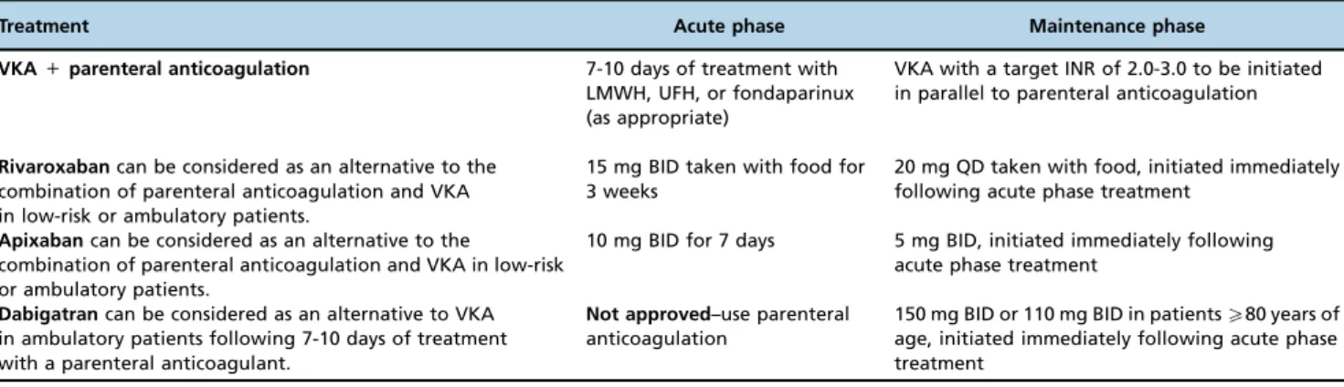
(dabigatran) is approved in Argentina, Brazil, Chile, Colombia and Mexico, and Eliquiss (apixaban) is currently approved in Argentina, Chile, Colombia, Mexico and Peru for this indication. Approval for all 3 agents in additional countries is expected in the near future. Guidelines for the use of rivaroxaban, apixaban, or dabigatran in the treatment of DVT or PE in ambulatory patients are presented in Table 3. At the time of writing, Savaysat(edoxaban) has not been approved for this indication in any Latin American country or in Europe and has yet to be included in the guidelines. However, prescribing information from the United States, where edoxaban was recently approved for the treatment of DVT and PE, states that edoxaban (60 mg once daily for patients with creatinine clearance450 top95 mL/min, reduced to 30 mg once daily
in patients with creatinine clearance 15–50 mL/min, who weigh
p60 kg, or who are taking specific concomitant P-glycoprotein
inhibitor medications) can be used following 5 to 10 days of initial therapy with a parenteral anticoagulant (72).
Barriers to widespread direct oral anticoagulant use Access to the healthcare system can present a significant barrier to care in general, with WHO estimates indicating that more than 40% of the population of some countries does not have effective access to healthcare for reasons ranging from language barriers and a lack of education to a lack of health infrastructure and lack of access to other public services, including electricity and sanitation, which thus impairs adequate provision of healthcare (73). Where access to the healthcare system is available, one of the major barriers to appropriate anticoagulation in Latin America is economic; the high pharmacy cost of outpatient anticoagulation treatment can make it inaccessible to patients who must pay drug costs out of pocket (73) and when funded publicly, the use of more expensive drugs may be restricted or controlled centrally (13). Consequently, ambulatory patients who would be suitable for outpatient treatment with new oral anticoagulants may be admitted to the hospital to receive the first few days of
Table 1-PESI and sPESI: prognostic scores for severity in patients with acute symptomatic pulmonary embolism.
PESI Score Simplified PESI Score
Variable Points Variable Points
Age 1/year Age480 years 1
Male sex 10 Cancer 1
Cancer 30 Chronic cardiopulmonary disease 1
Heart failure 10 Heart rateX110 beats/min 1
Chronic lung disease 10 Systolic blood pressureo100 mm Hg 1
Heart rateX110 beats/min 20 O2saturationo90% 1
Systolic blood pressureo100 mm Hg 30
Respiratory rateX30 breaths/min 20
Temperatureo36o
C 20
Altered mental status 60
O2saturationo90% 20
Interpretation:
Class I (very low risk):o65 points
Class II (low risk): 66-85 points
Class III (intermediate risk): 86-105 points Class IV (high risk): 106-125 points Class V (very high risk):4125 points
Interpretation: Low risk: 0 points High risk:X1 point(s)
treatment as inpatients. In some institutions, patients may be admitted to receive unfractionated heparin via continuous infu-sion pump to avoid the high cost of LMWH (13,26), despite the greater overall cost to the healthcare system of doing so (74,75). Thus, while it may be theoretically possible for 80% of patients with DVT to be managed on an outpatient basis (76,77), the actual rate of outpatient care is much lower and both institutional and healthcare system costs are increased.
Across many parts of Latin America there is a lack of knowl-edge regarding the pathology of VTE, leading to late consulta-tion. Furthermore, even if a clinician has adequate evidence to suspect a diagnosis requiring anticoagulation, therapy is occa-sionally not started until the diagnosis has been confirmed, which in some institutions results in an additional delay of several days while waiting for confirmation by ultrasound.
In some regions of Latin America, particularly rural areas, access to an anticoagulation clinic may be difficult. Therefore, in many countries, patients may be managed by general physicians (rather than anticoagulation specialists), who may lack detailed knowledge of how to evaluate anticoagulated patients. Non-specialists are also less likely to be aware of the range of available treatments and when oral anticoagulation is prescribed, may choose traditional anticoagulation, such as a VKA, based on familiarity, despite the additional burden of monitoring.
Emergency reversal of anticoagulant effects Evidence suggests that 4-factor prothrombin complex con-centrates may be as useful for managing bleeding related to
Figure 2 -Diagnosis and treatment pathway for a patient presenting with symptomatic PE who is normotensive. CT, computed tomography;
DOAC, direct oral anticoagulant (e.g., rivaroxaban, apixaban, dabigatran or edoxaban); DVT, deep vein thrombosis; PE, pulmonary embolism; PESI, Pulmonary Embolism Severity Index; sPESI, simplified PESI; VKA, vitamin K antagonist. *D-dimer cut-off should be age adjusted (age 10mg/L) in patients450 years of age.wFor patients with renal failure, allergy to contrast dye, pregnant patients, or other contraindications to
factor Xa (FXa) inhibitors and direct thrombin inhibitors as they are for bleeding related to VKAs (78-81); however, the use of DOACs has been limited by the lack of a specific active reversal agent or antidote for use in bleeding management or prior to an emergency procedure. Idarucizumab (Praxbind), an antibody fragment–based reversal agent for dabigatran, was approved in late 2015 (82) and results from phase III studies of andexanet alfa, a recombinant modified FXa for the reversal of FXa
inhibitors, are positive (83). Also under development is PER977, a synthetic small molecule that may eventually become a wide-range reversal agent for anticoagulants. PER977 exhibits com-plete reversal of FXa inhibition, direct thrombin inhibition, and the anticoagulant effects of LMWH, unfractionated heparin, and fondaparinux both in vitro and in preclinical animal models and, more recently, for the reversal of edoxaban, a FXa inhibitor, in healthy subjects (84).
Table 2-Summary of key clinical trials of novel oral anticoagulants in the treatment of venous thromboembolism and pulmonary embolism.
Study Patient population Study treatment Key findings
Dabigatran RE-COVER/ RE-COVER II (64) (66)
Patients with acute symptomatic VTE Heparin or LMWH for 8-11 days, followed by dabigatran 150 mg BID or warfarin given in doses adjusted to an INR of 2.0-3.0 for 6 months
Dabigatran was noninferior to warfarin for the prevention of recurrent or fatal VTE and had a similar rate of major bleeding but a lower risk of any bleeding events. RE-MEDY
(65)
Patients completing at least 3 months of VTE treatment who were considered at increased risk of recurrent VTE
Dabigatran 150 mg or warfarin (adjusted to an INR of 2.0-3.0)
Dabigatran was noninferior to warfarin (adjusted to an INR of 2.0-3.0) for the prevention of recurrent symptomatic and objectively verified VTE or death associated with VTE.
RE-SONATE (65)
Patients completing at least 6 months of treatment for VTE.
Dabigatran 150 mg BID or placebo Dabigatran significantly reduced the rate of recurrent VTE but with a significantly higher rate of major or CRNM bleeding (5.3%vs. 1.8%).
Rivaroxaban EINSTEIN-DVT (67)
Patients with acute symptomatic DVT in the deep veins of the knee or thigh, but without any symptoms of PE
Rivaroxaban or standard therapy (enoxaparin followed by VKA) for 3, 6, or 12 months
Rivaroxaban had non-inferior efficacy with respect to the primary outcome, with similar rates of major and CRNM bleeding. EINSTEIN-PE
(68)
Patients with acute symptomatic PE with or without symptomatic DVT
Rivaroxaban (15 mg BID for 3 weeks, followed by 20 mg QD) or standard therapy (enoxaparin followed by VKA) for 3, 6, or 12 months
Rivaroxaban was non-inferior to enoxaparin/VKA therapy for the prevention of recurrent VTE, with similar rates of total and CRNM bleeding. However, rivaroxaban was associated with a statistically significant reduction in major bleeding events (HR, 0,49 [0,31-0,79]) in patients with PE.
EINSTEIN-EXT (67)
Patients who had previously completed 6 to 12 months of treatment with a VKA for an acute episode of VTE or had participated in the EINSTEIN-DVT or EINSTEIN-PE trials
Rivaroxaban 20 mg QD or placebo for 6 to 12 months
Rivaroxaban demonstrated superiority to placebo for the primary outcome. Efficacy and safety results were consistent across all prespecified subgroups.
Apixaban AMPLIFY (69)
Patients presenting with acute DVT or PE Apixaban 10 mg BID for 7 days followed by 5 mg BID for 6 months or subcutaneous enoxaparin for 5 days followed by dose-adjusted warfarin for 6 months
There were no significant differences in the rates of the primary efficacy outcome of recurrent symptomatic VTE or VTE-related death (2.3%vs. 2.7%, respectively) between treatments, and apixaban was associated with significantly fewer major bleeding and CRNM bleeding events compared with conventional therapy (events occurred in 4.3%vs. 9.7% of patients, respectively).
AMPLIFY-EXT (70)
Patients with DVT/PE for whom there was clinical uncertainty about whether to continue oral anticoagulation after 6-12 months of routine treatment with a VKA
Placebo, apixaban 2.5 mg BID, or apixaban 5 mg BID for 12 months
Compared with placebo, both doses of apixaban reduced the risk of recurrent fatal or nonfatal VTE, while rates of major bleeding were low and comparable to those in the placebo group.
Edoxaban HOKUSAI-VTE (71)
Patients who presented with DVT or PE 5-7 days of heparin followed by edoxaban 30 or 60 mg QD or warfarin for 3-12 months
Edoxaban was noninferior to warfarin with respect to the primary efficacy outcome of recurrent symptomatic VTE, with less major or CRNM bleeding compared with the warfarin group.
Monitoring of anticoagulant effect
The use of DOACs has also been limited by the lack of a readily available standardized assay for anticoagulant activity. Despite the lack of a requirement for routine monitoring with DOAC administration, situations may arise where monitoring is desirable. The standard clotting assays used with heparin derivatives and VKAs cannot be used to quantitatively assess anticoagulant activity with DOACs, but other assays are becoming available to facilitate point-of-care testing. A throm-bin inhibitor assay suitable for use with dabigatran is now commercially available, although its real value in the clinical setting remains controversial (85). Tests based on chromogenic FXa-specific inhibitor assays with appropriate calibrators are now commercially available to assess the anticoagulant activity of anti-FXa drugs such as rivaroxaban and apixaban, but their clinical relevance has yet to be evaluated (86,87).
Adherence to treatment
Although the lack of a requirement to routinely monitor anticoagulation with DOACs may be advantageous in some situations, it may actually increase the likelihood of patients stopping their anticoagulant treatment earlier than recom-mended. Adherence may be particularly problematic where health literacy is relatively low, such as in some Latin American regions, and thorough education is needed to ensure that patients adhere to their treatment regimens as prescribed.
Increased drug costs
Drug purchase costs for DOACs are significantly greater than those for generic warfarin. Cost is an important limitation to the use of DOACs in Latin America, where many patients incur high out-of-pocket health costs, with estimates for the average percentage of household income spent on healthcare varying by country from 2.0% to 6.9% (73). At present, the increased drug cost may be partially offset by a reduction in the costs associated with regular monitoring of the anticoagulant effect, as well as potentially reduced costs relating to bleeding events and hospitalizations. In addition, as DOACs become more widely used, competition among the different agents may result in lower prices over time. The higher drug costs associated with DOACs compared with VKA therapy could be a factor in a
patient’s decision to discontinue anticoagulant therapy earlier than would be advised by a healthcare provider.
Clinical experience
Finally, a lack of clinical experience in a specific patient population compared with standard anticoagulant therapies may also be a barrier to treatment. Clinical trial populations may not accurately reflect the patient population in real-world clinics, who are subject to multiple comorbidities (78), polypharmacy (79) and other potential modifiers that would have resulted in the exclusion of these patients from the original trials. In addition to comorbidities and polypharmacy asso-ciated with an increased risk of bleeding and worse clinical outcomes (79 81), the presence of additional risk factors for bleeding may make physicians more cautious about prescrib-ing anticoagulants in general, particularly those with which they have limited clinical experience. Over time, more data regarding DOAC use in real-world patient populations will become available, and as clinical experience with these agents increases, this barrier to DOAC use is likely to be lowered.
VTE is a significant health problem in Latin America and is complicated by various region-specific issues. In some regions, awareness of diagnostic criteria for VTE is low. Many patients do not receive appropriate anticoagulation even after being diagnosed with VTE/PE. Clear guidance to facilitate the diagnosis of VTE and provide appropriate anticoagulation for patients once diagnosed may offer significant benefits in the region. Heparin-based anticoagulants are likely to remain the first choice for inpatients with VTE at high risk of bleeding or with additional complications for which the doctor may desire a precise level of control over anticoagulation. However, DOACs may be particularly beneficial for outpatients requir-ing anticoagulation, particularly in situations in which regular monitoring may not be feasible.
’ ACKNOWLEDGMENTS
Professional medical writing and editorial assistance were provided by Andy Shepherd and Nicole Draghi at Caudex Medical, funded by Bristol-Myers Squibb Company and Pfizer Inc.
’ FINANCIAL SUPPORT
Dr. Ceresetto is on the Advisory Board for Bristol-Myers Squibb in Argentina.
Table 3-Anticoagulation therapy for patients with DVT or patients with PE in the absence of hypotension or shock.
Treatment Acute phase Maintenance phase
VKA+parenteral anticoagulation 7-10 days of treatment with
LMWH, UFH, or fondaparinux (as appropriate)
VKA with a target INR of 2.0-3.0 to be initiated in parallel to parenteral anticoagulation
Rivaroxabancan be considered as an alternative to the combination of parenteral anticoagulation and VKA in low-risk or ambulatory patients.
15 mg BID taken with food for 3 weeks
20 mg QD taken with food, initiated immediately following acute phase treatment
Apixabancan be considered as an alternative to the
combination of parenteral anticoagulation and VKA in low-risk or ambulatory patients.
10 mg BID for 7 days 5 mg BID, initiated immediately following acute phase treatment
Dabigatrancan be considered as an alternative to VKA in ambulatory patients following 7-10 days of treatment with a parenteral anticoagulant.
Not approved–use parenteral anticoagulation
150 mg BID or 110 mg BID in patientsX80 years of age, initiated immediately following acute phase treatment
See prescribing information for individual agents for further information on contraindications or dosage adjustments in certain patient groups. All recommendations below are subject to local regulatory approval of these agents for this indication.
’ SPONSORSHIP
Professional medical writing and editorial assistance were provided by Andy Shepherd and Nicole Draghi at Caudex Medical, funded by Bristol-Myers Squibb Company and Pfizer Inc. Dr. Ceresetto is on the Advisory Board for Bristol-Myers Squibb in Argentina and has received support for this manuscript from Bristol-Myers Squibb Company and Pfizer Inc.
’ REFERENCES
1. White RH. The epidemiology of venous thromboembolism. Circulation. 2003;107(23 Suppl 1):14-8, http://dx.doi.org/10.1161/01.cir.0000078468. 11849.66.
2. Martínez-Zubieta R. [Venous thromboembolism risk and prophylaxis in the acute hospital care setting (ENDORSE II study): results of a Mexican national cross-sectional study]. Cir Cir. 2010;78(4):333-41.
3. Andrade EO, Bindá FA, Silva AM, Costa TD, Fernandes MC, Fernandes MC. Risk factors and prophylaxis for venous thromboembolism in hos-pitals in the city of Manaus, Brazil. J Bras Pneumol. 2009;35(2):114-21, http://dx.doi.org/10.1590/S1806-37132009000200003.
4. Vazquez FJ, Posadas-Martinez ML, Vicens J, González Bernaldo de Quirós F, Giunta DH. Incidence rate of symptomatic venous thromboembolic disease in patients from a medical care program in Buenos Aires, Argentina: a prospective cohort. Thromb J. 2013 Aug 1;11(1):16, http:// dx.doi.org/10.1186/1477-9560-11-16.
5. Mazzei JA, Campos AL, Melero MJ. [Frequency and incidence of venous thromboembolism in a general hospital]. Medicina (B Aires). 2005;65(4):289-94. 6. Volschan A, Albuquerque D, Tura BR, Knibel M, Esteves JP, Bodanese LC, et al. Predictors of hospital mortality in hemodynamically stable patients with pulmonary embolism. Arq Bras Cardiol. 2009;93(2):135-40. 7. Carvalho Bricola SA, Paiva EF, Lichtenstein A, Gianini RJ, Duarte JG,
Shinjo SK, et al. Fatal pulmonary embolism in hospitalized patients: a large autopsy-based matched case-control study. Clinics. 2013;68(5):679-85, http://dx.doi.org/10.6061/clinics/2013(05)16.
8. Majluf-Cruz A, Castro MG, Herrera Cornejo MA, Liceaga-Cravioto G, Espinosa-Larrañaga F, Garcia-Chavez J. Awareness regarding venous thromboembolism among internal medicine practitioners in Mexico: a national cross-sectional study. Intern Med J. 2012;42(12):1335-41, http:// dx.doi.org/10.1111/j.1445-5994.2011.02646.x.
9. Dennis RJ, Roa JH, Villadiego J, Méndez F, Vieda E, Restrepo H. [Venous thromboembolism prophylaxis in Colombian surgical and medical patients: results for Colombia of the ENDORSE study]. Biomedica. 2011;31(2):200-8, http://dx.doi.org/10.7705/biomedica.v31i2.304. 10. Languasco A, Galante M, Marin J, Soler C, Lopez Saubidet C, Milberg M.
Adherence to local guidelines for venous thromboprophylaxis: a cross-sectional study of medical inpatients in Argentina. Thromb J. 2011; 9:18, http://dx.doi.org/10.1186/1477-9560-9-18.
11. Caiafa JS, de Bastos M, Moura LK, Raymundo S. Managing venous throm-boembolism in Latin American patients: emerging results from the Brazilian Registry. Semin Thromb Hemost. 2002;28(Suppl 3):47-50, http://dx.doi.org/ 10.1055/s-2002-34076.
12. Deheinzelin D, Braga AL, Martins LC, Martins MA, Hernandez A, Yoshida WB, et al. Incorrect use of thromboprophylaxis for venous thromboembolism in medical and surgical patients: results of a multicentric, observational and cross-sectional study in Brazil. J Thromb Haemost. 2006;4(6):1266-70, http:// dx.doi.org/10.1111/j.1538-7836.2006.01981.x.
13. Fuzinatto F, Wajner A, Waldemar FS, Hopf JL, Schuh JF, Barreto SS. Venous thromboembolism prophylaxis in a general hospital. J Bras Pneumol. 2011;37(2):160-7.
14. Martinez-Zubieta R. [Venous thromboembolism risk and prophylaxis in the acute hospital care setting (ENDORSE II study): results of a Mexican national cross-sectional study]. Cir Cir. 2010;78(4):333-41.
15. Melero MJ, Pagotto VL, Mazzei JA. [Venous thromboembolism preven-tion in non-surgical adult patients admitted in a general hospital]. Med-icina (B Aires). 2012;72(5):361-6.
16. Diogo-Filho A, Maia CP, Diogo DM, Fedrigo Ldos S, Diogo PM, Vasconcelos PM. [Study of epidemiological surveillance of venous thromboem-bolism prophylaxis in surgical specialties of a school tertiary referral hospital]. Arq Gastroenterol. 2009;46(1):9-14, http://dx.doi.org/10.1590/ S0004-28032009000100007.
17. Bennett D, Abate J, Abrahamson PE. Characteristics of patients with venous thromboembolism and atrial fibrillation in Venezuela. BMC Public Health. 2011;11:415, http://dx.doi.org/10.1186/1471-2458-11-415. 18. de Bastos M, Barreto SM, Caiafa JS, Bogutchi T, Rezende SM. Assessment of
characteristics associated with pharmacologic thromboprophylaxis use in hospitalized patients: a cohort study of 10,016 patients. Blood Coagul Fibri-nolysis. 2013;24(7):691-7, http://dx.doi.org/10.1097/MBC.0b013e328360a52c. 19. Vazquez F, Watman R, Tabares A, Gumpel C, Baldessari E, Vilaseca AB, et al.
Risk of venous thromboembolic disease and adequacy of prophylaxis in
hospitalized patients in Argentina: a multicentric cross-sectional study. Thromb J. 2014;12:15, http://dx.doi.org/10.1186/1477-9560-12-15. 20. Cohen AT, Tapson VF, Bergmann JF, Goldhaber SZ, Kakkar AK,
Deslandes B, et al. Venous thromboembolism risk and prophylaxis in the acute hospital care setting (ENDORSE study): a multinational cross-sectional study. Lancet. 2008;371(9610):387-94, http://dx.doi.org/10.1016/ S0140-6736(08)60202-0.
21. ISTH Steering Committee for World Thrombosis Day. Thrombosis: a major contributor to the global disease burden. J Thromb Haemost. 2014;12(10):1580-90, http://dx.doi.org/10.1111/jth.12698.
22. International Society for Thrombosis and Haemostasis. Tracking aware-ness of venous thromboembolism among the general population. www. thd.org.tr/thdData/userfiles/file/VTE-Awareness-Study.pptx Published September 3, 2014. Accessed 23 October 2014.
23. Martinez-Murillo C, Guilar-Arteaga ML, Velasco-Ortega E, Alonso-Gon-zález R, Castellanos-Sinco H, Romo-Jiménez A, et al. [Clinical guideline for diagnosis and treatment of the thromboembolic venous disease]. Rev Med Inst Mex Seguro Soc. 2011;49(4):437-49.
24. Terra-Filho M, Menna-Barreto SS. [Recommendations for the management of pulmonary thromboembolism, 2010]. J Bras Pneumol. 2010;36 Suppl 1: S1-68, http://dx.doi.org/10.1590/S1806-37132010001400002.
25. Ubaldini J, Chertcoff J, Sampo E, Casey M, Ceresetto JM, Boughen R, et al. Consenso de enfermedad tromboembólica. Consenso Argentino de la Sociedad Argentina de Cardiología. Revista Argentina de Cardiología. 2009;77(5):412-26.
26. Fuzinatto F, Waldemar FS, Wajner A, Elias CA, Fernandez JF, Hopf JL, et al. A clinical decision support system for venous thromboembolism prophylaxis at a general hospital in a middle-income country. J Bras Pneumol. 2013;39(2): 138-46, http://dx.doi.org/10.1590/S1806-37132013000200004.
27. Rocha AT, Paiva EF, Araujo DM, Cardoso DN, Pereira AC, Lopes AA, et al. [Impact of a program for venous thromboembolism prophylaxis in hospi-talized patients in four hospitals in Salvador]. Rev Assoc Med Bras. 2010; 56(2):197-203, http://dx.doi.org/10.1590/S0104-42302010000200019. 28. Goldhaber SZ. Preventing pulmonary embolism and deep vein thrombosis:
a‘call to action’for vascular medicine specialists. J Thromb Haemost. 2007; 5(8):1607-9, http://dx.doi.org/10.1111/j.1538-7836.2007.02651.x.
29. Khorana AA, Kuderer NM, Culakova E, Lyman GH, Francis CW. Development and validation of a predictive model for chemotherapy-associated thrombosis. Blood. 2008;111(1):4902-7, http://dx.doi.org/ 10.1182/blood-2007-10-116327.
30. Caprini JA. Risk assessment as a guide for the prevention of the many faces of venous thromboembolism. Am J Surg. 2010;199(1 Suppl):S3-10, http://dx.doi.org/10.1016/j.amjsurg.2009.10.006.
31. Kearon C, Akl EA, Comerota AJ, Prandoni P, Bounameaux H, Goldhaber SZ, et al. Antithrombotic therapy for VTE disease: Antithrombotic Ther-apy and Prevention of Thrombosis, 9th ed: American College of Chest Physicians Evidence-Based Clinical Practice Guidelines. Chest. 2012;141 (2 Suppl):e419S-94S.
32. Lamontagne F, McIntyre L, Dodek P, Heels-Ansdell D, Meade M, Pem-berton J, et al. Nonleg venous thrombosis in critically ill adults: a nested prospective cohort study. JAMA Intern Med. 2014;174(5):689-96, http:// dx.doi.org/10.1001/jamainternmed.2014.169.
33. Mello TB, Orsi FL, Montalvao SA, Ozelo MC, de Paula EV, Annichinno-Bizzachi JM. Long-term prospective study of recurrent venous throm-boembolism in a Hispanic population. Blood Coagul Fibrinolysis. 2010; 21(7):660-5, http://dx.doi.org/10.1097/mbc.0b013e32833ceaef. 34. Ribeiro DD, Lijfering WM, Barreto SM, Lopes FD, Pires Gde S, Rosendaal
FR, et al. Risk of recurrent venous thrombosis related to past provoking risk situations: follow-up of a cohort study. Blood Coagul Fibrinolysis. 2013;24(5):562-6, http://dx.doi.org/10.1097/MBC.0b013e32835fad32. 35. Bates SM, Jaeschke R, Stevens SM, Goodacre S, Wells PS, Stevenson MD,
et al. Diagnosis of DVT: Antithrombotic Therapy and Prevention of Thrombosis, 9th ed: American College of Chest Physicians Evidence-Based Clinical Practice Guidelines. Chest. 2012;141(2 Suppl): e351S-418S. 36. Munoz FJ, Mismetti P, Poggio R, Valle R, Barrón M, Guil M, et al. Clinical outcome of patients with upper-extremity deep vein thrombosis: results from the RIETE Registry. Chest. 2008;133(1):143-8, http://dx.doi.org/ 10.1378/chest.07-1432.
37. Hingorani A, Ascher E, Hanson J, Scheinman M, Yorkovich W, Lorenson E, et al. Upper extremity versus lower extremity deep venous thrombosis. Am J Surg. 1997;174(2):214-7, http://dx.doi.org/10.1016/S0002-9610(97) 00088-3.
38. Hingorani A, Ascher E, Lorenson E, DePippo P, Salles-Cunha S, Schein-man M, et al. Upper extremity deep venous thrombosis and its impact on morbidity and mortality rates in a hospital-based population. J Vasc Surg. 1997;26(5):853-60, http://dx.doi.org/10.1016/S0741-5214(97)70100-9. 39. Hingorani AP, Ascher E, Markevich N, Schutzer RW, Kallakuri S, Mutyala
M, et al. Prospective evaluation of combined upper and lower extremity DVT. Vasc Endovascular Surg. 2006;40(2):131-4, http://dx.doi.org/10.1177/ 153857440604000207.
treatment of deep vein thrombosis of the upper extremity. Thromb Hae-most. 1999;82(3):1008-10.
41. Grant JD, Stevens SM, Woller SC, Lee EW, Kee ST, Liu DM, et al. Diagnosis and management of upper extremity deep-vein thrombosis in adults. Thromb Haemost. 2012;108(6):1097-108, http://dx.doi.org/10.1160/TH12-05-0352.
42. Jaff MR, McMurtry MS, Archer SL, Cushman M, Goldenberg N, Gold-haber SZ, et al. Management of massive and submassive pulmonary embolism, iliofemoral deep vein thrombosis, and chronic thromboembolic pulmonary hypertension: a scientific statement from the American Heart Association. Circulation. 2011;123(16):1788-830, http://dx.doi.org/ 10.1161/CIR.0b013e318214914f.
43. Piazza G, Goldhaber SZ. Management of submassive pulmonary embolism. Circulation. 2010;122(11):1124-9, http://dx.doi.org/10.1161/CIRCULA-TIONAHA.110.961136.
44. Langan CJ, Weingart S. New diagnostic and treatment modalities for pulmonary embolism: one path through the confusion. Mt Sinai J Med. 2006;7(3):528-41.
45. Goldhaber SZ, Visani L, De RM. Acute pulmonary embolism: clinical outcomes in the International Cooperative Pulmonary Embolism Registry (ICOPER). Lancet. 1999;353(9162):1386-89, http://dx.doi.org/10.1016/ S0140-6736(98)07534-5.
46. Kucher N, Rossi E, De RM, Goldhaber SZ. Massive pulmonary embolism. Cir-culation. 2006;113(4):577-82, http://dx.doi.org/10.1161/CIRCULATIONAHA. 105.592592.
47. Cohen AT, Agnelli G, Anderson FA, Arcelus JI, Bergqvist D, Brecht JG, et al. Venous thromboembolism (VTE) in Europe. The number of VTE events and associated morbidity and mortality. Thromb Haemost. 2007;98 (4):756-64, http://dx.doi.org/10.1160/th07-03-0212.
48. Scarvelis D, Wells PS. Diagnosis and treatment of deep-vein thrombosis. CMAJ. 2006;175(9):1087-92, http://dx.doi.org/10.1503/cmaj.060366. 49. van Belle A, Buller HR, Huisman MV, Huisman PM, Kaasjager K,
Kamphuisen PW, et al. Effectiveness of managing suspected pulmonary embolism using an algorithm combining clinical probability, D-dimer testing, and computed tomography. JAMA. 2006;295(2):172-9, http://dx.doi.org/ 10.1001/jama.295.2.172.
50. Le GG, Righini M, Roy PM, Sanchez O, Aujesky D, Bounameaux H, et al. Prediction of pulmonary embolism in the emergency department: the revised Geneva score. Ann Intern Med. 2006;144(3):165-71, http://dx.doi. org/10.7326/0003-4819-144-3-200602070-00004.
51. Jimenez D, Aujesky D, Moores L, Gómez V, Lobo JL, Uresandi F, et al. Simplification of the pulmonary embolism severity index for prog-nostication in patients with acute symptomatic pulmonary embolism. Arch Intern Med. 2010;170(15):1383-9, http://dx.doi.org/10.1001/ archinternmed.2010.199.
52. Aujesky D, Obrosky DS, Stone RA, Auble TE, Perrier A, Cornuz J, et al. Derivation and validation of a prognostic model for pulmonary embo-lism. Am J Respir Crit Care Med. 2005;172(8):1041-6, http://dx.doi.org/ 10.1164/rccm.200506-862OC.
53. Konstantinides SV, Torbicki A, Agnelli G, Danchin N, Fitzmaurice D, Galiè N, et al. 2014 ESC Guidelines on the diagnosis and management of acute pulmonary embolism: The Task Force for the Diagnosis and Management of Acute Pulmonary Embolism of the European Society of Cardiology (ESC) Endorsed by the European Respiratory Society (ERS). Eur Heart J. 2014;35 (43):3033-80, http://dx.doi.org/10.1093/eurheartj/ehu283.
54. Lozano F, Trujillo-Santos J, Barron M, Gallego P, Babalis D, Santos M, et al. Home versus in-hospital treatment of outpatients with acute deep venous thrombosis of the lower limbs. J Vasc Surg. 2014;59(5):1362-7, http:// dx.doi.org/10.1016/j.jvs.2013.11.091.
55. Korin J, Ferro H, Posse Cobarcos J, Barazzutti L, Tartas N, Sánchez Avalos JC. Tratamiento ambulatorio de la trombosis venosa. Acta Bioquím. Clín. Latinoam. 2014;1(PO22):42.
56. Hylek EM, Skates SJ, Sheehan MA, Singer DE. An analysis of the lowest effective intensity of prophylactic anticoagulation for patients with non-rheumatic atrial fibrillation. N Engl J Med. 1996;335(8):540-6, http://dx. doi.org/10.1056/NEJM199608223350802.
57. Healey J, Oldgren J, Parekh A, Commerford A, Avezum P, Pais J, et al. Abstract 9174: Global Variation in the Etiology and Management of Atrial Fibrillation: Results from a Global Atrial Fibrillation Registry. Circulation. 2011;124:A9174.
58. Connolly SJ, Ezekowitz MD, Yusuf S, Eikelboom J, Oldgren J, Parekh A, et al. Dabigatran versus warfarin in patients with atrial fibrillation. N Engl J Med. 2009;361(12):1139-51, http://dx.doi.org/10.1056/NEJMoa0905561. 59. Matchar DB, Samsa GP, Cohen SJ, Oddone EZ, Jurgelski AE. Improving the quality of anticoagulation of patients with atrial fibrillation in man-aged care organizations: results of the managing anticoagulation services trial. Am J Med. 2002;113(1):42-51, http://dx.doi.org/10.1016/S0002-9343 (02)01131-2.
60. Ceresetto JM, Bottaro F, Marti A, Casey M, Meschengieser S, Casais P, et al. Evaluación del Tiempo en Rango Terapéutico con antagonistas de la vita-mina K en pacientes con Fibrilación Auricular en Argentina. Estudio mul-ticéntrico TERRA. Acta bioquím. Clín. Latinoam. Supl No
1(OC5) 2014;19.
61. Schulman S, Crowther MA. How I treat with anticoagulants in 2012: new and old anticoagulants, and when and how to switch. Blood. 2012;119 (13):3016-23, http://dx.doi.org/10.1182/blood-2011-10-378950. 62. van der Hulle T, Kooiman J, den Exter PL, Dekkers OM, Klok FA, Huisman
MV. Effectiveness and safety of novel oral anticoagulants as compared with vitamin K antagonists in the treatment of acute symptomatic venous thromboembolism: a systematic review and meta-analysis. J Thromb Haemost. 2014;12(3):320-8, http://dx.doi.org/10.1111/jth.12485.
63. Yeh CH, Gross PL, Weitz JI. Evolving use of new oral anticoagulants for treatment of venous thromboembolism. Blood. 2014;124(7):1020-8, http://dx.doi.org/10.1182/blood-2014-03-563056.
64. Schulman S, Kakkar AK, Goldhaber SZ, Schellong S, Eriksson H, Mismetti P, et al. Treatment of acute venous thromboembolism with dabigatran or warfarin and pooled analysis. Circulation. 2014;129(7):764-72, http://dx. doi.org/10.1161/CIRCULATIONAHA.113.004450.
65. Schulman S, Kearon C, Kakkar AK, Schellong S, Eriksson H, Baanstra D, et al. Extended use of dabigatran, warfarin, or placebo in venous thromboembolism. N Engl J Med. 2013;368(8):709-18, http://dx.doi.org/ 10.1056/NEJMoa1113697.
66. Schulman S, Kearon C, Kakkar AK, Mismetti P, Schellong S, Eriksson H, et al. Dabigatran versus warfarin in the treatment of acute venous thromboembolism. N Engl J Med. 2009;361(24):2342-52, http://dx.doi. org/10.1056/NEJMoa0906598.
67. Bauersachs R, Berkowitz SD, Brenner B, Buller HR, Decousus H, Gallus AS, et al. Oral rivaroxaban for symptomatic venous thromboembolism. N Engl J Med. 2010;363(26):2499-510, http://dx.doi.org/10.1056/NEJMoa1007903. 68. Buller HR, Prins MH, Lensin AW, Decousus H, Jacobson BF, Minar E,
et al. Oral rivaroxaban for the treatment of symptomatic pulmonary embolism. N Engl J Med. 2012;366(14):1287-97, http://dx.doi.org/ 10.1056/NEJMoa1113572.
69. Agnelli G, Buller HR, Cohen A, Curto M, Gallus AS, Johnson M, et al. Oral apixaban for the treatment of acute venous thromboembolism. N Engl J Med. 2013;369(9):799-808, http://dx.doi.org/10.1056/NEJMoa1302507. 70. Agnelli G, Buller HR, Cohen A, Curto M, Gallus AS, Johnson M, et al.
Apixaban for extended treatment of venous thromboembolism. N Engl J Med. 2013;368(8):699-708, http://dx.doi.org/10.1056/NEJMoa1207541. 71. The Hokusai-VTE Investigators. Edoxaban versus warfarin for the
treat-ment of symptomatic venous thromboembolism. N Engl J Med. 2013;369 (15):1406-15, http://dx.doi.org/10.1056/NEJMoa1306638.
72. Daiichi Sankyo. SAVAYSATM(edoxaban tablets). Prescribing information.
http://www.accessdata.fda.gov/drugsatfda_docs/label/2015/206316lbl.pdf. Accessed 1 April 2015.
73. PAN AMERICAN HEALTH ORGANIZATION. Health in the Americas. Scientific and Technical Publication No. 622. 2007. Washington, D.C., U.S.A. Available from: http://iris.paho.org/xmlui/bitstream/handle/123456789/ 3009/health-americas-2007-vol-1.pdf?sequence=2.
74. Deitelzweig SB, Becker R, Lin J, Benner J. Comparison of the two-year outcomes and costs of prophylaxis in medical patients at risk of venous thromboembolism. Thromb Haemost. 2008;100(5):810–20, http://dx.doi. org/10.1160/th08-04-0248.
75. Leykum L, Pugh J, Diuguid D, Papadopoulos K. Cost utility of sub-stituting enoxaparin for unfractionated heparin for prophylaxis of venous thrombosis in the hospitalized medical patient. J Hosp Med. 2006;1(3): 168–76, http://dx.doi.org/10.1002/jhm.97.
76. Wells PS, Kovacs MJ, Bormanis J, Forgie MA, Goudie D, Morrow B, et al. Expanding eligibility for outpatient treatment of deep venous thrombosis and pulmonary embolism with low-molecular-weight heparin: a com-parison of patient self-injection with homecare injection. Arch Intern Med. 1998;158(16):1809-12, http://dx.doi.org/10.1001/archinte.158.16.1809. 77. Schwarz T, Schmidt B, Beyer J, Schröder HE, Schellong SM. Eligibility for
home treatment of deep vein thrombosis: a prospective study in 202 consecutive patients. Vasc Surg. 2001;34(6):1065-70, http://dx.doi.org/ 10.1067/mva.2001.118821.
78. Escolar G, Fernandez-Gallego V, Arellano-Rodrigo E, Roquer J, Reverter JC, Sanz VV, et al. Reversal of apixaban induced alterations in hemostasis by different coagulation factor concentrates: significance of studies in vitro with circulating human blood. PLoS One. 2013;8(11):e78696, http:// dx.doi.org/10.1371/journal.pone.0078696.
79. Escolar G, Arellano-Rodrigo E, Lopez-Vilchez I, Molina P, Sanchis J, Reverter JC, et al. Reversal of rivaroxaban-induced alterations on hemostasis by different coagulation factor concentrates–in vitro studies with steady and circulating human blood. Circ J. 2015;79(2):331-8, http:// dx.doi.org/10.1253/circj.CJ-14-0909.
80. Lindahl TL, Wallstedt M, Gustafsson KM, Persson E, Hillarp A. More efficient reversal of dabigatran inhibition of coagulation by activated prothrombin complex concentrate or recombinant factor VIIa than by four-factor prothrombin complex concentrate. Thromb Res. 2015;135 (3):544-7, http://dx.doi.org/10.1016/j.thromres.2014.12.019.
82. Boehringer Ingelheim Pharmaceuticals, Inc. PRAXBINDs
(idarucizumab) injection. Full prescribing information. http://docs.boehringer-ingelheim. com/Prescribing%20Information/PIs/Praxbind/Praxbind.pdf?DMW_ FORMAT=pdf. Accessed 22 December 2015.
83. Siegal DM, Curnutte JT, Connolly SJ, Lu G, Conley PB, Wiens BL, et al. Andexanet alfa for the reversal of factor Xa inhibitor activity. N Engl J Med. 2015;373(25):2413-24, http://dx.doi.org/10.1056/NEJMoa1510991. 84. Ansell JE, Bakhru SH, Laulicht BE, Steiner SS, Grosso M, Brown K, et al.
Use of PER977 to reverse the anticoagulant effect of edoxaban. N Engl J Med. 2014;371(22):2141-2, http://dx.doi.org/10.1056/NEJMc1411800. 85. SamošM, Stancˇiakova´ L, Ivankova´ J, Stasˇko J, Kova´rˇ F, Dobrotova´ M,
et al. Monitoring of dabigatran therapy using Hemoclots
Thrombin Inhib-itor assay in patients with atrial fibrillation. J Thromb Thrombolysis. 2015;39(1):95-100, http://dx.doi.org/10.1007/s11239-014-1125-y.
86. Hillarp A, Gustafsson KM, Faxalv L, Strandberg K, Baghaei F, Fagerberg Blixter I, et al. Effects of the oral, direct factor Xa inhibitor apixaban on routine coagulation assays and anti-FXa assays. J Thromb Haemost. 2014;12(9):1545-53, http://dx.doi.org/10.1111/jth.12649.
87. Mani H, Rohde G, Stratmann G, Hesse C, Herth N, Schwers S, et al. Accurate determination of rivaroxaban levels requires different calibrator sets but not addition of antithrombin. Thromb Haemost. 2012;108(1): 191-8, http://dx.doi.org/10.1160/TH11-12-0832.
88. Tsai J, Grant AM, Soucie JM, Helwig A, Yusuf HR, Boulet SL, et al. Clustering patterns of comorbidities associated with in-hospital death in hospitalizations of US adults with venous thromboembolism. Int J Med Sci. 2013;10(10):1352-60, http://dx.doi.org/10.7150/ijms.6714.
89. Leiss W, Méan M, Limacher A, Righini M, Jaeger K, Beer HJ, et al. Polypharmacy is associated with an increased risk of bleeding in elderly patients with venous thromboembolism. J Gen Intern Med. 2015;30(1): 17-24, http://dx.doi.org/10.1007/s11606-014-2993-8.
90. Ng AC, Chow V, Yong AS, Chung T, Kritharides L. Prognostic impact of the Charlson comorbidity index on mortality following acute pulmonary embo-lism. Respiration. 2013;85(5):408-16, http://dx.doi.org/10.1159/000342024. 91. Tsai J, Abe K, Boulet SL, Beckman MG, Hooper WC, Grant AM. Predictive
accuracy of 29-comorbidity index for in-hospital deaths in US adult hospitalizations with a diagnosis of venous thromboembolism. PLoS One. 2013;8:e70061, http://dx.doi.org/10.1371/journal.pone.0070061. 92. Wells PS, Anderson DR, Rodger M, Forgie M, Kearon C, Dreyer J, et al.