ISSN 0100-879X
BIOMEDICAL SCIENCES
AND
CLINICAL INVESTIGATION
www.bjournal.com.br
www.bjournal.com.br
Volume 44 (11) 1070-1193 November 2011
Institutional Sponsors
The Brazilian Journal of Medical and Biological Research is partially financed by
Faculdade de Medicina de Ribeirão Preto Campus
Ribeirão Preto
Ex plor e H igh - Pe r for m a n ce M S Or bit r a p Te ch n ology I n Pr ot e om ics & M e t a bolom ics
analit icaw eb.com .br S C I E N T I F I C
Braz J Med Biol Res, November 2011, Volume 44(11) 1156-1163
doi: 10.1590/S0100-879X2011007500136
Protective effects of organoselenium compounds against
methylmercury-induced oxidative stress in mouse brain
mitochondrial-enriched fractions
Protective effects of organoselenium compounds
against methylmercury-induced oxidative stress in
mouse brain mitochondrial-enriched fractions
D.F. Meinerz
2, M.T. de Paula
2, B. Comparsi
2, M.U. Silva
3,
A.E. Schmitz
3,
H.C. Braga
3, P.S. Taube
2, A.L. Braga
2, J.B.T. Rocha
2,
A.L. Dafre
3, M. Farina
3, J.L. Franco
1,2and T. Posser
1,21Campus São Gabriel, Universidade Federal do Pampa, São Gabriel, RS, Brasil 2Centro de Ciências Naturais e Exatas, Universidade Federal de Santa Maria, Santa Maria, RS, Brasil
3Centro de Ciências Biológicas, Universidade Federal de Santa Catarina, Florianópolis, SC, Brasil
Abstract
We evaluated the potential neuroprotective effect of 1-100 µM offour organoselenium compounds: diphenyl diselenide, 3’3-ditri-fluoromethyldiphenyl diselenide, p-methoxy-diphenyl diselenide, and p-chloro-diphenyl diselenide, against methylmercury-induced mitochondrial dysfunction and oxidative stress in mitochondrial-enriched fractions from adult Swiss mouse brain. Methylmercury (10-100 µM) significantly decreased mitochondrial activity, assessed by MTT reduction assay, in a dose-dependentmanner, which occurred in parallel with increased glutathione oxidation, hydroperoxide formation (xylenol orange assay) and lipid per-oxidation end-products (thiobarbituric acid reactive substances, TBARS). The co-incubation with diphenyl diselenide (100 µM) completely prevented the disruption of mitochondrial activity as well as the increase in TBARS levels caused by methylmercury. The compound 3’3-ditrifluoromethyldiphenyl diselenide provided a partial but significant protection against methylmercury-induced mitochondrial dysfunction (45.4 ± 5.8% inhibition of the methylmercury effect). Diphenyl diselenide showed a higher thiol peroxidase activity compared to the other three compounds. Catalase blocked methylmercury-induced TBARS, pointing to hydrogen peroxide as a vector during methylmercury toxicity in this model. This result also suggests that thiol peroxidase activity of organoselenium compounds accounts for their protective actions against methylmercury-induced oxidative stress. Our results show that diphenyl diselenide and potentially other organoselenium compounds may represent important molecules in the search for an improved therapy against the deleterious effects of methylmercury as well as other mercury compounds.
Key words: Methylmercury; Mitochondria; Oxidative stress; Organoselenium compounds; Diphenyl diselenide
Introduction
Correspondence: T. Posser and/or J.L. Franco, Campus São Gabriel, Universidade Federal do Pampa, 97300-000 São Gabriel, RS, Brasil and/or Centro de Ciências Naturais e Exatas, Universidade Federal de Santa Maria, 97105-900 Santa Maria, RS, Brasil. E-mail: thaisposser@unipampa.edu.br and/or jefersonfranco@unipampa.edu.br
Received January 13, 2011. Accepted September 21, 2011. Available online October 14, 2011. Published November 14, 2011. Reactive oxygen/nitrogen species (ROS/RNS) such as
superoxide anion, hydrogen peroxide and nitric oxide induce damage to key biological components and cell membranes. In order to counteract the deleterious effects of reactive spe-cies, cells developed a specialized machinery of antioxidant defense. Cellular defense against ROS involves enzymes such as catalase, superoxide dismutase and glutathione
peroxidase, which play a central role in the detoxification
of reactive species (1,2).
Seleno-organic compounds such as ebselen and di-phenyl diselenide (DD) (3,4) have a catalytic activity similar to that of the enzyme glutathione peroxidase involving the
reduction of peroxides at the expense of thiol compounds (2,5) and represent important molecules whose protective and antioxidant properties against experimental oxidative stress conditions have been reported (6-8). These studies stimulated the search for new organoselenium compounds with catalytic properties similar to those of ebselen and DD, which could provide antioxidant and protective effects in biological systems.
Methylmercury (MeHg) has been recognized as a ubiq-uitous environmental toxicant whose toxicity is associated
with neurological and developmental deficits in animals and
Organoselenium compounds block methylmercury neurotoxicity 1157
gold mining activity has been associated with intense environmental and human contamination with mercury compounds (10,11). Among the mechanisms involved in MeHg neurotoxicity, oxidative stress (12,13) appears to play a central role. MeHg-induced oxidative stress seems to be related to the direct oxidative properties of MeHg toward endogenous thiols (14) and to its inhibitory effects toward antioxidant enzymes like glutathione peroxidase (6,15,16). Mitochondria appear to be important cellular organelles targeted by MeHg (17), which is known to accumulate in mitochondria, where it can change mitochondrial mem-brane permeability and cause disruption of mitochondrial membrane potential (18,19). Considerable efforts have been made in the search for new drugs that counteract mercury toxicity. However, until now, no effective treat-ments are available to completely abolish the toxic effects of MeHg (20).
In previous studies, the organoselenium compounds DD and ebselen demonstrated potential protective effects
against MeHg toxicity (6,21). Moreover, in in vivo studies,
DD demonstrated lower toxicity than ebselen (5) and DD
reversed MeHg-induced oxidative stress in vivo and in
corti-cal slices (22,23). These data support the potential use of organoselenium compounds against MeHg poisoning.
The aim of the present study was to investigate the
potential protective effects of DDand three novel selenium
compounds: 3’3-ditrifluoromethyldiphenyl diselenide (DFD),
p-methoxy-diphenyl diselenide (MD) and p-chloro-diphenyl
diselenide (CLD) against MeHg-induced mitochondrial dysfunction.
Material and Methods
Chemicals
Glutathione reductase (G3664), reduced glutathione
(GSH), oxidized glutathione (GSSG), t-butyl-hydroperoxide
(t-bOOH), 5,5’-dithiobis(2-nitrobenzoic acid (DTNB),
meth-ylmercury (II) chloride, xylenol orange salt, and
methylthi-azolyldiphenyl-tetrazolium bromide(MTT) were purchased
from Sigma-Aldrich (USA). All other chemicals used in this study were of the highest analytical grade available.
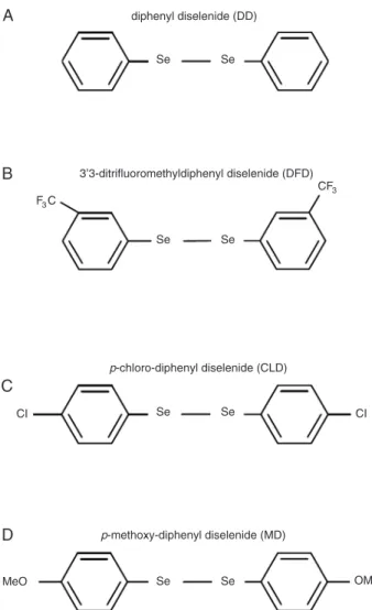
Synthesis and preparation of organoselenium compounds
All organoselenium compounds (Figure 1) tested in the present study were prepared as previously described (4). Analysis of the hydrogen-1 nuclear magnetic resonance
(1H NMR) and carbon-13 NMR (13C NMR) spectra showed
that all compounds presented analytical and spectroscopic data in full agreement with their assigned structures (data
not shown). The diselenides were purified by flash chro
-matography on silica gel (hexane) and identified by (1H
NMR), 13C NMR and gas chromatography-mass
spec-trometry (GCMS), which revealed homogeneous product (data not shown). Compounds were dissolved in ethanol
immediately prior to use in each assay. The final ethanol
in each experiment was 0.1%, and did not affect any of the parameters analyzed when compared to a control sample without ethanol (data not shown). We also investigated the basal activity of the compounds tested for the parameters
analyzed in this study. We did not find significant changes
when comparing the effects of compounds alone in all experiments performed (data not shown).
Preparation of mouse brain mitochondrial-enriched fractions
All procedures involving animals were performed ac-cording to the Animal Care Guidelines from the National Institutes of Health of the United States of America, and all procedures were approved by the Universidade Federal de Santa Catarina Ethics Committee for animal use (313/ CEUA; 23080.026023/2004-39/UFSC). Mouse brain
chondrial-enriched fractions were prepared as described
previously (24). Briefly, adult (8-10 weeks) male Swiss mice were sacrificed by decapitation. The whole brain (minus
the cerebellum) was removed and homogenized on ice in 10 volumes of isolation medium (10 mM HEPES buffer, pH 7.0, containing 220 mM mannitol, 68 mM sucrose, 10 mM KCl, and 0.1% serum albumin) and the homogenate was
centrifuged at 4°C for 10 min at 1000 g. The supernatant
was then centrifuged at 17,500 g for 10 min at 4°C,
provid-inga myelin-rich supernatant and a pellet (P2) consisting
of synaptosomes and free (extra-synaptosomal) mito-chondria. The supernatant was discarded, and the pellet was suspended in the isolation medium without albumin. The samples were kept on ice until the experiments were performed, usually within 10-15 min.
Incubations
P2 (2 mg protein) was incubated with different concentra-tions of MeHg (0, 10, 30, and 100 µM) diluted in incubation buffer, and/or selenium compounds (1, 10, 30, and 100 µM) in a incubation medium containing 10 mM HEPES buffer, pH 7.0, 220 mM mannitol, 68 mM sucrose, and 10 mM KCl (total incubation volume = 300 µL). In previous studies, we found a concentration of about 10 µM Hg in the brain of mice treated orally with 40 mg/L MeHg in drinking water (24). Considering the high amount of protein (2 mg), in the present study we used concentrations of MeHg up to 100 µM in order to obtain clear detection of all parameters
tested during the in vitro assays. Incubations were carried
out at 25°C. After incubation, mitochondrial dehydrogenase activity, GSH content, total hydroperoxides, and lipid per-oxidation (TBARS) were determined. Parallel experiments with the presence of catalase (200 U) were also carried
out in order to test the role of hydrogen peroxide (H2O2)
in the mechanisms of toxicity and protection of MeHg and organoselenium compounds, respectively.
Assessment of mitochondrial activity
Mitochondrial activity was assessed by the conversion
of the MTT dye to formazan (17). Thisassay is based on
the ability of mitochondrial enzymes to metabolize MTT
into formazan, a reaction thattakes place only in
function-ally intact mitochondria. Briefly, samples (300 µL) were
incubated for 30 min at 25°C. Thepurple formazan
crys-tals were pelleted by centrifugation, andthe supernatant
was discarded. The pellets were dissolved in DMSOand
the formazan was quantified spectrophotometrically by
absorbance measurements at 550 nm. Data are reported as percentage of control. Selenium compounds alone did not interfere with the MTT method described here (data not shown).
Assessment of glutathione andhydroperoxide
content and lipid peroxidation
Glutathione content was measured as nonprotein thiols
according to a method previously described (25). After treat-ment with different concentrations (10, 30, and 100 µM) of MeHg for 30 min, 300 µL 10% trichloroacetic acid was added to the samples (300 µL). After centrifugation (4000
g at 4°C for 10 min), the protein pellet was discarded and
free thiol groups were determined in the clear supernatant (which was neutralized with 0.1 M NaOH) by the method of Ellman (25). The total hydroperoxide content was assessed using the xylenol orange method (24) that allows the detec-tion of hydrogen peroxide as well as lipid hydroperoxides.
Briefly, samples were incubated in the medium described
above for 60 min at 25 ± 1°C in the presence or absence of MeHg and/or selenium compounds. Then, the xylenol
orange reagent, containing 0.25 mM Fe(NH4)2(SO4)2,
0.25 mM xylenol orange, and 110 mM perchloric acid, was added to the incubation medium. After 30 min, absorbance was recorded at 560 nm and compared to a hydrogen/ cumene peroxide standard curve. Selenium compounds alone did not interfere with the method described here (data not shown).
The lipid peroxidation end-products were determined by the TBARS assay originally described by Ohkawa et al. (26). After 60 min of incubation as described for the xylenol orange method, samples were incubated with 0.45 M acetic acid/HCl buffer, pH 3.4, 0.28% thiobarbituric acid, 1.2% SDS, and thereafter at 95°C for 60 min to promote color reaction, measured at 532 nm. Malondialdehyde (0 to 3 nmol/mL) was used as a standard. Protein concentra-tion was determined by the method of Bradford (27) using bovine serum albumin as standard.
Assessment of glutathione peroxidase (GPx)-like activity of organodiselenides
The GPx-like activity of the organoselenium compounds was determined using the coupled assay described by Wendel (28), which indirectly monitors the consumption of NADPH at 340 nm. The GPx/GPx-like compounds use GSH to reduce tert-butylhydroperoxide, producing GSSG, which is readily reduced to GSH by excess glutathione reductase (GR), thus consuming NADPH.
Statistical analysis
Statistically significant differences among groups were
analyzed by one-way ANOVA followed by the Duncan multiple range test when appropriate. Differences were
considered to be statistically significant when P < 0.05.
Results
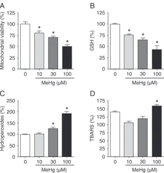
MeHg induces oxidative stress and reduction of mitochondrial metabolic activity
The toxicity of MeHg has been demonstrated in in
Organoselenium compounds block methylmercury neurotoxicity 1159
of GSH levels in the cell (15,16). Moreover, mitochondria are an important cellular target for MeHg toxicity (17). In the present study, as shown in Figure 2A, using the MTT reduction test, MeHg decreased the mitochondrial activ-ity in a concentration-dependent manner. This effect was
significantly different from control at concentrations as low
as 10 µM and a reduction of about 50% in cell viability was
verified at the highest concentration (100 µM). This effect
was followed by a concentration-dependent decrease in mitochondrial GSH levels (Figure 2B). In parallel, a
sig-nificant increase in total-hydroperoxide production (Figure
2C) and increased lipid peroxidation were observed with 100 µM MeHg (Figure 2D). The following experiments were performed using 100 µM MeHg, which affected all parameters analyzed.
Protective effects of organoselenium compounds against MeHg-induced mitochondrial oxidative stress
We investigated the potential protective effects of organoselenium compounds against the decrease in mitochondrial activity promoted by MeHg, using the MTT reduction assay. As observed in Figure 3A, DD at concen-trations of 30 and 100 µM was able to partially and totally
reverse the effect of 100 µM MeHg on mitochondrial activity, respectively. Only the highest concentration of DFD (100 µM) caused a slight reversal of the MeHg-induced reduc-tion of mitochondrial activity (Figure 3B), while CLD and MD did not prevent the effects of MeHg on mitochondrial function (Figure 3C and D).
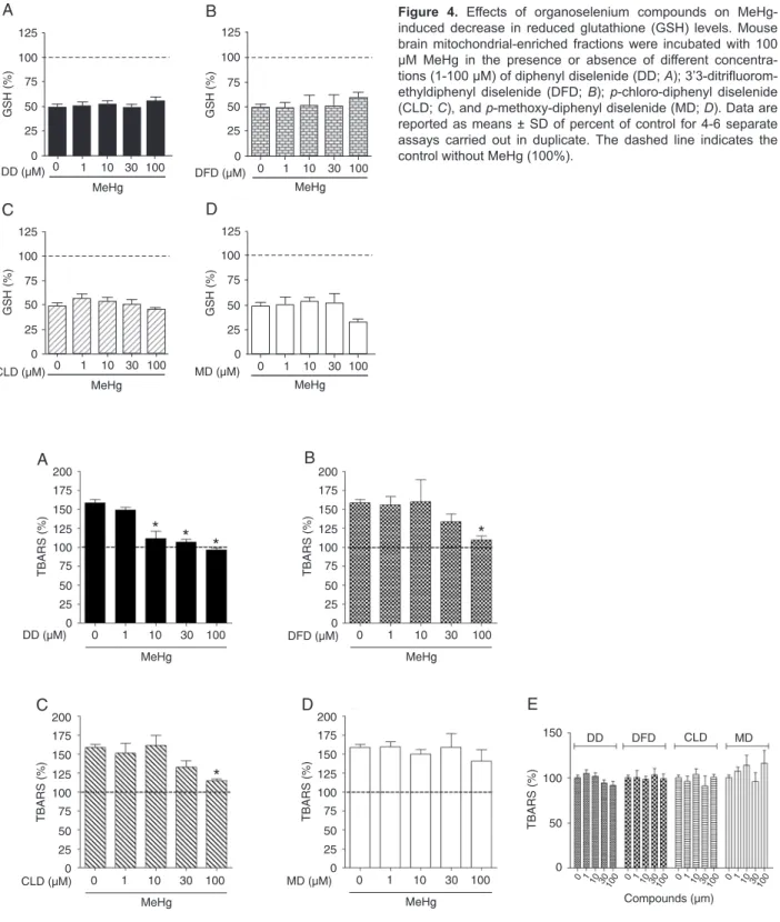
Considering the significant decrease in GSH levels
promoted by MeHg treatment, we investigated whether co-incubation of MeHg in the presence of organoselenium compounds would be able to modulate the decrease in GSH levels caused by this metal. As observed in Figure 4 (A-D), none of the compounds tested was able to protect against the depletion of GSH levels caused by MeHg. In fact, when MD and MeHg were co-administered (Figure 4D), the decrease of GSH levels was greater than in the presence of MeHg alone, although this effect was not
sta-tistically significant (P = 0.08).
The antioxidant potential of the different organoselenium compounds against lipid peroxidation induced by MeHg was investigated by determining TBARS levels. As observed in Figure 5A, DD totally blocked the increase in lipid peroxi-dation induced by MeHg at a concentration as low as 10
Figure 2. Effects of MeHg on brain mitochondria. Mouse brain mitochondrial-enriched fractions were isolated and incubated with different concentrations (0, 10, 30, and 100 µM) of MeHg for 30 min (MTT test and GSH measurement) or 60 min (total-hydroperoxide and TBARS content). Data are reported as means ± SD of percent of control for 4-6 separate assays carried outin duplicate. *P < 0.05 compared to control (without MeHg; ANOVA and Duncan post hoc test). A, Mitochondrial activity; B, GSH lev-els; C, total-hydroperoxides content; D, production of thiobarbitu-ric acid reactive substances (TBARS).
Figure 3. Effects of organoselenium compounds on MeHg-induced decrease in mitochondrial activity. Mouse brain mito-chondrial-enriched fractions were incubated with 100 µM MeHg in the presence or absence of different concentrations (1-100 µM) of diphenyl diselenide (DD; A); 3’3-ditrifluoromethyldiphenyl
diselenide (DFD; B); p-chloro-diphenyl diselenide (CLD; C), and
Figure 4. Effects of organoselenium compounds on MeHg-induced decrease in reduced glutathione (GSH) levels. Mouse brain mitochondrial-enriched fractions were incubated with 100 µM MeHg in the presence or absence of different concentra-tions (1-100 µM) of diphenyl diselenide (DD; A); 3’3-ditrifluorom -ethyldiphenyl diselenide (DFD; B); p-chloro-diphenyl diselenide (CLD; C), and p-methoxy-diphenyl diselenide (MD; D). Data are reported as means ± SD ofpercent of control for 4-6 separate assays carried outin duplicate. The dashed line indicates the control without MeHg (100%).
Organoselenium compounds block methylmercury neurotoxicity 1161
µM. However, DFD and CLD (Figure 5B and C) blocked the increase in lipid peroxidation promoted by MeHg only at the highest concentration (100 µM), while MD did not demonstrate a protective potential against lipid peroxidation
induced by MeHg (Figure 5D). There was no significant
TBARS induction by compounds alone (Figure 5E).
Glutathione peroxidase-like activity of organoselenium compounds
In a previous report (24), our group demonstrated that
H2O2 generation represents a relevant event in
MeHg-mediated oxidative stress in mouse brain mitochondria. GPx (EC 1.11.1.9) is a main cellular antioxidant responsible for the removal of peroxides in the brain (29). Thus, consider-ing that GPx-like activity of organoselenium compounds is potentially involved in their antioxidant properties, we
investigated the in vitro GPx-like activity of each compound.
As observed in Figure 6A, DD demonstrated a higher GPx-like activity when compared to the other compounds tested. The order of magnitude for GPx-like activity was DD > DFD > CLD > MD. In parallel experiments, in order to
confirm whether the anti-peroxidative activity of DD is linked
to its peroxide removal ability, we incubated mouse brain mitochondrial-enriched fractions with MeHg in the presence or absence of DD and catalase, an enzyme involved in the
clearance of H2O2. As shown in Figure 6B, MeHg-induced
lipid peroxidation was completely reversed by catalase
(200 U) as well as 100 µM DD, confirming that removal of
peroxides is an important mechanism responsible for the
protective effects of DD in our study model.
Discussion
In the present study, we used isolated brain mitochondria as a model to investigate MeHg toxicity, since this organelle represents a major target for MeHg in cells and plays a piv-otal role in the initiation of biochemical cascades that lead to cell death (30,31). The effects of MeHg on mitochondrial function are associated with loss of the regular organiza-tion of the cristae (32) and dissipaorganiza-tion of mitochondrial membrane potential (18,17,33).
The acute treatment of mitochondrial-enriched frac-tions from mouse brain with MeHg caused a decrease in mitochondrial activity, in agreement with previously reported results for kidney, brain and striatal mitochondrial fractions (17,24,34). This effect occurred in parallel to an increase in lipid peroxidation and GSH depletion. The relationship between ROS formation and mitochondrial damage after MeHg exposure is not fully understood. ROS can cause oxidative damage to mitochondria, leading to compromised mitochondrial function (35,36). On the other hand, ROS can also be produced by the mitochondria via leakage of electrons from the electron transport chain to molecular
O2, forming superoxide anion radicals (O2.-). The O2.- is
converted to H2O2 by the mitochondrial enzyme manganese
superoxide dismutase (MnSOD) (37). Although this process
occurs normally at a low rate in intact mitochondria, O2
.-production can be dramatically increased if mitochondria
are challenged by toxicants (38). It was previously dem-onstrated that treatment with MeHg causes an increase in
H2O2 generation as well inhibition of GPx activity (15,16,39),
which could be responsible in part for the increase in lipid
peroxidation observed in our model. We confirmed the
participation of H2O2 formation in MeHg toxicity when we
incubated the brain mitochondria with catalase. This H2O2
detoxifying enzyme was able to ameliorate the increase in lipid peroxidation promoted by MeHg, which points to
an involvement of H2O2 in the lipid peroxidation promoted
by MeHg and suggests that the GPx-like activity of DD is involved in the antioxidant effect against MeHg.
DD (30 and 100 µM) protected mouse brain mitochon-dria against MeHg toxicity by reversing the MeHg-induced loss of mitochondrial activity/viability. Among the novel organoselenium compounds tested here, only DFD partially reversed the effect of MeHg on mitochondrial activity at the highest concentration (100 µM). The co-treatment with DD completely blocked TBARS production by MeHg. This effect was observed from 10 µM up to 100 µM of this compound. The novel organodiselenides DFD and CLD were able to reverse the increase in lipid peroxidation promoted by
MeHg only at 100 µM, emphasizing a higher efficiency of
DD as a protective antioxidant, as demonstrated in previ-ous studies (6-8).
GPx mimetic compounds can degrade hydroperox-ides, consuming thiol reserves. Such ability confers to these compounds the capacity of protecting cells against oxidative stress conditions (7). The GPx-like activity of DD
was significantly higher than the activity of DFD, CLD and
MD, an effect possibly related to the higher antioxidant and protective effects of this compound. This result was
confirmed by the fact that incubation of samples with
catalase, which removes H2O2, avoided TBARS formation
induced by MeHg exposure. These data suggest that the protective and antioxidant actions of DD are linked to its ability to remove peroxides. MeHg is known to increase
H2O2 formation by mitochondria (24,34). In this regard,
the GPx mimetic activity of DD may represent a promis-ing tool against the cytotoxic effects of this environmental neurotoxin. The lack of protective effect of the
organo-diselenides DFD, MD and CLD may be related to their lower peroxidase-like activity compared to DD. A recent study from our group has shown the central role of GPx in the toxicity of MeHg (40). In that study, we showed that MeHg was able to decrease GPx activity in cell and animal models. In addition, the inhibition of GPx activity with mercaptosuccinic acid increased cell susceptibility to the toxic effects of methylmercury. On this basis, it seems plausible that DD, which showed higher thiol peroxidase activity than the other three substituted diselenides tested here, had the most prominent protective effects against methylmercury-induced oxidative stress and loss of
mi-tochondrial activity in vitro. In addition to the antioxidant
properties of selenium compounds, the ability to bind Hg ions may represent an important mechanism for cytopro-tection. In fact, a recent study from our group showed that DD is able to remove Hg from tissues (22), reinforcing the therapeutic potential of selenium compounds against Hg intoxication.
The data reported herereinforce the antioxidant and
pro-tective potential of DD when comparing to other substituted
organodiselenides. Our findings support the fact that com -pounds with glutathione peroxidase-like activity are potent blockers of mercurial-induced neurotoxic actions. In addition, our data indicate that depending on the chemical substitutions made on DD, its GPx-like activity may be impaired, which is crucial for the protective capacity of the compound. Con-sidering that oxidative stress has been implicated in MeHg toxicity and that there are no effective treatments available to counteract the toxic effects of MeHg, the use of DD may represent an important therapeutic approach.
Acknowledgments
Research supported by a CNPq grant to T. Posser, a FAPERGS grant to J.L. Franco, CNPq grants to D.F. Meinerz and M.T. de Paula (#140030/2011-5 and #556081/2010-2, respectively), a FAPERGS-PRONEX grant to J.B.T. Rocha, CNPq and FAPESC grants to M. Farina and A.L. Dafre, and FINEP research grants “Rede Instituto Brasileiro de Neurociência (IBN-Net)”.
References
1. Finkel T, Holbrook NJ. Oxidants, oxidative stress and the biology of ageing. Nature 2000; 408: 239-247.
2. Mugesh G, Singh H. Synthetic organoselenium compounds as antioxidants: glutathione peroxidase activity. Chem Soc Rev 2000; 29: 347-357.
3. Arteel GE, Sies H. The biochemistry of selenium and the glutathione system. Environ Toxicol Pharmacol 2011; 10: 153-158.
4. Paulmier C. Selenium reagents and intermediates in organic
synthesis. Oxford: Pergamon Press; 1986.
5. Nogueira CW, Zeni G, Rocha JB. Organoselenium and
organotellurium compounds: toxicology and pharmacology.
Chem Rev 2004; 104: 6255-6285.
6. Farina M, Frizzo ME, Soares FA, Schwalm FD, Dietrich MO, Zeni G, et al. Ebselen protects against methylmercury-induced inhibition of glutamate uptake by cortical slices from adult mice. Toxicol Lett 2003; 144: 351-357.
7. Posser T, Franco JL, dos Santos DA, Rigon AP, Farina M, Dafre AL, et al. Diphenyl diselenide confers neuroprotection against hydrogen peroxide toxicity in hippocampal slices.
Brain Res 2008; 1199: 138-147.
Organoselenium compounds block methylmercury neurotoxicity 1163
Nogueira CW, et al. Antioxidant effect of diphenyl diselenide against sodium nitroprusside (SNP) induced lipid peroxida-tion in human platelets and erythrocyte membranes: an in
vitro evaluation. Chem Biol Interact 2006; 164: 126-135.
9. Clarkson TW, Magos L, Myers GJ. The toxicology of mer-cury - current exposures and clinical manifestations. N Engl
J Med 2003; 349: 1731-1737.
10. Malm O. Gold mining as a source of mercury exposure in the Brazilian Amazon. Environ Res 1998; 77: 73-78. 11. Myers GJ, Davidson PW, Strain JJ. Nutrient and methyl
mercury exposure from consuming fish. J Nutr 2007; 137: 2805-2808.
12. Aschner M, Syversen T, Souza DO, Rocha JB, Farina M. Involvement of glutamate and reactive oxygen species in methylmercury neurotoxicity. Braz J Med Biol Res 2007; 40: 285-291.
13. Ou YC, White CC, Krejsa CM, Ponce RA, Kavanagh TJ, Faustman EM. The role of intracellular glutathione in methylmercury-induced toxicity in embryonic neuronal cells.
Neurotoxicology 1999; 20: 793-804.
14. Shanker G, Syversen T, Aschner JL, Aschner M. Modulatory effect of glutathione status and antioxidants on methylmer-cury-induced free radical formation in primary cultures of cerebral astrocytes. Brain Res Mol Brain Res 2005; 137: 11-22.
15. Farina M, Cereser V, Portela LV, Mendez A, Porciúncula LO, Fornaguera J, et al. Methylmercury increases S100B content in rat cerebrospinal fluid. Environ Toxicol Pharmacol
2005; 19: 249-253.
16. Mori N, Yasutake A, Hirayama K. Comparative study of activ-ities in reactive oxygen species production/defense system in mitochondria of rat brain and liver, and their susceptibility to methylmercury toxicity. Arch Toxicol 2007; 81: 769-776. 17. Dreiem A, Gertz CC, Seegal RF. The effects of
methylmer-cury on mitochondrial function and reactive oxygen species formation in rat striatal synaptosomes are age-dependent.
Toxicol Sci 2005; 87: 156-162.
18. Araragi S, Kondoh M, Kawase M, Saito S, Higashimoto M, Sato M. Mercuric chloride induces apoptosis via a mitochondrial-dependent pathway in human leukemia cells.
Toxicology 2003; 184: 1-9.
19. Shenker BJ, Guo TL, Insung O, Shapiro IM. Induction of apop-tosis in human T-cells by methyl mercury: temporal relation-ship between mitochondrial dysfunction and loss of reductive reserve. Toxicol Appl Pharmacol 1999; 157: 23-35.
20. Tchounwou PB, Ayensu WK, Ninashvili N, Sutton D. Environ-mental exposure to mercury and its toxicopathologic implica-tions for public health. Environ Toxicol 2003; 18: 149-175. 21. Moretto MB, Funchal C, Santos AQ, Gottfried C, Boff B, Zeni
G, et al. Ebselen protects glutamate uptake inhibition caused by methyl mercury but does not by Hg2+. Toxicology 2005; 214: 57-66.
22. de Freitas AS, Funck VR, Rotta MS, Bohrer D, Morsch-bacher V, Puntel RL, et al. Diphenyl diselenide, a simple organoselenium compound, decreases methylmercury-induced cerebral, hepatic and renal oxidative stress and mercury deposition in adult mice. Brain Res Bull 2009; 79: 77-84.
23. Roos DH, Puntel RL, Santos MM, Souza DO, Farina M,
No-gueira CW, et al. Guanosine and synthetic organoselenium compounds modulate methylmercury-induced oxidative stress in rat brain cortical slices: involvement of oxidative stress and glutamatergic system. Toxicol In Vitro 2009; 23: 302-307.
24. Franco JL, Braga HC, Stringari J, Missau FC, Posser T, Mendes BG, et al. Mercurial-induced hydrogen peroxide generation in mouse brain mitochondria: protective effects of quercetin. Chem Res Toxicol 2007; 20: 1919-1926. 25. Ellman GL. Tissue sulfhydryl groups. Arch Biochem Biophys
1959; 82: 70-77.
26. Ohkawa H, Ohishi N, Yagi K. Assay for lipid peroxides in animal tissues by thiobarbituric acid reaction. Anal Biochem
1979; 95: 351-358.
27. Bradford MM. A rapid and sensitive method for the quantita-tion of microgram quantities of protein utilizing the principle of protein-dye binding. Anal Biochem 1976; 72: 248-254. 28. Wendel A. Glutathione peroxidase. Methods Enzymol 1981;
77: 325-333.
29. Dringen R, Pawlowski PG, Hirrlinger J. Peroxide detoxifica-tion by brain cells. J Neurosci Res 2005; 79: 157-165. 30. Aschner M, Syversen T. Methylmercury: recent advances in
the understanding of its neurotoxicity. Ther Drug Monit 2005; 27: 278-283.
31. Atchison WD, Hare MF. Mechanisms of methylmercury-induced neurotoxicity. FASEB J 1994; 8: 622-629.
32. O’Kusky J. Methylmercury poisoning of the developing nervous system: morphological changes in neuronal mito-chondria. Acta Neuropathol 1983; 61: 116-122.
33. Stavrovskaya IG, Kristal BS. The powerhouse takes control of the cell: is the mitochondrial permeability transition a vi-able therapeutic target against neuronal dysfunction and death? Free Radic Biol Med 2005; 38: 687-697.
34. Lund BO, Miller DM, Woods JS. Studies on Hg(II)-induced H2O2 formation and oxidative stress in vivo and in vitro in rat kidney mitochondria. Biochem Pharmacol 1993; 45: 2017-2024.
35. Galindo MF, Jordan J, Gonzalez-Garcia C, Cena V. Reactive oxygen species induce swelling and cytochrome c release but not transmembrane depolarization in isolated rat brain mitochondria. Br J Pharmacol 2003; 139: 797-804. 36. Radi R, Cassina A, Hodara R, Quijano C, Castro L.
Peroxyni-trite reactions and formation in mitochondria. Free Radic Biol Med 2002; 33: 1451-1464.
37. Shanker G, Aschner JL, Syversen T, Aschner M. Free radical formation in cerebral cortical astrocytes in culture induced by methylmercury. Brain Res Mol Brain Res 2004; 128: 48-57.
38. Halliwell B, Gutteridge JMC. Free radicals in biology and
medicine. New York: Oxford University Press; 1999.
39. Carvalho MC, Franco JL, Ghizoni H, Kobus K, Nazari EM, Rocha JB, et al. Effects of 2,3-dimercapto-1-propanesulfonic acid (DMPS) on methylmercury-induced locomotor deficits and cerebellar toxicity in mice. Toxicology 2007; 239: 195-203.