Urethroplasty using a bovine
pericardium graft: an experimental
study using normal urethras from dogs
1Departamento de Urologia, Faculdade de Medicina do Triângulo Mineiro,
Uberaba, MG, Brasil
2Divisão de Urologia, Faculdade de Medicina, Universidade de São Paulo,
São Paulo, SP, Brasil R.C. Lara1,
A.M. Lucon2
and S. Arap2
Abstract
The use of bovine pericardium as a urethral patch to substitute a ventral segment of canine urethras was studied. Healing, epithelial growth, urethral permeability, fistulas, and calcification were ana-lyzed. Thirty male mongrel dogs of medium and large size underwent resection of a ventral segment of the medial urethra measuring 2.0 x 0.5 cm, which was replaced with a bovine pericardium graft, treated with buffered glutaraldehyde and preserved in formaldehyde. Two running sutures of polygalactin 5-0 were applied, one on each side of the patch. The corpus spongiosum was closed with uninterrupted suture and the skin with interrupted suture of polygalactin 5-0. Six months later, the animals were examined and sacrificed under anesthe-sia. Retrograde urethrograms showed that the urethral healing was complete in six of the 30 animals, without stenosis, fistulas or dila-tions. Microscopic examination showed complete epithelization of these six urethras. The remaining 24 animals presented urethrocutane-ous fistulas without stenosis, demonstrated by urethral catheterism using a 10-Fr plastic catheter. These data show that a successful urethral reconstruction of the penile urethra was possible in only 20% of the operated animals. Infection and leakage may be the cause of the urethrocutaneous fistulas present in 80% of cases. Further studies are necessary to determine whether such fistulas are avoidable. If they are, the bovine pericardium may well be an option in the treatment of urethral lesions in dogs.
Correspondence
R.C. Lara
Rua Constituição, 788, Sala 203 38026-280 Uberaba, MG Brasil
Fax: +55-34-3312-8181 E-mail: [email protected] Publication supported by FAPESP.
Received October 15, 2002 Accepted November 6, 2003
Key words
•Urethra •Urethral stricture •Bovine pericardium
Introduction
Several kinds of tissue have been used to correct complex urethral lesions: vein (1), vermiform appendix (2), uterine tube (3), ureter (4), skin (5), bladder mucosa (6), buc-cal mucosa (7), dura mater (8), peritoneum (9), tunica vaginalis (10), and preputial is-land flap (11). The best results have been
achieved with preputial island flaps in one-stage surgeries for the construction of neourethras (12,13). Nevertheless, no ideal tissue for urethral replacement has been iden-tified.
results and without immunological rejection or infection. Bovine pericardium may also be a good option for urinary tract reconstruc-tion because of its availability and low cost. In the present investigation we studied its use in reconstructions of urethral defects experimentally induced in dogs.
Material and Methods
The study was approved by the Ethics Committee of the University of São Paulo School of Medicine. Thirty male mongrel dogs of medium and large size were operated upon.
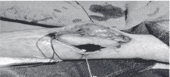
The animals were anesthetized with 3% sodium pentobarbital administered in frac-tionated doses as needed through a cannu-lated vein of the dog. Each animal received 400,000 IU procaine penicillin, 600,000 IU benzathine penicillin G, plus 40 mg of gen-tamicin. A 10-Fr plastic catheter was intro-duced into the bladder through the urethra. An incision was made in the ventral surface of the penis, the medial urethra was exposed and a segment of 2.0 x 0.5 cm was resected. A patch of Fisics/INCOR® (Fisics Biofísica
Aplicada, S/A, Água Branca, SP, Brazil) bovine pericardium was washed in 0.9% saline and used to correct the defect. Two running sutures of polygalactin 5-0 were
applied, one on each side of the patch (Fig-ure 1). The corpus spongiosum was closed with uninterrupted suture and the skin with interrupted suture of polygalactin 5-0.
The urethral catheter was cut at the end of the penis, fixed with two propylene 3-0 stitches, and removed 10 days later. During this period the animals received 40 mg gen-tamicin a day. They were divided into groups of 5 animals per cage in the animal facility and fed once a day.
After six months, 29 animals were exam-ined under anesthesia and retrograde ure-thrograms were performed on the dogs with no macroscopic urethral defects. Next, the animals were killed and the penis was resected for examination. The dogs with ure-thral defects and urine leakage had their urethras gently catheterized with 10-Fr cath-eters to determine urethral permeability. They were killed and had their penis resected for examination. The specimens were stained with hematoxylin and eosin for microscopic study. One of the animals died of a respira-tory infection four months after the surgery, when a retrograde urethrogram was per-formed followed by penis study.
Results
Retrograde urethrograms showed normal
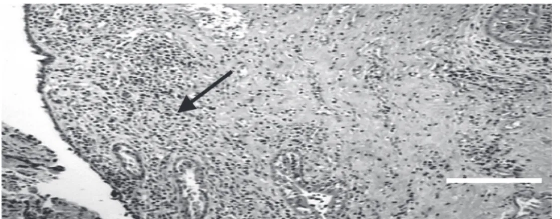
urethras without stenosis, leakage, or dila-tions in six animals. The dog that died four months after surgery was included in this group. A small amount of the iodine contrast injected into the urethras was observed in the corpus spongiosum of all these six dogs. The microscopic study of these urethras showed complete epithelial growth over the graft site with infiltration of mononuclear inflammatory cells in the mucosa. No granu-lomas or calcification were observed (Figure 2). It was only possible to identify the graft in one of these six animals.
Twenty-four dogs had urethral fistulas. The urethral catheterization of these animals was performed easily using 10-Fr catheters. The penises showed fistulas connecting the urethras to the skin of the patch graft. Histol-ogy revealed a normal epithelium with mi-gration of inflammatory cells. There was no evidence of a residual pericardial graft, ac-tive granulation, or calcification in any of the six animals studied.
Discussion
The search for an ideal urethral substitute is a challenge. Several attempts have been made to correct urinary tract system defects using various materials. Memmelaar (19) was the first to describe the use of bladder mucosa to create a neourethra in 1947, and many studies using bladder mucosa were
published since then with varying sucess rates. Complications occur in 12 to 76% of cases. The major ones are meatal and anasto-motic stenosis and urethral fistulas (6,20-22).
Buccal mucosa has also been used for urethral reconstruction. Humby (23) was the first to use buccal mucosal grafts for urethro-plasty in 1941. Buccal mucosa may be used as a patch graft with good results in 89 to 100% of cases, with urethral strictures oc-curring in 11% and fistulas in 6% of the patients. Tubularized buccal mucosal grafts produce successful results in 55 to 60% of the reconstructions, urethral strictures in 40 to 45%, and fistulas in 6 to 45% (7,24-26).
Tunica vaginalis has been used for ure-thral reconstructions in humans, with disap-pointing results. Urethral stricture is present in 60% of the patch grafts and in all tubularized cases (27).
Dura mater has been used for urethral reconstructions and the results vary among series (8,28). Villavicencio et al. (8) reported treatment of 131 patients using dura mater. A patch graft was used in 124 cases and a tubular graft in seven. Average follow-up was 56.6 months and success was obtained in 41% of cases. In the seven patients in which a tubular graft was performed there was a recurrence of the stricture.
The bovine pericardium used for bladder substitution is biodegradable and allows
plete epithelization within 4 to 8 weeks (29-32). The question of bladder muscle regen-eration is controversial. Several reports have shown incomplete muscle regeneration (30,32,33), while others have found no re-generation (29,31).
In the present study the bovine pericar-dium worked as a matrix for normal epithe-lial growth in the urethra of 6/30 dogs. In five of these cases it was found that the urethras were normal. It was only possible to identify the graft in one of these animals. Eighty percent of the cases 24/30 had urethrocuta-neous fistulas. The method used in the pres-ent study permitted no conclusion as to the reason for this occurrence. The use of bovine pericardium in bladder substitution does not result in such a high incidence of complica-tions (29,33-36). Local infection of the ure-thras may be the cause of the fistulas. An-other possibility is that a vesicostomy may be a better technique for urinary diversion than a urethral catheter because it prevents urinary infiltration through the suture lines. In 5 of the 6 successful cases with no fistula there was no residual pericardial graft, which confirms the absorption of the bovine pericardium (29,33-36). Emphasis should be
placed on the fact that the grafts were used to correct urethral defects in normal urethras with good surrounding tissue, which is not the case in urethral strictures.
Urethral reconstruction represents an area of clinical urologic surgery in which the search for an ideal tissue continues. Atala (37) has been carrying out tissue engineering studies. A biopsy of tissue is obtained from the host, and the cells are dissociated and expanded in vitro, attached to a matrix, and
reimplanted in the same host. Studies are being conducted on animals using these tech-niques for the substitution of urethras, blad-ders and ureters (37). Our result of 20% success employing biodegradable materials suggests that this approach is viable but more studies are required.
Acknowledgments
I would like to thank Prof. Dr. Vicente de Paula Antunes Teixeira for contributing to the pathology studies and Luiz Gonzaga Silveira Filho for performing the urethro-grams, both from Faculdade de Medicina do Triângulo Mineiro, Uberaba, MG, Brazil.
References
1. Kahveci R, Kahveci Z, Sirmali S & Özcan M (1995). Urethral recon-struction with autologous vein graft: an experimental study. British Journal of Plastic Surgery, 48: 500-503.
2. Sheldon CA & Gilbert A (1992). Use of the appendix for urethral reconstruction in children with congenital anomalies of the bladder. Surgery, 112: 805-811.
3. Barroso UJ, Duel B, Barthold JS & González R (1999). Orthotopic urethral substitution in female patients using the Mitrofanoff prin-ciple. Journal of Urology, 161: 251-253.
4. Mitchell ME, Adams MC & Rink RC (1988). Urethral replacement with ureter. Journal of Urology, 139: 1282-1285.
5. Valla JS, Takvorian P, Galifer RB, Chavrier Y, Aubert D, Morrisson-Lacombe G, Montupet P, Reinberg O & Dyon JF (1991). Single-stage correction of posterior hypospadia (178 cases). Comparison of three techniques: free skin graft, free bladder mucosal graft, transverse pedicle preputial graft. European Journal of Pediatric Surgery, 1: 287-290.
6. Kinkead TM, Borzi PA, Duffy PG & Ransley PG (1994). Long-term follow-up of bladder mucosa graft for male urethral reconstruction. Journal of Urology, 151: 1056-1058.
7. Andrich DE & Mundy AR (2001). Substitution urethroplasty with buccal mucosal-free grafts. Journal of Urology, 165: 1131-1133. 8. Villavicencio H, Moreno RP, Sariol JC, Briones JR, Bordes AR &
Rodríguez JV (1998). Experience with lyophilized human dura mater for urethral strictures. Journal of Urology, 160: 1310-1311. 9. Shaul DB, Xie HW, Diaz JF, Mahnoviski V & Hardy BE (1996). Use of
tubularized peritoneal free grafts as urethral substitutes in the rab-bit. Journal of Pediatric Surgery, 31: 225-228.
10. Theodorescu D, Balcom A, Smith CR, Mclorie GA, Churchill BM & Khoury AE (1998). Urethral replacement with vascularized tunica vaginalis: defining the optimal form of use. Journal of Urology, 159: 1708-1711.
11. Wiener JS, Sutherland RW, Roth DR & Gonzales Jr ET (1997). Comparison of onlay and tubularized island flaps of inner preputial skin for the repair of proximal hypospadias. Journal of Urology, 158: 1172-1174.
12. Borer JG & Retik AB (1999). Current trends in hypospadias repair. Urologic Clinics of North America, 26: 15-37.
Philadel-phia, PA, USA, 2093-2119.
14. Pomerantzeff PMA, Brandão CMA, Cauduro P et al. (1997). Biopróteses de pericárdio bovino Fisics-Incor: 15 anos. Revista Brasileira de Cirurgia Cardiovascular, 12: 359-366.
15. Grimsley BR, Wells JK, Pearl GJ, Garrett WV, Shutze WP, Talkington CM, Gable DR, Smith BL & Thompson JE (2001). Bovine pericardial patch angioplasty in carotid endarterectomy. American Surgeon, 67: 890-895.
16. Del Campo C & Fonseca A (2001). Replacement of the left common iliac vein with a custom-made bovine pericardium tubular graft. Texas Heart Institute Journal, 28: 39-41.
17. Filippi R, Schwarz M, Voth D, Reisch R, Grunert P & Perneczky A (2001). Bovine pericardium for duraplasty: clinical results in 32 patients. Neurosurgical Review, 24: 103-107.
18. Egydio PH (2000). Correção da deformidade na doença de Peyronie com incisão circunferencial incompleta em duplo y na placa não tubularizada e enxerto de pericárdio bovino. Doctoral thesis, Facul-dade de Medicina, UniversiFacul-dade de São Paulo, São Paulo, SP, Brazil. 19. Memmelaar J (1947). Use of bladder mucosa in one-stage repair of
hypospadias. Journal of Urology, 58: 68-73.
20. Coleman JW, Mcgovern JH & Marshall VF (1981). The bladder mucosal graft technique for hypospadias repair. Urologic Clinics of North America, 8: 457-462.
21. Koyle MA & Ehrlich RM (1987). The bladder mucosal graft for urethral reconstruction. Journal of Urology, 138: 1093-1095. 22. Mollard P, Mouriquand P, Bringeon G & Bugmann P (1989). Repair
of hypospadias using bladder mucosal graft in 76 cases. Journal of Urology, 142: 1548-1550.
23. Humby GA (1941). A one-stage operation for hypospadias. British Journal of Urology, 24: 84-92.
24. Morey AF & McAninch JW (1996). When and how to use the buccal mucosal grafts in adult bulbar urethroplasty. Urology, 48: 194-198. 25. Fichtner J, Fisch M, Filipas D, Thuroff JW & Hohenfellner R (1998).
Refinements in buccal mucosal grafts urethroplasty for hypospa-dias repair. World Journal of Urology, 16: 192-194.
26. Pansadoro V, Emiliozzi P, Gaffi M & Scarpone P (1999). Buccal mucosa urethroplasty for the treatment of bulbar urethral strictures. Journal of Urology, 161: 1501-1503.
27. Joseph DB & Perez LM (1999). Tunica vaginalis onlay urethroplasty as a salvage repair. Journal of Urology, 162: 1146-1147.
28. Valor PC, Garcia-Matres MJ, Alonso-Dorrego JM, Cardoso JVG & Martinez-Piñeiro JA (1995). Uretroplastia con duramadre humana liofilizada. Archivos Españoles de Urología, 48: 681-683.
29. Agishi T, Nakazono M, Kiraly RJ, Picha G & Nosé Y (1975). Biode-gradable material for bladder reconstruction. Journal of Biomedical Materials Research Symposium, 6: 119-131.
30. Novick AC, Straffon RA, Banowsky LH, Nose Y, Levin H & Stewart BH (1977). Experimental bladder substitution using biodegradable graft of natural tissue. Urology, 10: 118-127.
31. Kambic H, Kay R, Chen JF, Matsushita M, Harasaki H & Zilber S (1992). Biodegradable pericardial implants for bladder augmenta-tion: a 2.5 year study in dogs. Journal of Urology, 148: 539-543. 32. Amaro JL, Fabri V, Curi PR & Trindade JCS (2000). Alterações
histológicas na ampliação vesical com dura-máter e pericárdio bovino: estudo comparativo em coelhos. Jornal Brasileiro de Patolo-gia, 36: 178-184.
33. Goldstein MB, Gualtiere V & Getzoff PL (1970). Expansion mechan-isms of the partially cystectomized bladder: an experimental study in rabbits. Journal of Urology, 164: 413-417.
34. Anwar H, Dave B & Seebode JJ (1984). Replacement of partially resected canine urethra by polytetrafluoroethylene. Urology, 24: 583-586.
35. Olsen L, Bowald S, Busch C, Carlsten J & Eriksson I (1992). Urethral reconstruction with a new synthetic absorbable device. Scandina-vian Journal of Nephrology, 26: 323-326.
36. Da Silva EA & Zungri Telo E (2000). Urethral substitution with synthetic material. Actas Urológicas Españolas, 24: 235-242. 37. Atala A (2000). Tissue engineering of artificial organs. Journal of