Stenosis after Jatene Operation for Transposition of Great Arteries
Prevalence and Surgical Approach of Supravalvular Pulmonary Stenosis after Jatene Operation for Transposition
of Great Arteries
Marcelo Biscegli Jatene
1,2, Ieda Biscegli Jatene
2, Patrícia Marques de Oliveira
2, Rafael Aon Moysés
2, Luis Carlos Bento de
Souza
2, Valmir Fontes
2, Nana Miura
1, Antonio Augusto Lopes
1, Miguel Barbero Marcial
1, Adib Domingos Jatene
1,2 Instituto do Coração (InCor) do Hospital das Clínicas da Faculdade de Medicina da Universidade de São Paulo1, Hospital do Coração da Associação do Sanatório Sírio2, São Paulo, SP - BrazilSummary
Background:The Transposition of the Great Arteries is the most frequent congenital cyanogenic cardiopathy in the neonatal period, corresponding to 7% of all congenital cardiopathies. Among the operations for surgical treatment, the Jatene operation,
with arterial correction, is the treatment of choice. During the late postoperative evolution, some problems were observed, with the most common being the occurrence of supravalvular stenosis at the neopulmonary, regardless of the type of surgical
technique used.
Objective:To study and analyze the prevalence of stenosis, as well as describe the surgical treatment and propose technical maneuvers to prevent its onset.
Methods: Of the 553 patients that underwent surgery, 409 were discharged from the hospital and 281 had late follow-up; 59 (20.9%) presented different degrees of supravalvular pulmonary stenosis and 21 had a mean gradient > 60 mmHg, needing
surgical treatment. Depending on the location and anatomy of the stenosis, the surgical treatment consisted of the use of
different techniques, such as the enlargement of stenosis areas with bovine pericardium patches, resection of stenotic areas
and termino-terminal anastomosis, replacement of retracted patches and synthetic tubes.
Results:Twenty patients presented good evolution and only one patient died.
Conclusion:It can be concluded that the supravalvular pulmonary stenosis, post-Jatene operation for Transposition of Great
Arteries, had a prevalence of 20.9%; once identified and with indication for treatment, it can be treated surgically with low mortality levels, through different surgical techniques; to prevent the occurrence of stenosis, ample dissection and release
of the pulmonary branches, double anastomoses, large patches of autologous pericardium and careful reconstruction of the
aorta are proposed, which prevents the compression of the neopulmonary. (Arq Bras Cardiol 2008;91(1):17-23)
Key words:Pulmonary valve stenosis; heart defects, congenital; Jatene’s surgery; transposition of great vessels.
Mailing Address: Marcelo Biscegli Jatene •
Rua João Moura, l535 – Jd. Das Bandeiras - 05412-003 – São Paulo, SP - Brazil
E-mail: [email protected], [email protected]
Manuscript received July 25, 2007; revised received October 28, 2007; accepted December 05, 2007..
Introduction
The transposition of the great arteries (TGA) is the most frequently observed congenital cyanogenic cardiopathy in the neonatal period, corresponding to approximately 7% of all congenital cardiopathies1-4. It is a cardiopathy with a lethal
evolution which, before the appearance of the correction techniques, led 50% of the children to death within the first month of life and more than 90% within the first year2.
The surgeries at atrial level proposed by Senning and Mustard5,6, were the first with satisfactory results. Other
proposals for the correction of TGA7-9 had been described,
until Jatene10 performed for the first time, successfully, the
correction at arterial level in a case of TGA with interventricular communication (IVC).
Under the technical point of view, the concept of Jatene operation aims at inverting the base vessels, making the left ventricle (LV) be in communication with the aorta and the right ventricle (RV) with the pulmonary trunk (PT), in addition to the translocation of the coronary arteries from the aorta to the PT (or neoaorta).
Among the main aspects to be considered during the evolution of patients submitted to Jatene operation, those related to the surgical technique are the noteworthy ones.
The neopulmonary reconstruction is the technical aspect that most commonly presents complications during the evolution, more specifically, pulmonary supravalvular stenosis. Its incidence can vary from 3 to 30%11, with a generally
progressive incidence12.
the present study, the prevalence of this type of complication, as well as aspects related to its surgical correction and the postoperative evolution.
Patients and methods
From April 1975 to December 2000, at the Hospital da Real e Benemérita Sociedade Portuguesa de Beneficência, Instituto Dante Pazzanese de Cardiologia, Hospital do Coração da Associação do Sanatório Sírio and Instituto do Coração (The Heart Institute) of the School of Medicine of the University de São Paulo, 553 children with TGA were submitted to surgical treatment through Jatene operation. The two last institutions were responsible for 96.4% of the operations, respectively: 50.7% and 45.7%.
Of the 409 children (74%) who attained hospital discharge, 281 (68.7%) could be followed during the late evolution, through routine medical visits at the institutions where they were operated or in the medical office of the reference cardiologist.
Of the children that were followed, 59 (20.9%) presented, during the postoperative evolution, varied degrees of pulmonary supravalvular stenosis; considering the 409 children that survived the operation, the incidence was 14.4%. Through the echocardiographic assessment, the systolic gradients between the RV and the PT were considered slight when < 39 mmHg, moderate when between 40 and 59 mmHg and severe when > 60 mmHg. In 22 (37.3%) of the cases, the stenosis was slight, moderate in 14 (23.8%0 and severe in 23 (38.9%).
After the clinical and image assessment (echocardiography and pulmonary angiography through cardiac catheterism), 21 children were submitted to surgical treatment to correct the stenosis and constitute the group to be assessed in the present study.
Six children (28.5%) has associated residual lesions, with IVC in two cases (one with stenosis of the ostium of the left coronary artery) and in the other cases, occlusion of the left coronary, important tricuspid failure, pseudo aneurism of the ascending aorta and interatrial communication (IAC).
In six cases, during the cardiac catheterism, the stenosis dilation by a balloon catheter was attempted without success; these cases were then referred to surgical treatment. A stent was not used in any of the cases during the attempted dilation. The operations for correction of the supravalvular stenosis were carried out from September 1981 to October 2002.
The criteria for surgical indication were: RV/superior pulmonary arteries gradient > 60 mmHg or, when this gradient was lower, there was indication for the correction of another defect. Cases considered and resolved by interventionist catheterism were not included in the present study. The patients that did not meet the criteria for surgical indication remained under clinical follow-up, with periodical assessments to evaluate the progression or not of the stenosis.
Regarding the surgical technique, in 17 of the 21 cases (80,9%) the base vessels were positioned side by side, with the neopulmonary positioned to the right of the neoaorta in 16 cases and, in 1 case, the neopulmonary was positioned
to the left of the neoaorta; in 4 cases (19.1%) the Lecompte maneuver was used, with anteriorization of the PT.
The preoperative RV/PT gradient was 73.2mmHg and varied from 49 to 154mmHg. The mean time between the Jatene operation and the correction of the supravalvular pulmonary stenosis was 109.1 months, varying from 36 and 228 months; in the 17 cases with base vessels located side by side, the time was 114.9 months (37 to 228 months) and in the four cases with Lecompte maneuver, it was 48.6 months (36 to 56 months).
The surgical correction of the supravalvular stenosis was carried out, through different techniques, aiming at the stenosis relief. The techniques to be employed aim at the resection of the stenosed segment and reconstruction with termino-terminal anastomosis and the enlargement of the narrowed site with patches; the eventual use of other techniques to attain stenosis relief will be individualized for each particular case. The surgical access, in all cases, was through the median sternotomy, with release of the adherences caused by the previous operation; the isolation of the neoaorta and neopulmonary and its branches was carried out carefully. The preparation of the surgical field, with the positioning of the extracorporeal circulation (ECC) tubes before opening the sternum, was carried out in all cases.
A membrane oxygenator and moderate hypothermia (between 25o and 28o C) were used in all cases; myocardial
protection with crystalloid cardioplegia was used in the first 8 cases and from 1996 on, a hypothermic blood cardioplegic solution was used.
Regarding the surgical procedures performed, the techniques used for the correction of the supravalvular stenosis can be summarized in four main types:
• enlargement of the areas of stenosis with bovine pericardium patches;
• resection of the stenotic area and termino-terminal anastomosis;
• replacement of a synthetic tube for another with a larger diameter;
• replacement of retracted patches by new ones. Additionally, some maneuvers were carried out concomitantly to the described techniques, such as stretching of the aorta by the interposition of the synthetic tube in the neoaorta or interposition of the patch to increase the length of the neoaorta, specifically in cases with decrease in the retroaortic space, when the vessels were reconstructed in the side by side position. The different types of surgical techniques for the correction of the supravalvular pulmonary stenosis are shown in Table 1.
The associated defects were corrected during the same operation during which the supravalvular pulmonary stenosis was corrected, in a sequence defined by the surgeon in charge of the operation.
Table 1 – Surgical techniques for the correction of supravalvular pulmonary stenosis.
Side by
side Lecompte
Pulmonary trunk enlargement 4 1
Pulmonary trunk and right pulmonary
artery enlargement 2
Pulmonary trunk and left pulmonary
artery enlargement 3
Pulmonary trunk, left and right
pulmonary artery enlargement 3 2
Left pulmonary artery enlargement 3
Resection and termino-terminal
anastomosis 1
Tube replacement 1
Patch replacement 1
Tube or patch in neoaorta 4
Chart 1 – Reoperation-free incidence. Chart 2 – neopulmonary reconstruction.Reoperation-free incidence according to the type of
Statistical analysis
The curves of the reoperation rates were determined through the Kaplan-Meier method and the Log-Rank test was used for the comparison between the curves of events. A significance level was set at 0.05. The numerical data were presented as means and SD, whereas the event incidence rates were presented with a confidence i8nterval of 68%.
Results
After the assessment of 281 patients that had a late follow-up, it was observed that 66% of them were free of reoperations in a period of up to 20 years of evolution (Chart 1).
When analyzing the patients separately by the type of reconstruction used for the base vessels in Jatene operation, it was observed that cases with side-by-side reconstruction presented a higher incidence of reoperations than the cases the Lecompte maneuver. However, there was no statistical significance due to some aspects: small number of cases with Lecompte maneuver that were reoperated, fewer cases that could be followed and shorter time of follow-up. The reoperation-free curves for the two types of base vessel
reconstruction are shown in Chart 2.
One patient (4.7%) died on the first day PO due to ventricular dysfunction and low cardiac output, caused by right coronary lesion during the opening; this patient was an eight year and six-month old child, with eight years of evolution, with segmental pulmonary valvular stenosis that encompassed from the valvular plane to the bifurcation of the pulmonary branches; the correction was carried out with interposition of a Dacron tube to replace the stenosed segment.
The other patients presented a satisfactory immediate postoperative evolution, without major complications; two patients needed reoperation due to postoperative bleeding, with resolution of the picture. During the hospital evolution, one patient presented mediastinal infection and was submitted to reoperation with resuturing of the sternum and mediastinal cleaning, with good evolution.
Regarding the associated procedures, a ventriculospetoplasty was performed in two cases with bovine pericardium patch; in one case, the enlargement of the ostium of the right coronary was performed with bovine pericardium patch; in one case, the myocardial revascularization in the left coronary artery was performed, before the bifurcation in the anterior interventricular and circumflex branches; in one case, the plasty of the tricuspid valve was carried out by the DeVega technique and in another case, the interposition of a Dacron tube was carried out to treat a pseudoaneurism in the ascending neoaorta.
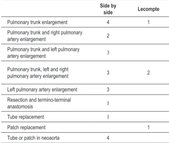
Among the surgical and angiographic findings observed, the occurrence of stenosis located immediately before the bifurcation between the right pulmonary artery (RPA) and the left pulmonary artery (LPA) was one of the types of obstruction. The surgical finding confirmed the angiographic image, reinforcing the impression of scar retraction and growth restriction at the level of the anastomosis. After the longitudinal opening, the correction was performed with the enlargement of the stenosis site with a bovine pericardium patch (Figure 1).
Figure 1 -Angiography with stenosis before the bifurcation and bovine pericardium patch enlarging the pulmonary trunk.
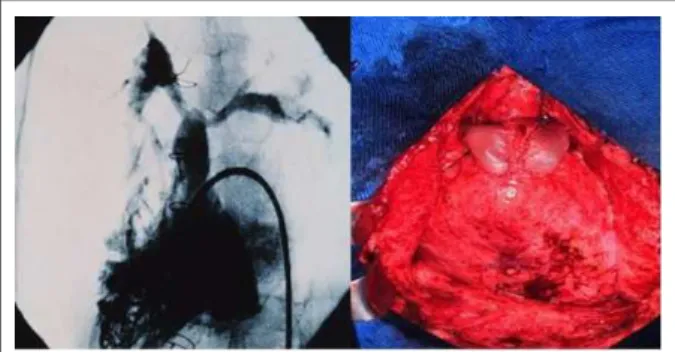
Figure 2 -Compression of the left pulmonary artery and pulmonary trunk by the
aorta and inal aspect after correction with 2 bovine pericardium patches.bovino. Figure 3 -branches and inal aspect after enlargement with two bovine pericardium patches.Angiography showing stenosis at the emergence of the pulmonary isolated plaques in different directions and sutured to each
other, amplifying the LPA in its upper side and the PT in its right anterolateral side (Figure 2). In the cases with Lecompte maneuver, when the pulmonary arteries were not adequately dissected and mobilized, extrinsic compression by the neoaorta can occur with excessive tension in the pulmonary arteries, leading to stenosis at its emergence from the PT. Figure 3 shows the angiographic study of the stenosis in both pulmonary arteries, in addition to a small narrowing in the PT. The surgical correction consisted of the release of the pulmonary arteries and enlargement of the RPA and LPA with isolated bovine pericardium patches (Figure 3).
During the postoperative follow-up period, all 20 patients that were discharged from the hospital remained asymptomatic, with no physical restrictions; from the immediate postoperative period on, assessments of the surgical correction outcome were carried out through annual echocardiographic evaluation in all patients and by nuclear magnetic resonance in 8 of the 20 patients (in the first year of postoperative evolution).
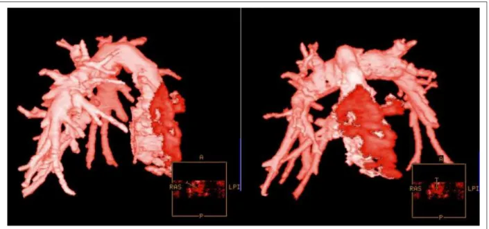
The gradients between the right ventricle and the neopulmonary, assessed by the echocardiogram, varied from 10 to 38 mmHg, with a mean of 24 mmHg, with the outcome maintenance, without progression of the gradient during the evolution. One aspect of the resonance of the patient submitted to the enlargement of the PT and RPA can be seen in Figure 4, where the adequate dimension of the PT and RPA can be observed, without residual stenoses.
Discussion
The original surgical technique described by Jatene, although still currently used by some surgeons, has given place to a pattern of correction that encompasses basically the use of Lecompte maneuver, translocation of the coronary arteries with large fragment of the aortic wall, use of technical
resources (trap-door) to aid the implantation of the coronaries and neopulmonary reconstruction with large fragments of synthetic tissue (Gore-tex), bovine or autologous pericardium (fresh or fixed)12-15.
Despite the currently less frequent use of the original technique, as described in details by Jatene, its use is mandatory in some situations, especially in cases of TGA with vessels located side by side, such as in Taussig Bing anomaly cases.
In this situation, if the intraventricular tunneling proposed by Kawashima et al16 cannot be used, the correction must be
carried out with the closing of the IVC, connecting the left ventricle to the PT and the right ventricle to the aorta and subsequently, the transposition of the great vessels.
Until the end of the 1981, when Lecompte described the maneuver of anteriorization of the PT in the TGA17, all cases
in our series were operated using the original technique, until the proposition by Castaneda et al18 to use the operation to
treat neonates became popular and standardized Lecompte maneuver.
In the first cases of our series, some aspects still needed to be improved, such as the height of the aortic and PT section; in the beginning, the greatest concern was to preserve the aorta, using long proximal and distal stumps to make the reconstruction. As a consequence, the stumps that were left to reconstruct the neopulmonary were short and the solution found was to use a synthetic tube to stretch the neopulmonary.
The price to be paid was the necessity to replace the tube after a variable period of time, as observed in the first case of our series that needed two tube replacements, one at five years and another at 12 years postoperatively, with good evolution. This patient is now in the 29th year of the postoperative period,
has a normal life, is married and has two children.
Fig. 4 -Ressonância nuclear magnética de paciente submetido à ampliação do TP e APD.
probably there will be no compression of the neopulmonary that is usually located to the right of the neoaorta.
On the other hand, if the neoaorta is shorter, in cases with Lecompte maneuver, there will be no compression of the neopulmonary. In cases of vessels positioned side by side, a very short neoaorta will compress the pulmonary artery, usually the LPA, and even the PT, if the retro-aortic space is very small. In our series, it was necessary to elongate the ascending part of the neoaorta in 3 cases, in addition to the correction of the stenosis itself, by the aforementioned techniques.
When the extrinsic compression is backward to forward in cases with the Lecompte maneuver, the positioning of the patches is also important; in our opinion, the enlargements of the RPA, LPA and PT must be carried out in the anterior side of neopulmonary.
If there is a possibility of compression of the enlarged site by the sternum, the patches can be positioned more laterally, to the right or to the left. Serraf, in 16 cases of supravalvular stenosis located at the suture line, describes as treatment the local enlargement with a circular patch of polytetrafluoroethylene, in an attempt not to position the patch at sites that are prone to extrinsic compression by other structures13.
Another important technical aspect refers to how the coronary ostia are removed and the consequent reconstruction of the neopulmonary. When the ostia are removed with a small tissue edge adjacent to the ostial orifice, the reconstruction is carried out with a circular patch occluding the orifice created by the removal of the ostium.
In this situation, almost the entire circumference of the proximal part of the neopulmonary consists of viable tissue and a small circular patch, with growth potential. In spite of that, it was observed in our series that, in three cases where the vessels were positioned side by side and the ostia were removed in this way, there was the development of supravalvular stenosis by patch retraction, associated or not to the adequate growth
of the neopulmonary.
The other technique to remove the coronary ostia is carried out by resecting all the tissue of the sinus of Valsalva where the coronary is located and substituting the sinus wall with a large patch, of slightly larger dimensions than the removed tissue. A study by Prifti et al14 reports the different techniques
of neopulmonary reconstruction, with double patch, single patch or direct anastomosis, with better outcome regarding the occurrence of neopulmonary stenosis in cases where the reconstruction was carried out with single patch. In another study, by Haas et al19, no difference was observed
among the different types of technique or tissue used in the neopulmonary reconstruction.
We believe the best way to reconstruct the neopulmonary after the removal of the ostia is to remove all tissue from the sinus of Valsalva and carry out the reconstruction with a large fresh autologous pericardium patch, removed immediately before the transplantation; we believe that there is growth potential in fresh pericardium, in addition to the large dimensions of the patch, which would leave a large anastomosis with less chance of stenosis occurrence.
Although our series showed a higher incidence of supravalvular stenosis in the cases positioned side by side in comparison with those with Lecompte maneuver, we believe this aspect is due to some factors, in addition to the aforementioned technical-surgical aspects.
Among these factors, we cite the fact that a large number of late stenosis cases occurred in the beginning of the series, when the concepts and surgical tactics were consolidating and all the cases were operated with the vessels positioned side by side, without the Lecompte maneuver, which was not described until 198117.
A multicentric study, published by Williams et al12,
that in addition to instituting worse outcomes, the initial experience is relevant concerning the incidence of stenosis of the PT or branches. As early as in the first reports of mid-term and long-term evolution, Jatene et al20 and Wernovski et al21
reported the occurrence of neopulmonary stenosis, during the follow-up of initial experiences, which varied from 10 to 28% of the operated cases.
A more recent experience described by Haas et al19, in
children operated until 1997, did not show any influence of the type of technique used for the reconstruction of the PT on the occurrence of supravalvular stenosis, with an incidence of around 15%, when most of the cases used the Lecompte maneuver.
Another aspect observed in our series was the occurrence of stenosis in the neopulmonary, predominantly at the supravalvular level, with no reference of subvalvular stenosis and only one case where the pulmonary valve was stenotic, which was not confirmed intraoperatively. The retraction of the tissue used to reconstruct the neopulmonary was observed, which brought together the commissural posts, giving the impression of valvular stenosis; with the resection of the tissue and its replacement by new patches, there was an enlargement of the site and the stenosis was resolved.
A similar experience was reported by Gandhi et al22, who
observed that of 21 patients that developed neopulmonary stenosis, 16 were at the supravalvular level. A study carried out by Weteer et al23 evaluated the development of the neoaorta
and the neopulmonary, in its initial segment, demonstrating a tendency to reduction in the size of the neopulmonary root, with no associated clinical consequences.
We believe it is mandatory to perform a comprehensive intraoperative inspection, trying to analyze the anatomy of the coronary arteries, in addition to the spatial association of the great arteries, aiming a better programming regarding the type of technique to be employed.
In cases where the base vessels need to be positioned side by side, we suggest the following measures:
• adequate release of the pulmonary arteries, performing an ample dissection;
• sectioning and suturing of the ductus arteriosus.
• careful reconstruction of the neoaorta, making it longer to prevent decrease in the retro-aortic space;
• performing ample anastomosis in the neopulmonary, using fresh autologous pericardium patches to reconstruct the proximal stump;
In situations where the Lecompte maneuver is used, with anteriorization of the neopulmonary, we suggest the following measures:
• adequate release of the pulmonary arteries, performing an ample dissection;
• sectioning and suturing of the ductus arteriosus. • approach and effective release of the LPA after the sectioning of the ductus arteriosus;
• careful reconstruction of the neoaorta, making it shorter to prevent backward to forward compression of the neopulmonary.
We can conclude that the pulmonary supravalvular stenosis after the Jatene operation for TGA had a prevalence of 20.9% and that it is related to several technical surgical aspects at the moment of the operation. Different degrees of stenosis were observed; however, we consider its treatment to be indicated in cases with a gradient > 60 mmHg. Operations were carried out with low mortality, through different surgical techniques; in an attempt to prevent the occurrence of stenosis, we propose ample dissection and release of the pulmonary branches, large anastomoses, use of large autologous pericardium patches and careful reconstruction of the neoaorta to prevent compression of the neopulmonary.
Potential Conflict of Interest
No potential conflict of interest relevant to this article was reported.
Sources of Funding
There were no external funding sources for this study.
Study Association
This study is not associated with any graduation program.
Referências
1. Carlgren LE. The incidence of congenital heart disease in children Born in Gothenburg 1941-1950. Br Heart J. 1959; 21 (1): 40-50.
2. Liebman J, Cullum L, Belloc NB. Natural history of transposition of the great arteries. Anatomy and birth and death characteristics. Circulation. 1969; 40 (2): 237-62.
3. Fyler DC. Report of the New England Regional Infant Cardiac Program. Pediatrics. 1989; 65 (Suppl): 376-461.
4. Ferencz C, Rubin JD, McCarter RJ, Brenner JI, Neill CA, Perry LW, et al. Congenital heart disease: prevalence at livebirth. The Baltimore-Washington Infant Study. Am J Epidemiol. 1985; 121 (1): 31-6.
5. Senning A. Surgical correction of transposition of the great vessels. Surgery. 1959; 45: 966-80.
6. Mustard WT. Successful two-stage correction of transposition of the great
vessels. Surgery. 1964; 55: 469-72.
7. Bailey CP, Cookson BA, Downing DF, Neptune WB. Cardiac surgery under hypothermia. J Thorac Cardiovasc Surg. 1954; 27: 73-91.
8. Idriss FS, Goldstein IR, Grana L, French D, Potts WH. A new technic for complete correction of transposition of the great vessels. Circulation. 1961; 24: 5-11.
9. Kay EB, Cross FS. Surgical treatment of transposition of the great vessels. Surgery. 1955; 38: 712-6.
10. Jatene AD, Fontes VF, Paulista PP, de Souza LC, Neger F, Galantier M, et al. Successful anatomic correction of transposition of the great vessels: a preliminary report. Arq Bras Cardiol. 1975; 28: 461-4.
12. Williams WG, Quaegebeur JM, Kirklin JW, Blackstone EH. Outflow obstruction after the arterial switch operation: a mutiinstitutional study; Congenital Heart Surgeons Society. J Thorac Cardiovasc Surg. 1997; 114 (6): 975-87.
13. Serraf A, Roux D, Lacour-Gayet F, Touchot A, Bruaniaux J, Sousa-Uva M, et al. Reoperation after the arterial switch operation switch operation for transposition of the great arteries. J Thorac Cardiovasc Surg. 1995; 110 (4): 892-9.
14. Prifti E, Crucean A, Bonacchi M, Bernabei M, Murzi SVL, Vanini V. Early and long term outcome of the arterial switch operation for transposition of the great arteries: predictors and functional evaluation. Eur J Cardiothorac Surg. 2002; (22): 864-73.
15. Hraska V, Podnar T, Kunovsky P, Kovacikova L, Kaldararova M, Horvathova E, et al. Is a learning curve for arterial switch operation in small countries still acceptable? Model for cooperation in Europe. Eur J Cardiothorac Surg. 2003; (24): 352-7.
16. Kawashima Y, Matsuda H, Yagihara T, Shimazaki Y, Yamamoto F, Nishigaki K, et al. Intraventricular repair for Taussig-Bing anomaly. J Thorac Cardiovasc Surg. 1993; 105: 591-7.
17. Lecompte Y, Zannini L, Hazan E, Jarreau MM, Bex JP, Tu TV, et al. Anatomic correction of transposition of the great arteries: a new technique with out use
of prosthetic conduit. J Thorac Cardiovasc Surg. 1981; 82 (4): 629-31. 18. Castaneda AR, Norwood WI, Jonas RA, Colon SD, Sanders SP, Lang P.
Transposition of the great arteries and intact ventricular septum: anatomical repair in the neonate. Ann Thorac Surg. 1984; 38: 438-43.
19. Haas F, Wottke M, Poppert H, Meisner H. Long term survival and functional follow-up in patients after the arterial switch operation. Ann Thorac Surg. 1999; 68: 1692-7.
20. Jatene FB, Bosisio IBJ, Jatene M, Souza LCB, Barbero Marcial ML, Jatene AD. Late results (50 to 182 months) of the Jatene Operation. Eur J Cardiothorac Surg. 1992; (6): 575-8.
21. Wernovsky G, Hougen TJ, Walsh EP, Sholler GF, Colan Sd, Sanders SP, et al. Midterm results after the arterial switch operation for transposition of the great arteries with intact ventricular septum: clinical, hemodynamic, echocardiographic, and electrophysiologic data. Circulation. 1998; 77 (6): 1333-44.
22. Gandhi SK, Pigula FA, Siewers R. Successful late reintervention after the arterial switch procedure. Ann Thorac Surg. 2002; 73: 88-95.