ISSN 0100-879X
BIOMEDICAL SCIENCES
AND
CLINICAL INVESTIGATION
www.bjournal.com.br
www.bjournal.com.br
Volume 43 (01) 1-123 January 2010
Braz J Med Biol Res, January 2010, Volume 43(1) 36-42
Mechanism of the transforming growth factor-β induction of
fibronectin expression in hepatic stem-like cells
W. Cui, H.B. Jin and Z.W. Li
Faculdade de Medicina de Ribeirão Preto Campus
Ribeirão Preto
Institutional Sponsors
Mechanism of the transforming growth
factor-β induction of fibronectin
expression in hepatic stem-like cells
W. Cui
1,2, H.B. Jin
1and Z.W. Li
11Department of Infectious Diseases, Shengjing Hospital Affiliated to
China Medical University, Shenyang, China
2Department of Pharmacology, Shenyang Pharmaceutical University College of Life Science
and Biopharmaceutical, Shenyang, China
Abstract
Transforming growth factor-β1 (TGF-β1) plays an important role in the fibrogenic process in the liver. The aim of the present study was to explore the action of TGF-β1 on fibronectin expression in rat hepatic stem-like cells and the underlying mechanisms. The level of fibronectin expression was determined in hepatic stem-like cells (WB cells) before and after TGF-β1 stimulation by RT-PCR and Western blot methods. Using immunogold transmission electron microscopy and the Western blot method, we observed the result of the expression and the distribution of cAMP, phosphorylated Smad3 and Smad7 before and after TGF-β1 treatment. The levels of fibronectin expression in both mRNA and protein increased 4- to 5-fold after TGF-β1 stimula
-tion, reaching an optimum level after 8 h and then gradually falling back. Similarly, TGF-β1 stimulation resulted in an increase of cAMP in WB cells, peaking at 8 h. After treatment with TGF-β1 for 24 h, the expression of cAMP gradually decreased. In addition, we found that TGF-β1 treatment also contributed to the increased expression and to changes in cellular distribution
of phosphorylated Smad3 (translocation from the cytoplasm to the nucleus) and Smad7 (translocation from the nucleus to
the cytoplasm) in WB cells. The present study demonstrates that TGF-β is involved in the fibrogenic process in hepatic stem cells through up-regulation of fibronectin expression, and the mechanisms underlying this process may be associated with the
activation of cAMP and Smad pathways.
Key words: TGF-β; Fibronectin; Fibrogenesis; Hepatic stem cell; cAMP; Smad
Introduction
Correspondence: Z.W. Li, Department of Infectious Diseases, Shengjing Hospital Affiliated to China Medical University, Shenyang, 110004 China. Fax: +86-024-2389-2617. E-mail: [email protected]
Received June 26, 2009. Accepted November 4, 2009. Available online November 23, 2009. Published January 11, 2010.
The remarkable regenerative capacity of the human liver is usually provided by the presence of hepatocytes and cholangiocytes (1). Sometimes, when the ability of hepato -cytes to divide and replace damaged tissue is compromised,
bipotent progenitor cells will be activated, giving rise to both new hepatocytes and biliary epithelial cells.
While the function and nomenclature of stem cells lo-cated in the liver is still an area of dispute, it is nevertheless generally agreed that liver oval cells are ‘stem-like’ cells descending from a population of progenitor cells involved in liver regeneration (2,3). More importantly, recent reports hint
that hepatic stellate cell (HSC) activation may contribute to hepatic stem cell behavior (4). Thus, ‘stem-like’ cells in the liver may play a significant role in hepatic fibrogenesis.
It is firmly established that the fibrogenic process in the liver is prominently regulated by transforming growth
factor-β1 (TGF-β1) (5). TGF-β1 signaling is initiated fol
-lowing ligand binding to the TGF-β type II receptor, TβR-II. This allows the recruitment of the TGF-β type I receptor, TβR-I, into a heteromeric complex, resulting in transphos
-phorylation of TβR-I by TβR-II. Following phos-phorylation of Smad2 or Smad3 by the activated TβR-I, a heteromeric
complex is formed with Smad4, resulting in translocation of the complex to the nucleus where the complex can
di-rectly or indidi-rectly, by interactions with other transcription
factors, regulate the involvement of gene transcription in
fibrosis (5-7). Besides HSC, which are induced by TGF-β to transdifferentiate to myofibroblasts and to synthesize an array of extracellular matrix molecules such as fibronectin (FN), hepatocytes are also increasingly recognized as an important cell type involved in the TGF-β-regulated fibro
Hepatic stem-like cell and fibronectin response to TGF-β 37
www.bjournal.com.br Braz J Med Biol Res 43(1) 2010
regulates the fibrogenic process in hepatic stem cells. The rat liver epithelial stem-like WB-F344 cell line, isolated from the liver of an adult male Fischer 344 rat, is considered to be an in vitro model of bipotent oval cells since it shares
their phenotype (8,9). In the present study, using the
WB-F344 cell line, we demonstrate that TGF-β is involved in the fibrogenic process by regulating FN in hepatic stem
cells, and show that the cAMP and Smad pathways may
be required for this process.
Material and Methods
Cell culture
The rat hepatic stem-like cell line WB-F344 was gen
-erously provided by Dr. Han Rui (Chinese Academy of Medical Sciences, Beijing) and maintained in Dulbecco’s modified Eagle’s medium (Life Technologies, Inc., USA) supplemented with 10% FCS, 200 mM glutamine, and 0.05% gentamicin.
Western blot analysis
Cells were treated with 5 ng/mL TGF-β1 for various times (0, 2, 4, 8, 12, and 24 h) followed by incubation in lysis buffer on ice for 30 min. Denatured proteins were then separated by SDS-PAGE, transferred to PVDF mem
-branes, and incubated at room temperature for 1 h or at 4°C overnight with a rabbit-anti-rat FN antibody (1:100; Santa Cruz Biotechnology, USA), or a rabbit monoclonal anti-rat P-Smad3 antibody (1:500; Santa Cruz Biotech
-nology), or a rabbit monoclonal anti-rat Smad7 antibody (1:500; Santa Cruz Biotechnology). After washing in Tris-buffered saline with 0.05% Tween 20 (TBS-Tween), the blots were reacted with alkaline phosphorylated-linked goat anti-rabbit IgG (1:500) at room temperature for 2
h. Pre-stained molecular mass was used to estimate the position of various proteins on the gel. Blots were stripped
and incubated with a monoclonal antibody against β-actin (1:1000; Santa Cruz Biotechnology) to confirm that an equal amount of protein was loaded. The target was then stained with 5-bromo-4-chloro-indolyl phosphate/ nitroblue tetrazodium and the bands were analyzed with the Tanon-Gis system.
RNA extraction and reverse transcription-PCR After treating WB cells with TGF-β1 (5 ng/mL) for 0, 4,
8, and 24 h, total RNA was extracted from the cells with the TRIzol Reagent (Invitrogen-Life Technologies, Inc., USA) according to manufacturer instructions. RNA was
reverse-transcribed with Superscript II Transcriptase (Invitrogen-Life
Technologies, Inc.) in the presence of oligo-dT and random primers. Primer sequences and PCR conditions are shown
in Table 1. PCR products were analyzed with 1% agarose gels stained with ethidium bromide.
Immunogold transmission electron microscopy After treatment with TGF-β1, WB cells were first col
-lected and prefixed with 2.5% glutaraldehyde in PBS for 120 min, washed three times with 0.1 M PBS, postfixed in 1% osmium tetroxide fixative (Millonigs buffer, pH 7.3) for
30 min and dehydrated in an ethanol series. They were then
saturated and embedded in epoxy resin (Emitech, Ashford, Kent, UK) overnight. The cell blocks were subsequently
cut on a Leica Ultracut machine to an ultrathin thickness of 70 nm. The ultrathin sections were loaded on nickel
nets, which were then blocked for 30 min with 1% bovine serum albumin (BSA) and washed with 0.1 M PBS. The primary antibodies (rabbit monoclonal anti-rat cAMP, 1:80; P-Smad3, 1:80; Smad7, 1:60) were incubated in incubation buffer [1% goat serum (British Biocell International, UK),
0.1% Tween 20 (British Biocell International) and 1% BSA
(British Biocell International) in PBS, adjusted to pH 8.2 by
the addition of sodium hydroxide and hydrochloric acid] for
2 h, and then washed twice with the incubation buffer. Next, the cells were incubated with the secondary gold-labelled antibodies (gold-conjugated goat anti-rabbit IgG 1:25, 6 nm) in the incubating buffer (as above) for 1.5 h and then washed three times in PBS. Finally, the ultrathin sections
were stained with uranyl acetate and areas were selected
for viewing by transmission electron microscopy (TEM).
Results
TGF-β induces fibronectin expression in WB cells In order to investigate the role of TGF-β in the fibrogenic process in hepatic stem cells, we first detected the expres
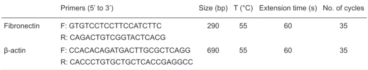
-Table 1. Primers, amplicon size, and protocol details for PCR.
Primers (5’ to 3’) Size (bp) T (°C) Extension time (s) No. of cycles
Fibronectin F: GTGTCCTCCTTCCATCTTC 290 55 60 35
R: CAGACTGTCGGTACTCACG
β-actin F: CCACACAGATGACTTGCGCTCAGG 690 55 60 35
R: CACCCTGTGCTGCTCACCGAGGCC
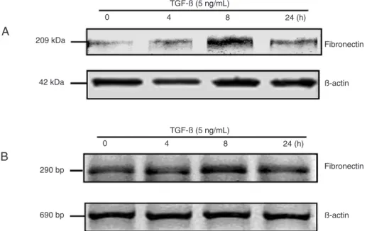
sion of FN, an extracellular matrix glycoprotein, at 0, 4, 8, and 24 h after TGF-β stimulation by Western blot analysis. The results showed that FN levels increased gradually and
reached their optimum yield (5-fold increasing) after 8 h,
decreasing thereafter (Figure 1A). Next, to ascertain the up-regulation of FN transcription induced by TGF-β, we
performed RT-PCR. The results showed a pattern similar to
that of the immunoblot data (Figure 1B). When combined, the results demonstrated that TGF-β1 stimulation can promote FN expression at both the levels of mRNA and protein. Involvement of Smad signaling in TGF-β-induced up-regulation of FN
To determine whether stem cells signal in response
to TGF-β and to explore the potential mechanism of FN
up-regulation, we examined Smad signaling using
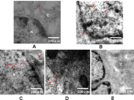
immuno-gold TEM and the Western blot method. The immunoimmuno-gold TEM data showed that, after TGF-β1 treatment, WB cells exhibited a substantial increase in phospho-Smad3 at 4 h (Figure 2B), especially in the nucleus, indicating that stem cells contain sufficient quantities of receptors and Smads to signal in response to TGF-β. Smad3 phosphorylation peaked in WB cells at 8 h, and began to decrease after 24 h (Figure 2C,D). The Western blot results showed a pattern similar to that of immunogold TEM data (Figure 3).
The phospho-Smad3 content increased within the initial 2 h
following TGF-β stimulation and then fell to baseline by 24
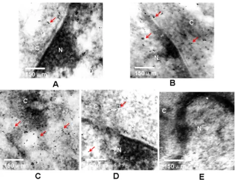
h. In addition, we also observed alteration of Smad7 after TGF-β stimulation in WB cells. There was low expression of Smad7 in WB cells before TGF-β treatment (Figure 4A).
The cytoplasmic expression of Smad7 was increased in WB
cells after TGF-β treatment for 4 h (Figure 4B). Smad7 began to diminish in WB cells at 24 h (Figure 4D). To confirm the data described above, we performed a Western blot. The
content of Smad7 increased within the initial 4 h following
TGF-β stimulation and then fell by 24 h, in agreement with the results of immunogold TEM (Figure 3).
cAMP involvement in TGF-β-induced up-regulation of FN
cAMP is a second messenger that is important in
many biological processes. To determine whether cAMP is involved in the FN up-regulation induced by TGF-β, we determined cAMP expression using immunogold TEM. As shown in Figure 5A, there was low expression of cAMP in the cytoplasm and nucleus of WB cells before TGF-β1
treatment. In contrast, when WB cells were exposed to
TGF-β1 for 4 h, the expressions of cAMP in cytoplasm and nucleus increased significantly (Figure 5B) and peaked after 8 h, especially in the cytoplasm (Figure 5C). After treatment with TGF-β1 for 24 h, the expression of cAMP gradually decreased in WB cells (Figure 5D). The altera
-tions of cAMP induced by TGF-β1 in WB cells had a pattern similar to that of FN.
Figure 1. Effects of stimulation with transforming growth factor-beta (TGF-β) for various times on the expression of fibronectin (FN) in
hepatic stem-like cells. A, Immunoblot analysis of FN protein. FN levels increased at 4 h, reached a peak near 8 h, and decreased after
Hepatic stem-like cell and fibronectin response to TGF-β 39
www.bjournal.com.br Braz J Med Biol Res 43(1) 2010
Figure 3. Effects of stimulation with transforming growth factor-beta (TGF-β) for various times on the expression of phosphorylated
Smad3 and Smad7 in hepatic stem-like cells determined by immunoblot. For phosphorylated Smad3, the content increased within the initial 2 h following TGF-β stimulation and then fell to baseline by 24 h. For Smad7, the expression level increased within the initial 4 h following TGF-β stimulation and then fell by 24 h. The primary antibodies of rabbit monoclonal anti-rat P-Smad3 and anti-rat-Smad7
were diluted five hundred times, and the secondary antibodies of monoclonal antibody against β-actin was diluted one thousand times.
β-actin was used to normalize protein levels.
Figure 2. Electron micrographs of phosphorylated Smad3 expression and cellular distribution before and after stimulation with trans
-forming growth factor-beta (TGF-β) in hepatic stem-like cells. A, Before TGF-β stimulation, weak nuclear and cytoplasmic staining. B,
TGF-β stimulation for 4 h, strong nuclear staining with mediumcytoplasm staining. C, TGF-β stimulation for 8 h, strong nuclear staining
with medium cytoplasm staining. D, TGF-β stimulation for 24 h, with weak cytoplasm staining. E, Negative control, negative nuclear and
cytoplasmic staining. The primary antibody of rabbit monoclonal anti-rat P-Smad3 was diluted eighty times, and the secondary antibody of goat anti-rabbit IgG (6 nm) was diluted twenty-five times. Red arrows indicate positive staining. Magnification: 4000X. N = nucleus; C
Figure 4. Electron micrographs of Smad7 expression and cellular distribution before and after stimulation with transforming growth
factor-beta (TGF-β) in hepatic stem-like cells. A, Before TGF-β stimulation, weak nuclear and cytoplasm staining. B, TGF-β stimulation
for 4 h, strong cytoplasm staining. C, TGF-β stimulation for 8 h, strong cytoplasm staining. D, TGF-β stimulation for 24 h, weak nuclear
and cytoplasm staining. E, Negative control, negative nuclear and cytoplasm staining. The primary antibody of rabbit monoclonal anti-rat Smad7 was diluted sixty times, and the secondary antibody of goat anti-rabbit IgG (6 nm) was diluted twenty-five times. Red arrows
indicate positive staining. Magnification: 4000X. N = nucleus; C = cytoplasm.
Figure 5. Electron micrographs of cAMP expression and cellular distribution before and after stimulation with transforming growth
factor-beta (TGF-β) in hepatic stem-like cells. A, Before TGF-β stimulation, weak nuclear and cytoplasm staining. B, TGF-β stimulation
for 4 h, middle cytoplasm and nuclear staining. C, TGF-β stimulation for 8 h, strong cytoplasm staining. D, TGF-β stimulation for 24 h,
Hepatic stem-like cell and fibronectin response to TGF-β 41
www.bjournal.com.br Braz J Med Biol Res 43(1) 2010
Discussion
Coordinated regulation of the production and turnover
of components of the extracellular matrix (ECM) is essential for normal tissue homeostasis. The composition of the ECM can influence cell growth, state of differentiation and specific gene induction. FN, a major component of the ECM, plays
an important role during development and wound healing
by promoting cell adhesion, migration and cytoskeletal
organization (10,11). Overproduction of matrix components
including FN is the main pathological finding in tissue fibrosis (12). Decreased FN production, however, is often observed
following oncogenic transformation, leading to decreased
adhesion and increased metastatic potential (11). TGF-β, which is released by degranulating platelets at a wound site, has been shown to potently induce FN expression at both the mRNA and protein levels (5,13,14).
Recent research has shown that TGF-β induces liver fibrosis by promoting FN expression. Ha et al. (15) reported that high glucose levels may increase FN expression in peritoneal mesothelial cells, leading to ECM deposition (15). Kitamura and Ninomiya (16) showed that TGF-β1 causes liver fibrosis by promoting Smad 2/3, 4, and 7 expression in
HSC. Schnur et al. (17) used transgenic mice to study the
relationship between TGF-β and liver fibrosis and proved that TGF-β is somehow related to liver fibrosis. These stud -ies demonstrate that HSC and hepatocytes are the major
cells which produce ECM including FN. TGF-β causes HSC and hepatocytes to produce FN and, as a result, leads to liver fibrosis. But it is not known whether TGF-βcan cause
hepatic stem cells to express FN.
Here, we used TGF-βto stimulate hepatic stem-like cells
in vitro and measured FN protein and mRNA expression at different intervals after exposure to TGF-β1. Our results indicated that the expression of FN increased gradually and peaked at 8 h after TGF-βexposure. It is suggested that
TGF-βcan cause ECM deposition by promoting not only HSC and hepatocytes, but also hepatic stem cells during the process of liver fibrosis.
In the presence of the TGF-βligand, the receptor-activated Smads (R-Smads), Smads2 and 3, are
phospho-rylated directly by the TGF-β receptor I kinase, bind to the
common mediator Smad, Smad4, and are translocated into the nucleus (6,7). In a recent experiment using microarrays
to compare gene expression profiles of fibroblasts taken
from adult Smad3-/- and Smad3+/+ mice, TGF-β did not
induce transcription in Smad3-/- fibroblasts (18). Thus, in
adult fibroblasts Smad3 is required for TGF-β-induced gene expression. On the basis of these findings, we measured the expression levels and cellular distribution of phospho -rylated Smad3 to identify the mechanism underlying the
TGF-β-induced up-regulation of FN. We observed that WB cells presented a substantial increase in phospho-Smad3 levels after 4 h, peaking at 8 h, before decreasing after 24 h. These results suggest that phospho-Smad3 might be
involved in the TGF-β-induced up-regulation of FN. Zhao et al. (19) reported that TGF-β induced-phospho-Smad3 reached a peak at 1.5 h and then quickly fell by 3 h in HSC.
Compared with these data, our result is relatively late and
persistent, indicating that the duration of response to TGF-β
is cell-type dependent.
In normal cells, TGF-β directly stimulates the transcrip
-tion of inhibitory Smad7. Thus, Smad7 induc-tion serves as a critical intracellular “brake” of TGF-β-induced responses.
The physiologic importance of this Smad7-mediated
autoin-hibitory feedback loop is highlighted by recent observations
with cultured cells in in vivo animal models. For example, tumor necrosis factor-α (TNF-α)-induced abrogation of TGF-β-dependent profibrotic responses was linked to the
induction of endogenous Smad7 (20). On the other hand, defective Smad7 induction is implicated in the
exagger-ated TGF-β responsiveness characteristic of hepatic cells and myofibroblasts from chronically injured livers (21,22). In the present study, we observed that the expression of Smad7 began to increase after TGF-β treatment for 4 h. These data agree well with previous results obtained for skin fibroblasts (23). In addition, our result showed an obvious Smad7 translocation from nucleus to cytoplasm after treatment with TGF-β for 4 h. In fact, Smad7 has been shown to inhibit TGF-β signaling by associating with the type I receptor and thereby interfering with Smad2/3 phosphorylation by the activated receptor complex in the cytoplasm (6,7). Thus, it is possible that the elevated levels
of Smad7 in the cytoplasm of stem cells provide a means
to inhibit the signaling response to the ligand, which can
prevent the phosphorylation and translocation to the nucleus
of Smad2/3, and consequently inhibit the TGF-β-induced expression of FN. This hypothesis is supported by the finding of the alteration process of phospho-Smad3 and Smad7 induced by TGF-β in WB cells.
In order to determine the mechanism underlying this
response to TGF-β, we also examined the levels of cAMP
in hepatic stem-like cells. cAMP is a second messenger that
has been implicated as a modulator of cell proliferation and cell growth in several fibroblastic cell types (24). Recently, it was demonstrated that increased cAMP levels inhibit cardiac myofibroblast formation and collagen synthesis (25). In addi
-tion, β-adrenergic receptor agonists, which stimulate cAMP production, attenuate the fibroblast-mediated contraction
of collagen gels (26). Nonetheless, in hepatic stem cells,
the role of cAMP in TGF-β-induced FN expression remains undefined. The present study indicates that when WB cells are exposed to TGF-β for 4 h, the expression of cAMP in cytoplasm and nucleus increases significantly and peaks at 8 h, especially in the cytoplasm. After treatment with TGF-β
for 24 h, the expression of cAMP gradually decreased in
-References
1. Taub R. Liver regeneration: from myth to mechanism. Nat Rev Mol Cell Biol 2004; 5: 836-847.
2. Lowes KN, Croager EJ, Olynyk JK, Abraham LJ, Yeoh GC.
Oval cell-mediated liver regeneration: Role of cytokines and growth factors. J Gastroenterol Hepatol 2003; 18: 4-12.
3. Walkup MH, Gerber DA. Hepatic stem cells: in search of.
Stem Cells 2006; 24: 1833-1840.
4. Friedman SL. Mechanisms of hepatic fibrogenesis. Gastro-enterology 2008; 134: 1655-1669.
5. Gressner AM, Weiskirchen R, Breitkopf K, Dooley S. Roles of TGF-beta in hepatic fibrosis. Front Biosci 2002; 7:
d793-d807.
6. Shi Y, Massague J. Mechanisms of TGF-beta signaling from cell membrane to the nucleus. Cell 2003; 113: 685-700.
7. Wells RG. Fibrogenesis. V. TGF-beta signaling pathways.
Am J Physiol Gastrointest Liver Physiol 2000; 279:
G845-G850.
8. Thorgeirsson SS, Grisham JW. Overview of recent experi -mental studies on liver stem cells. Semin Liver Dis 2003; 23:
303-312.
9. Tsao MS, Smith JD, Nelson KG, Grisham JW. A diploid
epithelial cell line from normal adult rat liver with phenotypic properties of ‘oval’ cells. Exp Cell Res 1984; 154: 38-52.
10. Hynes R. Molecular biology of fibronectin. Annu Rev Cell Biol 1985; 1: 67-90.
11. Kornblihtt AR, Pesce CG, Alonso CR, Cramer P, Srebrow A, Werbajh S, et al. The fibronectin gene as a model for splicing
and transcription studies. FASEB J 1996; 10: 248-257.
12. Border WA, Noble NA. Transforming growth factor beta in tissue fibrosis. N Engl J Med 1994; 331: 1286-1292.
13. Friedman SL. Cytokines and fibrogenesis. Semin Liver Dis
1999; 19: 129-140.
14. Gressner AM, Weiskirchen R. Modern pathogenetic con
-cepts of liver fibrosis suggest stellate cells and TGF-beta as
major players and therapeutic targets. J Cell Mol Med 2006;
10: 76-99.
15. Ha H, Yu MR, Lee HB. High glucose-induced PKC activation mediates TGF-beta 1 and fibronectin synthesis by peritoneal
mesothelial cells. Kidney Int 2001; 59: 463-470.
16. Kitamura Y, Ninomiya H. Smad expression of hepatic stellate
cells in liver cirrhosis in vivo and hepatic stellate cell line in vitro. Pathol Int 2003; 53: 18-26.
17. Schnur J, Olah J, Szepesi A, Nagy P, Thorgeirsson SS.
Thioacetamide-induced hepatic fibrosis in transforming growth factor beta-1 transgenic mice. Eur J Gastroenterol
Hepatol 2004; 16: 127-133.
18. Yang YC, Piek E, Zavadil J, Liang D, Xie D, Heyer J, et al. Hierarchical model of gene regulation by transforming growth factor beta. Proc Natl Acad Sci U S A 2003; 100:
10269-10274.
19. Zhao C, Chen W, Yang L, Chen L, Stimpson SA, Diehl AM. PPARgamma agonists prevent TGFbeta1/Smad3-signaling
in human hepatic stellate cells. Biochem Biophys Res Com-mun 2006; 350: 385-391.
20. Bitzer M, von Gersdorff G, Liang D, Dominguez-Rosales A,
Beg AA, Rojkind M, et al. A mechanism of suppression of
TGF-beta/SMAD signaling by NF-kappa B/RelA. Genes Dev
2000; 14: 187-197.
21. Tahashi Y, Matsuzaki K, Date M, Yoshida K, Furukawa F, Sugano Y, et al. Differential regulation of TGF-beta signal in hepatic stellate cells between acute and chronic rat liver
injury. Hepatology 2002; 35: 49-61.
22. Dooley S, Delvoux B, Lahme B, Mangasser-Stephan K,
Gressner AM. Modulation of transforming growth factor beta
response and signaling during transdifferentiation of rat
he-patic stellate cells to myofibroblasts. Hepatology 2000; 31:
1094-1106.
23. Mori Y, Chen SJ, Varga J. Expression and regulation of in
-tracellular SMAD signaling in scleroderma skin fibroblasts.
Arthritis Rheum 2003; 48: 1964-1978.
24. Takahashi H, Honma M, Miyauchi Y, Nakamura S, Ishida-Yamamoto A, Iizuka H. Cyclic AMP differentially regulates
cell proliferation of normal human keratinocytes through
ERK activation depending on the expression pattern of
B-Raf. Arch Dermatol Res 2004; 296: 74-82.
25. Swaney JS, Roth DM, Olson ER, Naugle JE, Meszaros JG, Insel PA. Inhibition of cardiac myofibroblast formation and collagen synthesis by activation and overexpression of
adenylyl cyclase. Proc Natl Acad Sci U S A 2005; 102:
437-442.
26. Mio T, Adachi Y, Carnevali S, Romberger DJ, Spurzem JR, Rennard SI. Beta-adrenergic agonists attenuate
fibroblast-mediated contraction of released collagen gels. Am J Physiol 1996; 270: L829-L835.
27. Ramirez A, Ramadan B, Ritzenthaler JD, Rivera HN, Jones
DP, Roman J. Extracellular cysteine/cystine redox potential controls lung fibroblast proliferation and matrix expression through upregulation of transforming growth factor-beta. Am J Physiol Lung Cell Mol Physiol 2007; 293: L972-L981.
tion of cAMP could result in the phosphorylation of cAMP
response element binding protein (CREB), a transcription factor known for its ability to stimulate FN expression (27). Thus, it is possible that TGF-β-induced up-regulation of FN occurs in part through the cAMP-CREB pathway. However, the detailed roles of cAMP in TGF-β-induced up-regulation
of FN in hepatic stem-like cells merit further investigation. The present study demonstrated for the first time that TGF-β is involved in the fibrogenic process in hepatic stem-like cells through up-regulation of FN expression, and the mechanisms underlying this process seem to be associated