MFG-E8, a clearance glycoprotein of apoptotic cells,
as a new marker of disease severity in chronic
obstructive pulmonary disease
S. Zhang
1*, J.G. Xie
3*, B.T. Su
1, J.L. Li
1, N. Hu
1, J. Chen
1, G.W. Luo
2and T.P. Cui
1 1Laboratory of Clinical Immunology, Wuhan No. 1 Hospital, Tongji Medical College, Huazhong University of Science and Technology, Wuhan, China 2
Department of Respiratory and Critical Care Medicine, Wuhan No. 1 Hospital, Tongji Medical College, Huazhong University of Science and Technology, Wuhan, China 3
Department of Respiratory and Critical Care Medicine, Tongji Hospital, Tongji Medical College, Huazhong University of Science and Technology, Wuhan, China
Abstract
Milk fat globule epidermal growth factor 8 (MFG-E8) is an opsonin involved in the phagocytosis of apoptotic cells. In patients with chronic obstructive pulmonary disease (COPD), apoptotic cell clearance is defective. However, whether aberrant MFG-E8 expression is involved in this defect is unknown. In this study, we examined the expression of MFG-E8 in COPD patients. MFG-E8, interleukin (IL)-1b and transforming growth factor (TGF)-blevels were measured in the plasma of 96 COPD patients (93 males, 3 females; age range: 62.12±10.39) and 87 age-matched healthy controls (85 males, 2 females; age range: 64.81±10.11
years) using an enzyme-linked immunosorbent assay. Compared with controls, COPD patients had a significantly lower plasma MFG-E8 levels (Po0.01) and significantly higher plasma TGF-blevels (P=0.002), whereas there was no difference in plasma IL-1blevels between the two groups. Moreover, plasma MFG-E8 levels decreased progressively between Global Initiative for Chronic Obstructive Lung Disease (GOLD) I and GOLD IV stage COPD. Multiple regression analysis showed that the forced expiratory volume in 1 s (FEV1% predicted) and smoking habit were powerful predictors of MFG-E8 in COPD (Po0.01 and P=0.026, respectively). MFG-E8
was positively associated with the FEV1% predicted and negatively associated with smoking habit. The area under the receiver
operating characteristic curve was 0.874 (95% confidence interval: 0.798–0.95; Po0.01). Ourfindings demonstrated the utility of
MFG-E8 as a marker of disease severity in COPD and that cigarette smoke impaired MFG-E8 expression in these patients. Key words: Apoptosis; COPD; MFG-E8; Phagocytosis; Smoking
Introduction
Chronic obstructive pulmonary disease (COPD) is a primary cause of chronic morbidity and mortality and has become one of the most serious diseases threatening human health throughout the world (1). Despite its prevalence, the precise pathogenesis of COPD is poorly understood. However, many studies have identified the pulmonary inflammatory response to cigarette smoke as the critical mechanism in COPD development. In addition, recent investigations have shown an association between COPD and both increased apoptosis and the reduced clearance of apoptotic cells (2).
Milk fat globule epidermal growth factor 8 (MFG-E8), a protein expressed mainly by activated macrophages and immature dendritic cells, is required for the phagocytosis of
apoptotic cells as it links externalized phosphatidylserine present on the surface of apoptotic cells with integrins expressed on phagocytes (3). MFG-E8 thus plays a critical role in maintaining homeostasis by mediating the removal of apoptotic cells. Mice deficient in MFG-E8 develop a severe autoimmune and inflammatory disease resembling human systemic lupus erythematosus (SLE) (4). The aberrant expression of MFG-E8 has been demonstrated in several diseases, including SLE, atherosclerosis, and Alzheimer’s disease (5–7). A common feature of these diseases is the
defective phagocytic clearance of apoptotic cells. A defi -ciency in the ability of macrophages from patients with COPD to phagocytose apoptotic cells has also been reported (8). However, neither the relationship between MFG-E8 and
Correspondence: Tian-Pen Cui:<[email protected]>.
*These authors contributed equally to this study.
defective efferocytosis in COPD nor the expression and function of MFG-E8 in COPD has been elucidated.
The aims of this study were to investigate the levels of plasma MFG-E8 in patients with COPD and to analyze the potential association between MFG-E8, disease severity, and smoking history.
Material and Methods
Ethics statements
This study was approved by the Ethics Committee of Wuhan No. 1 Hospital and Tongji Hospital, Tongji Medical College, Huazhong University of Science and Technology, Wuhan, China. All participants signed written informed consent, and the research complied with the Declaration of Helsinki and its amendments.
Subjects
The 96 COPD patients and 87 healthy controls were recruited from the Wuhan No. 1 Hospital and Tongji Hospital, between March 2010 and March 2011. COPD was diagnosed according to the Global Initiative for Chronic Obstructive Lung Disease (GOLD) guidelines (1) and classified according to the GOLD criteria as: GOLD I (mild): FEV1(forced expiratory volume in 1 s)/FVC (forced vital capacity) o70%, FEV1 X80% predicted; GOLD II (moderate): FEV1/FVC o70%, 50%p FEV1 o80% predicted, GOLD III (severe): FEV1/FVC o70%, 30%p FEV1 o50% predicted; GOLD IV (very severe): FEV1/FVC o70%, FEV1o30% predicted. The 87 age-matched healthy controls without a smoking history (never smokers) were recruited from the Department of Medical Examination. Participants with a history of bronchial asthma, bronchiectasis, coronary heart disease, hyper-lipidemia, cancer, autoimmune disease, serious brain, kidney or vascular disease, and inflammatory diseases were excluded.
The demographic and clinical characteristics of the patients and controls were obtained, including medical history and smoking habit (pack-years). Pulmonary func-tion parameters (FEV1, FVC, and FEV1/FVC) were measured using a clinical spirometer. Each participant
Statistical analysis
All statistical analyses were carried out using the SPSS software, version 18.0. The data are reported as the mean±SE. Two independent groups with normal and non-normal distributions were analyzed using Student’s t-test and the Mann-Whitney U-test, respectively. The w2 test was used to compare proportions in different groups. One-way ANOVA and the Kruskal-Wallis H test were used to compare more than two groups depending on their distribution status. Multiple linear regression was used to evaluate the relationship between the levels of plasma MFG-E8, different pulmonary function indicators, and confounding factors such as age, body mass index (BMI), and smoking history. A stepwise method was applied to derive the final model. The inclusion and exclusion criteria of independent variables were Pp0.05 and PX0.10, respectively. The receiver operating char-acteristic (ROC) curve for the various MFG-E8 cut-off values, as a predictor of COPD, was also obtained. Youden’s index was calculated (YI=sensitivity+specificity–1) to determine the
cut-off value with the maximum sensitivity and specificity.
Results
Clinical characteristics of the study participants Among the 96 COPD patients, 5 patients had GOLD I, 40 had GOLD II, 34 had GOLD III, and 17 had GOLD IV disease. The baseline characteristics of these patients and the 87 healthy controls are reported in Table 1. The two groups did not differ significantly with respect to age, gender, and BMI. However, the pulmonary function tests revealed a significantly lower FEV1, FVC, FEV1/FVC, and FEV1 % predicted in COPD patients than in the control group (Table 1).
Plasma levels of MFG-E8 in COPD patients vs healthy controls
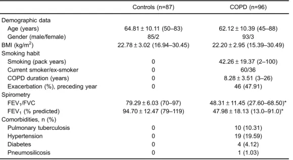
Table 1.Characteristics of the study groups.
Controls (n=87) COPD (n=96)
Demographic data
Age (years) 64.81±10.11 (50–83) 62.12±10.39 (45–88)
Gender (male/female) 85/2 93/3
BMI (kg/m2) 22.78±3.02 (16.94
–30.45) 22.20±2.95 (15.39–30.49) Smoking habit
Smoking (pack years) 0 42.26±19.37 (2
–100)
Current smoker/ex-smoker 0 60/36
COPD duration (years) 0 8.28±3.51 (3–26)
Exacerbation (%), preceding year 0 46 (47.91)
Spirometry
FEV1/FVC 79.29±6.03 (70–97) 48.31±11.45 (27.60–68.50)* FEV1(% predicted) 94.70±12.47 (79–119) 47.98±18.13 (13.0–91.0)* Comorbidities, n (%)
Pulmonary tuberculosis 0 10 (10.31)
Hypertension 0 19 (19.59)
Diabetes 0 4 (4.12)
Pneumosilicosis 0 1 (1.03)
Data are reported as the mean±SE (range) unless otherwise indicated. BMI: body mass index; FEV1: forced expiratory volume in 1 s; FVC: forced vital capacity. *Po0.05vscontrol group (Student’st-test was used to compare two groups and the chi-square test was used to compare proportions in two groups).
Variables associated with plasma MFG-E8
The associations between plasma MFG-E8 levels, pulmonary function indicators, and confounding factors such as BMI and smoking history were investigated using a stepwise multiple regression analysis. The results showed that only two variables, FEV1 % predicted and the amount of smoking, fit the regression equation (Po0.01 and P=0.026, respectively; Table 3). Moreover, plasma MFG-E8 levels were positively associated with the FEV1% predicted (Figure 2A) and negatively associated with smoking habit (Figure 2B). BMI and the pulmonary function indicators FEV1/FVC, FVC, and FEV1, as potentially confounding factors, had no effects on the plasma levels of MFG-E8 (PX0.1).
Plasma IL-1band TGF-blevels in COPD patientsvs healthy controls
MFG-E8 is thought to attenuate inflammation by clearing apoptotic cells, such that a reduction in MFG-E8 production should be associated with an increase in the pro-inflammatory response. To confirm this hypothesis, we measured the plasma levels of IL-1b and TGF-b, as
markers of inflammation, in patients with COPD. The results showed that the plasma levels of the pro-infl amma-tory cytokine IL-1b did not significantly differ between COPD patients and healthy controls (0.005±0.004 vs 0.004±0.002 ng/mL; P40.05) nor among COPD patients with different disease severity according to the GOLD criteria (P40.05; Table 2). Likewise, there was no significant difference among never smokers, current smokers, and ex-smokers (P40.05; Figure 3C). By contrast, plasma TGF-blevels were significantly higher in COPD patients than in healthy controls (0.018±0.0035vs 0.0073±0.0014 ng/mL, P=0.002; Figure 3B), in patients with GOLD III and IVvsGOLD I and II disease (Po0.01; Table 2), and in current and ex-smokers than in never smokers (Po0.01 and P=0.012, respectively; Figure 3D).
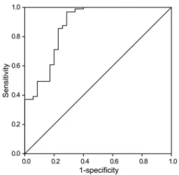
ROC curve analysis
In a ROC curve analysis, the area under the curve was 0.874 and the 95% confidence interval was 0.798–0.95
(Po0.01). The MFG-E8 cut-off value in predicting COPD was 5.27 ng/mL (sensitivity 96.9%, specificity 71.4%; Figure 4).
Table 2.Levels of plasma milk fat globule epidermal growth factor (MFG-E8), transforming growth factor (TGF)-b, and interleukin (IL)-1bin patients with chronic obstructive pulmonary disease differing in severity as measured by the Global Initiative for Chronic Obstructive Lung Disease (GOLD) criteria.
GOLD I GOLD II GOLD III GOLD IV
MFG-E8 (ng/mL) 4.79±0.59* 3.06±0.20* 2.58±0.17* 1.83±0.18 (min–max) (3.63–6.89) (0.89–6.39) (0.89–4.65) (0.16–3.14) TGF-b(ng/mL) 0.0079±0.0013 0.0094±0.0022 0.021±0.0016# 0.033±0.0027# (min–max) (0.00092–0.013) (0.0019–0.023) (0.0043–0.091) (0.0059–0.13) IL-1b(ng/mL) 0.0037±0.0011 0.0042±0.0012 0.0048±0.0021# 0.0054±0.0018# (min–max) (0.0015–0.0051) (0.0017–0.0054) (0.0014–0.0061) (0.0021–0.0077)
Data are reported as the mean±SE. Global Initiative For Chronic Obstructive Lung Disease (GOLD) criteria: GOLD I (mild); GOLD II (moderate); GOLD III (severe); GOLD IV (very severe). * Po0.05 vsGOLD IV. #
Discussion
Apoptosis, in which damaged or unwanted cells are removed, is critical to the maintenance of homeostasis. The recognition and removal of apoptotic cells is mediated by a variety of molecules, including receptors on the phagocyte surface and soluble bridging proteins such as MFG-E8, thrombospondin-1, and protein S. Both an increase in the number of apoptotic cells and defective efferocytosis are involved in the pathogenesis of COPD (8-10). However, their exact contributions are poorly understood. Hodge et al. (8) reported that a decrease in the expression of phagocyte surface receptors, such as CD31, CD91, and CD44, is responsible for defective efferocytosis in COPD (11). In the present work, we asked whether the altered expression of the soluble bridging protein MFG-E8 is associated with defective efferocytosis in COPD.
Our result showed that plasma MFG-E8 levels were distinctly lower in patients with COPD than in healthy controls. Moreover, the concentration of MFG-E8 in COPD patients decreased progressively from GOLD I to GOLD IV, suggesting a negative correlation between MFG-E8 and
the severity of COPD. This relationship was confirmed in a multiple regression analysis, which showed that plasma MFG-E8 levels correlated positively with FEV1% predicted. According to these data MFG-E8 may be a useful marker to evaluate disease severity in COPD. Moreover, the ROC curve analysis showed that the cut-off value for MFG-E8 was a good diagnostic indicator of COPD.
Consistent with our findings, the reduced expression of MFG-E8 in other diseases characterized by defective efferocytosis, such as atherosclerosis, has been reported. In the study of Dai et al. (6), plasma MFG-E8 levels were lower in patients with coronary atherosclerotic heart disease and MFG-E8 expression was reduced in atherosclerotic plaques. In another study, MFG-E8 was shown to facilitate the phagocytic clearance of apoptotic cells, and in atherosclerotic mice deficient in MFG-E8 there was both a substantial accumulation of apoptotic cells and an increase in athero-sclerotic lesion size (12). MFG-E8 has been implicated in Alzheimer’s disease as well, based on a significant reduction in its expression in the brain tissue of these patients (7). The same authors found that MFG-E8 was involved in the phagocytosis of amyloidb-peptide. Their results indicate that Figure 2. Correlation analysis between milk fat globule epidermal growth factor 8 (MFG-E8), forced expiratory volume in 1 s (FEV1) % pre-dicted, and smoking habit.
Figure 3.A, Plasma interleukin (IL)-1blevels in healthy controls and patients with chronic obstruc-tive pulmonary disease (COPD).B, Plasma trans-forming growth factor (TGF)-b levels in healthy controls and COPD patients. C, Plasma IL-1b levels among never, current, and ex-smokers.D, Plasma transforming growth factor (TGF)-blevels among never, current, and ex-smokers. *Po0.05 vscontrols;#P
alterations in MFG-E8 production contribute to the initiation of Alzheimer’s disease (7). Similarly, in COPD patients, the reduced expression of MFG-E8 may reflect its increased consumption during the removal of the large numbers of apoptotic cells present in the lungs and in bronchial tissue.
Because cigarette smoking is the major cause of COPD, a multiple regression analysis was performed to determine whether this relationship involved the decreased expression of MFG-E8. The results showed a negative correlation between MFG-E8 and the amount of smoking. Cigarette smoke impairs the clearance of apoptotic cells by phagocytes (13,14) and thus may inhibit the expression of macrophage surface molecules that recognize apoptotic cells (11). Accordingly, cigarette smoke may impair macrophage expression of the apoptosis bridging protein MFG-E8 in patients with COPD.
Inflammation is an important mechanism in the development of COPD and MFG-E8 could attenuate
inflammation indirectly by removing apoptotic cells. There is also an increasing body of evidence supporting a direct anti-inflammatory role of MFG-E8 in inflammatory dis-eases such as acute lung injury, sepsis, and colitis (15–17). MFG-E8 reduces the expression of pro-infl
am-matory cytokines, including IL-1, IL-6, and TNF-a. To determine whether the low-level expression of MFG-E8 in COPD is coupled to an increased inflammatory response, we measured the levels of plasma IL-1b and TGF-b in patients with COPD. While the levels of the anti-inflammatory cytokine TGF-b were markedly elevated, the difference in the levels of the pro-inflammatory cytokine IL-1b did not significantly differ from those of healthy controls. This observation is in agreement with a recent finding of increases in TGF-b and apoptotic T lymphocytes in the peripheral blood of COPD patients (18). The authors suggested that the increase in TGF-b was involved in inducing apoptosis by T cells and alveolar wall cells, via binding to the TGF-b receptor 1, thereby leading to the development of emphysema.
Our study had two major limitations. First, it may have been useful to measure the expression of MFG-E8 in lung tissue or in bronchoalveolar lavagefluid. Second, the effect of statins, corticosteroids, and other drugs on the expression of MFG-E8 was not determined. Nonetheless, our study is the first to report a decrease in plasma MFG-E8 levels in patients with COPD and to demonstrate a negative association between MFG-E8 and the severity of COPD. Thus, measuring the changes in plasma MFG-E8 may be a promising method for evaluating the severity of COPD.
Acknowledgments
This work was supported by the National Nature Science Foundation of China (#81170035), Wuhan Municipal Human Resources and Social Security Bureau (#2009-97), and The Program of Wuhan Scientific and Technological Research from Wuhan Scientific and Technological Bureau (#201260523191-3).
References
7. Boddaert J, Kinugawa K, Lambert JC, Boukhtouche F, Zoll J, Merval R, et al. Evidence of a role for lactadherin in Alzheimer’s disease.Am J Pathol2007; 170: 921–929. 8. Hodge S, Hodge G, Scicchitano R, Reynolds PN, Holmes
M. Alveolar macrophages from subjects with chronic obstructive pulmonary disease are deficient in their ability to phagocytose apoptotic airway epithelial cells. Immunol Cell Biol2003; 81: 289–296, doi: 10.1046/j.1440-1711.2003. t01-1-01170.x.
9. Vandivier RW, Henson PM, Douglas IS. Burying the dead: the impact of failed apoptotic cell removal (efferocytosis) on chronic inflammatory lung disease.Chest2006; 129: 1673–1682, doi: 10.1378/chest.129.6.1673.
10. Henson PM, Cosgrove GP, Vandivier RW. State of the art. Apoptosis and cell homeostasis in chronic obstructive pulmonary disease.Proc Am Thorac Soc2006; 3: 512–516, doi: 10.1513/pats.200603-072MS.
11. Hodge S, Hodge G, Ahern J, Jersmann H, Holmes M, Reynolds PN. Smoking alters alveolar macrophage recogni-tion and phagocytic ability: implicarecogni-tions in chronic obstruc-tive pulmonary disease.Am J Respir Cell Mol Biol2007; 37: 748–755.
12. Ait-Oufella H, Kinugawa K, Zoll J, Simon T, Boddaert J, Heeneman S, et al. Lactadherin deficiency leads to apoptotic cell accumulation and accelerated atherosclerosis in mice. Circulation 2007; 115: 2168–2177, doi: 10.1161/ CIRCULATIONAHA.106.662080.
13. Richens TR, Linderman DJ, Horstmann SA, Lambert C, Xiao YQ, Keith RL, et al. Cigarette smoke impairs clearance of apoptotic cells through oxidant-dependent activation of RhoA.Am J Respir Crit Care Med2009; 179: 1011–1021, doi: 10.1164/rccm.200807-1148OC.
14. Minematsu N, Blumental-Perry A, Shapiro SD. Cigarette smoke inhibits engulfment of apoptotic cells by macrophages through inhibition of actin rearrangement.Am J Respir Cell Mol Biol2011; 44: 474–482.
15. Aziz M, Matsuda A, Yang WL, Jacob A, Wang P. Milk fat globule-epidermal growth factor-factor 8 attenuates neutrophil infiltration in acute lung injury via modulation of CXCR2. J Immunol 2012; 189: 393–402, doi: 10.4049/jimmunol.1200262. 16. Miksa M, Wu R, Dong W, Das P, Yang D, Wang P. Dendritic
cell-derived exosomes containing milk fat globule epidermal growth factor-factor VIII attenuate proinflammatory responses in sepsis. Shock 2006; 25: 586–593, doi: 10.1097/01.shk. 0000209533.22941.d0.