ABSTRACT
Objectives: Percutaneous Renal Surgery (PRS) is a demanding procedure and success is mostly hampered by the lacking of training facilities. Thus, the purpose of the study was to evaluate a signiicantly improved pre-existing porcine kidney-training model for percutaneous renal access and PRS.
Materials and Methods: A biologic training model using porcine kidneys coated by a full-thickness porcine skin lap was prepared. The ureter was dissected, stones were placed into the collecting system using an 18F amplatz sheath, and a catheter was placed in the ureter for further irrigation with saline or contrast medium. For initial training with an easy access, a standard guide-wire was inserted in the ureter through the renal parenchyma. The kidney was punctured with radiographic or ultrasound guidance. Minimally invasive percutaneous nephrolithotomy (MIP) was then tested using the model under radiographic or ultrasound guidance. The model was then evaluated in MIP training courses, which are regularly held at The Hannover Medical School.
Results: All trainees were urologists with experience in endourologic surgery but lacked practice in PRS. In conclusion, all 36 participants attained access to the collecting system using models with readily placed guide-wires. Subsequently, PRS was successful in all cases. Percutaneous puncture under ultrasound guidance and following intrarenal surgery was successful in 30 (83.3%) cases. Therefore, all participants rated the model useful for simulating percutaneous renal surgery.
Conclusions: This new porcine kidney model is easy to build and is made cost effective by using readily available mate-rial. Moreover, it provides realistic and reproducible training model for PRS. The “organ” model mimics the retroperito-neum by having a full-thickness skin lap with a layer of subcutaneous fatty tissue.
Key words: kidney; minimally invasive surgical procedures; percutaneous nephrolithotomy animal models; kidney cal -culi
Int Braz J Urol. 2011; 37: 388-394
INTRODUCTION
The increasing incidence and prevalence of urolithiasis in Germany throughout the last de-cades (1) grew to be a socioeconomic burden with regard to diagnostics and treatment. Thus, new challenges in diagnostic and therapy have to be addressed. A modern treatment of a stone disease has to meet several requirements: It has to be fast and effective, providing a high stone free rate with minimal perioperative morbidity along with a low re-intervention rate. Due to diameter, the number and location of urinary tract stones, mostly ESWL,
ureterorenoscopy and percutaneous nephrolithoto-my (PCNL) are recommended in the current uro-logic guidelines (e.g. EAU, AUA Guidelines). The use of percutaneous nephrolithotomy was devel-oped in the 1970s as an alternative to open surgery, but with the introduction of the ESWL in the early 80s PCNL became less popular. This technical evolution, as well as, the improvement in the ield of semirigid and lexible ureterorenoscopy dur -ing the follow-ing three decades, has dramatically changed the operative management of urolithiasis. Nevertheless, PCNL was recommended for large renal calculi. Although, PCNL has been accepted
New Ex-vivo Organ Model for Percutaneous Renal Surgery
Florian Imkamp, Christoph von Klot, Udo Nagele, Thomas R.W. Herrmann
to be safe and effective, it also has signiicant perioperative morbidity (2,3). Therefore, urologist minimized their PCNL instruments in the past de-cade so to be less invasive and moreover reduce therapy-associated morbidity. This led to the de-velopment of new minimized instruments, where less invasive procedures such as Mini-PCNL (miniaturized PCNL) (4) and MIP (minimally in-vasive percutaneous nephrolithotomy) (5,6) take part. These techniques showed advantages in terms of shorter hospital stay and reduced postop-erative pain while maintaining the high stone free rates of former PCNL (7). These improvements in the ield of percutaneous surgery and the critical evaluation and discussion of the results of ESWL and ureterorensocopy with respect to stone free rates and complications led to a rising acceptance of minimally invasive percutaneous procedures throughout the previous years. Therefore, an in-creasing number of these procedures require sufi -cient structured training opportunities to maintain surgical effectiveness, with respect to stone free rates and patient safety.
Although miniaturization decreased the perioperative morbidity, percutaneous renal sur-gery is still one of the most advanced techniques in modern endourology. Frequently, urologists com-plain of lack of training experience due to scarce training facilities and high cost organ models. Consequently, operative expertise is mostly ac-quired in the operating theatre. Analysis of struc-tured learning curve of percutaneous nephrolithot-omy suggested that basic skills are achieved after > 20 procedures, surgical competence is achieved after 60 cases and surgical excellence after > 100 cases (8-10).
An ex-vivo PCNL organ training model described by Zhang et al. (11) was modiied in sig -niicant aspects. It was then evaluated during MIP training courses at The Hannover Medical School, and has proven eficiency in various teaching ses -sions taken place in our own department, in addi-tion to the training courses held for urologists. The model allows training of multiple steps of percuta-neous renal surgery such as; renal puncture, tract dilation, access sheath introduction, intrarenal endoscopic exploration and further intrarenal
sur-gery. It also provides a suficient tool to overcome the initial learning curve of the irst 10 procedures.
MATERIAL AND METHODS
The PCNL kidney model was built using a porcine kidney, a porcine full thickness skin lap with subcutaneous tissue, an indwelling catheter, artiicial renal calculi with a maximum diameter of 5 millimeters and a standard plastic tray.
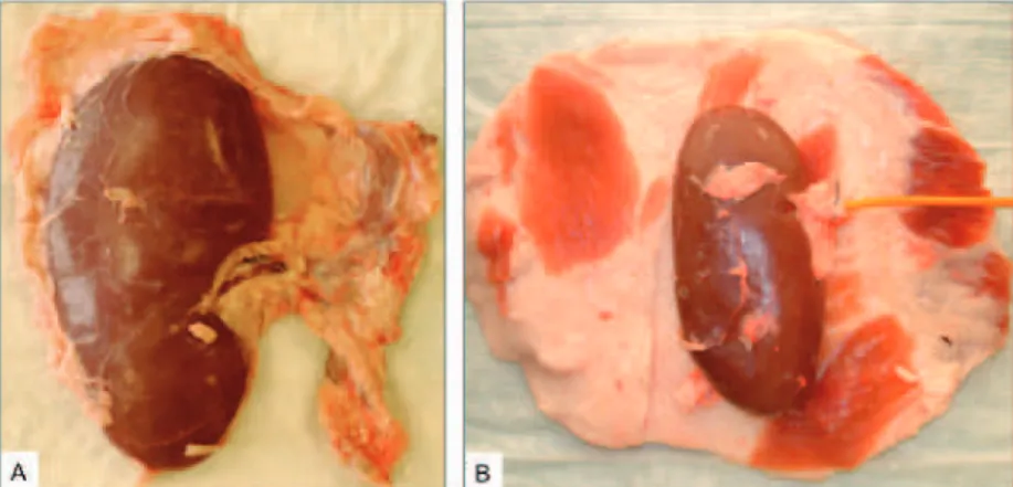
The kidneys and skin laps were obtained from freshly slaughtered adult pigs. For ureter preparation and catheterization, a minimum length of 10 cm ureter was preserved during kidney ex-traction (Figure-1). The full-thickness skin lap with subcutaneous tissue, in which was harvested from the abdominal wall of adult pigs, was ap-proximately 5-6 cm in thickness and 40-50 cm in diameter, enough to cover the entire kidney (Fig-ure-2).
The model was applied to percutane-ous renal surgery training under radiologic or ultrasound guidance. Either the X-ray unit (Phil-ips Uro Diagnost MRF, Netherlands) or the ul-trasound unit (BK Medical, Falcon Ulul-trasound
Scanner Type 2101, Copenhagen, Denmark) with a 3-6 MHz probe (BK Medical Type 8803, Copen-hagen, Denmark) was used during the procedure. Also, a radiologic contrast medium or normal saline was injected through the indwell-ing catheter to produce visual im-ages, in order to achieve sufficient artificial hydronephrosis for per-cutaneous renal puncture. Next, various surgical steps of PRS were performed such as; puncturing, guide-wire placement, single step dilation, insertion of the miniaturized 18F Am-platz sheath (Karl Storz, Tuttlingen, Germany), introduction of a 12F nephroscope (Karl Storz, Tuttlingen, Germany), intrarenal exploration, and minimally invasive percutaneous nephrolithoto-my (MIP). The organ model was than evaluated in MIP training courses, which are held in our department with basic questionnaire in terms of successful puncture of the collecting system, ac-cess to the renal pelvis and subsequent intrarenal surgery. Moreover, the preexisting surgical expe-rience in the field of percutaneous renal surgery of the participating trainees and the personal per-ception of this model were also evaluated as not useful, undetermined or helpful.
RESULTS
Based on the previously described organ model (11) a total number of 6 porcine kidneys were necessary to improve and modify this model for percutaneous renal surgery. The preparation of one single organ model acquired 10-15 minutes time, until it was ready for a percutaneous train-ing session.
Altogether, 36 urologists attended 6 uro-logic training courses for percutaneous renal sur-gery at the affiliated hospitals. All 36 participants were known to have experience in urologic endo-scopic surgery (defined as ureteroscopy, renosco-py) yet no experience in the field of percutaneous renal surgery (defined as minimally invasive per-cutaneous nephrolithotomy (MIP), perper-cutaneous Figure 1 - Freshly harvested porcine kidney (A) and full thickness skin lap
used to cover the kidney (B).
Figure 2 - Placement of artiicial calculi into the renal pelvis (A) and catheterized renal pelvis for saline or dye
nephrolithotomy (PCNL)). All 36 (100%) trainees were able to access the renal pelvis using the de-signed model, readily prepared with a guide wire. Furthermore, percutaneous surgery (MIP) was successful in all cases through this predefined ac-cess. 30 (83.3%) participants managed to achieve access to the collecting system by ultrasound or radiographic guidance. If surgical access was
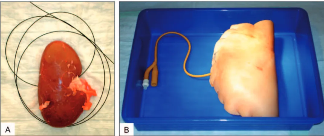
achieved, subsequent percutaneous transrenal sur-gery (MIP) was successful in 30 cases (100%). All participants practiced the percutaneous hands-on manipulatihands-ons hands-on this model under the direct guidance and surveillance of three experienced endourologists. By the end of this course, all at-tendants rated the porcine kidney model for simu-lation of percutaneous renal surgery as “helpful”. Figure 3 - Guide-wire for easy access to the renal pelvis (A) and wrapped organ model,
closed by running sutures, in the plastic tray for subsequent training (B).
Figure 4 - Puncture of the renal pelvis (A) and subsequent intrarenal surgery (B).
Alter-nate ultrasound guided puncture (a) and veriication of successful access to the renal pelvis
The low cost for this organ model was the result of use of inexpensive ureteral catheters for irrigation of the renal pelvis during percutaneous surgery, as well as, the use of standard sewing material. The use of reusable material and instru-ments improved the cost-value relation of this model. In summary, the total cost accounted ap-proximately 10€ for each prepared organ model. Nonetheless to be considered, that initial cost for the described reusable instruments in animal use only may occur establishing an organ model train-ing program for percutaneous renal surgery.
In conclusion, this percutaneous renal ac-cess and other intrarenal procedures proved feasi-ble and practical under radiographic or ultrasound guidance.
DISCUSSION
Learning percutaneous renal surgery is still demanding, although several virtual and bio-logic models have been published. But only vir-tual or laboratory training of percutaneous renal surgery provides the opportunity to overcome the initial learning curve. Thus, training models have to meet several requirements. These models must be cost effective, easy and fast to prepare with commonly available material and organs. They need to be realistic, provide the feeling of human tissue and simulate the retroperitoneal anatomy. Finally, they should provide easy access to the renal pelvis for subsequent renal surgery, along with a high success rate under training conditions. To address these challenging requirements, sev-eral training models have been published to date, consisting of virtual computer-based non-biologic training and organ-based models using porcine kidneys.
A nonbiologic computer-based simulator PERCMentor (Simbionix, Lod, Israel) has been published by Knudsen et al. in 2006, providing virtual reality skills. This might allow trainees to develop the basic skills necessary to perform percutaneous access to the renal collecting sys-tem (12). However, expenses of this sort of train-ing system are unclear for software and hardware costs, addition to time-consuming labor-intensive
training of urologists. The advantage of biologic models is the “tissue feeling”, an imitation of hu-man tissue while allowing a great variety of proce-dures of intrarenal surgery (13). To date 6 reports have been published providing feasible biologic models using porcine kidneys for percutaneous renal surgery training. Porcine kidneys have been wrapped in a foam layer, embedded in silicon, enclosed in chicken carcasses and in porcine tho-racic/abdominal walls. Radiologic and ultrasound guidance were applied for percutaneous renal ac-cess guidance (13-17).
A model published by Zhang et al. in 2008 was primarily the inspiration of building our own (11). This ex-vivo porcine kidney training model was significantly improved in meeting criterions of cost effectiveness, preparation, and simplified access to the collecting system in the initial train-ing phase and therefore, concludtrain-ing success rates in percutaneous renal surgery. Not to mention, im-proved cost effectiveness was achieved by dimin-ishing the use of expensive disposable materials. Moreover, the ureteral catheter was replaced by a 12F indwelling catheter and reusable plastic trays were used instead of wooden boards. The use of these plastic trays not only improves the hygiene, but it allows the collection of the saline irrigation during the procedures. The full-thickness skin was replaced by porcine entire abdominal wall of adult pigs including the subcutaneous tissue, providing enhanced ultrasound visualization for subsequent puncture, as well as improvement in realistic retroperitoneal circumstances with re-gard to consistency and resistance of the skin. This setting allows a realistic training of punc-turing, single step tract dilatation, amplatz sheath introduction and subsequent intrarenal surgery. The porcine kidney anatomy is similar to that of humans, facilitating intrarenal endoscopic prac-tice. The transureteral introduction of renal cal-culi through the 18F amplatz sheath into the renal pelvis significantly eased the preparation of the organ model.
In contrast to previous descriptions, our ex-perience using the depicted organ model supports that neither an immediate irrigation following the kidney harvest (15), nor the refrigeration of the or-gan model (11) is required to maintain the rigid-ity of the kidney tissue. It was demonstrated high success rates for optimal puncture of the collecting system, subsequent tract dilatation followed by in-trarenal surgery. Thus, this organ model can easily be prepared hours before a training session, reduc-ing labor and preparation while retainreduc-ing realistic scenario for training.
In contrast to other models (13,14), the kidney is not ixed to the surrounding structures in the skin-lap compartment, providing a suf -icient and realistic mobility of the kidney, mim -icking in-vivo circumstances. Comparing to organ models with porcine kidneys embedded in chicken carcasses (15), this model suficiently avoids in -terferences in x-ray or ultrasound based puncture, usually caused by the chicken skeleton. Thus, im-provement of success level in the initial training period was up to 10 percutaneous interventions in particular. This ex-vivo model lacks the simulation of ribs, simulating realistic conditions as in human anatomy. This could be developed to some extent by using part of the thoracic wall and supericial soft tissue to give an even better practice environ-ment (17). Such model might improve the training results creating further advanced urologic surgeons with basic skills in percutaneous surgery.
All attending trainees were urologists (36/100%) without experience in percutaneous re-nal surgery while previously experienced in trans-urethral endourology. It is intriguing, whether the reported high success rate accessing the renal pel-vis was due to the easy surgical access by a read-ily placed guide wire, or by pre-existing surgical skills in minimally invasive endourology. This organ model was used in 6 training sessions with 6 participants. An initial 90 minute didatic lesson with basic knowledge of percutaneous renal sur-gery was followed by a 3-hour hands-on-training at 3 training venues simultaneously. All trainees were personally instructed by three certiied urolo -gists with the opportunity of puncture practice, tract dilatation and percutaneous surgery at their
own organ models. This setting allowed individual instructions given to all trainees, to guarantee the reported high success rate. Moreover, the individu-al training with personindividu-al porcine organ models led to the acceptance of the model, in which was dem-onstrated by the evaluations.
CONCLUSIONS
This porcine kidney model previously de-scribed is simple, cost effective and easy to prepare with reusable material and instruments. It mimics the natural circumstance, and provides realistic and reproducible practice for percutaneous renal surgery in the training laboratory. Furthermore, it provides incremental training opportunities for trainees with various skills in the ield of percu -taneous surgery with an easier wired access to the renal pelvis for the initial training period. The ma-jority of the participating trainees evaluated this organ model as “helpful”. Hence, it is believed that this model will become an integral part of struc-tured training for minimally invasive percutaneous nephrolithomy (MIP) “in the near future”.
ABBREVIATIONS
PRS Percutaneous renal surgery
MIP Minimally invasive percutaneous nephro-lithotomy
ESWL Extracorporal shockwave lithotripsy PCNL Percutaneous nephrolithotomy EAU European Association of Urology AUA American Association of Urology F French
CONFLICT OF INTEREST
None declared.
REFERENCES
1. Hesse A, Brändle E, Wilbert D, Köhrmann KU, Al-ken P: Study on the prevalence and incidence of uro-lithiasis in Germany comparing the years 1979 vs. 2000. Eur Urol. 2003; 44: 709-13.
in percutaneous nephrolithotomy. Eur Urol. 2007; 51: 899-906; discussion 906.
3. Osman M, Wendt-Nordahl G, Heger K, Michel MS, Alken P, Knoll T: Percutaneous nephrolithotomy with ultrasonography-guided renal access: experi-ence from over 300 cases. BJU Int. 2005; 96: 875-8. 4. Lahme S, Zimmermanns V, Hochmuth A, Janitzki V:
Minimally invasive PCNL (mini-perc). Alternative treatment modality or replacement of conventional PCNL?. Urologe A. 2008; 47: 563-8.
5. Nagele U, Knoll T, Schilling D, Michel MS, Stenzl A: Lower pole calyceal stones. Urologe A. 2008; 47: 875-84.
6. Nagele U, Schilling D, Anastasiadis AG, Walcher U, Sievert KD, Merseburger AS, et al.: Minimally in-vasive percutaneous nephrolitholapaxy (MIP). Uro-loge A. 2008; 47: 1066, 1068-73.
7. Knoll T, Wezel F, Michel MS, Honeck P, Wendt-Nordahl G: Do patients beneit from miniaturized tubeless percutaneous nephrolithotomy? A compara-tive prospeccompara-tive study. J Endourol. 2010; 24: 1075-9.
8. Allen D, O’Brien T, Tiptaft R, Glass J: Deining the learning curve for percutaneous nephrolithotomy. J Endourol. 2005; 19: 279-82.
9. de la Rosette JJ, Laguna MP, Rassweiler JJ, Conort P: Training in percutaneous nephrolithotomy--a crit-ical review. Eur Urol. 2008; 54: 994-1001.
10. Tanriverdi O, Boylu U, Kendirci M, Kadihasanoglu M, Horasanli K, Miroglu C: The learning curve in the training of percutaneous nephrolithotomy. Eur Urol. 2007; 52: 206-11.
11. Zhang Y, Ou TW, Jia JG, Gao W, Cui X, Wu JT, et al.: Novel biologic model for percutaneous renal surgery learning and training in the laboratory. Urol-ogy. 2008; 72: 513-6.
12. Knudsen BE, Matsumoto ED, Chew BH, Johnson B, Margulis V, Cadeddu JA, et al.: A randomized,
con-trolled, prospective study validating the acquisition of percutaneous renal collecting system access skills using a computer based hybrid virtual reality surgi-cal simulator: phase I. J Urol. 2006; 176: 2173-8. 13. Strohmaier WL, Giese A: Ex vivo training model for
percutaneous renal surgery. Urol Res. 2005; 33: 191-3.
14. Earp PP: Percutaneous renal surgery--new model for learning and training. Int Braz J Urol. 2003; 29: 151-4.
15. Häcker A, Wendt-Nordahl G, Honeck P, Michel MS, Alken P, Knoll T: A biological model to teach percu-taneous nephrolithotomy technique with ultrasound- and luoroscopy-guided access. J Endourol. 2007; 21: 545-50.
16. Hammond L, Ketchum J, Schwartz BF: A new ap-proach to urology training: a laboratory model for percutaneous nephrolithotomy. J Urol. 2004; 172: 1950-2.
17. Strohmaier WL, Giese A: Improved ex vivo train-ing model for percutaneous renal surgery. Urol Res. 2009; 37: 107-10.
___________________ Accepted after revision:
December 15, 2010
______________________ Correspondence address: Dr. Florian Imkamp
Clinic for Urology and Urologic Oncology Hannover Medical School
Carl-Neuberg-Str. 1
Hannover, 30625, Germany Fax: + 49 511 532-3481