DOI: 10.5935/2359-4802.20170019
Introduction
Sleep duration is one of the determining factors of cardiovascular health. Studies have demonstrated that a shorter time allocated to sleep is closely associated with increased prevalence and incidence of cardiovascular diseases, such as hypertension.1 Among the possible mechanisms, an increased sympathetic activity and catabolism is evidenced by increased levels of catecholamine and cortisol/corticosterone2-6 which leads to molecular and morphological changes7 that may negatively affect cardiac tissue.
In this scenario, resistance training stands out as a possible tool to minimize of even reverse the impact of sleep debt on cardiovascular health. In addition to reducing resting arterial pressure, left ventricular systolic pressure and heart rate,8,9 resistance training can also increase papillary muscle isometric strength and its contractile performance.10 Also, due to its effect on endocrine axes, resistance training can mitigate the catabolic effect of sleep deprivation.11
Despite studies showing the impact of sleep debt on cardiovascular health,4,12 little is known about its effects
Mailing Address: Profa. Dra. Hanna Karen Moreira Antunes
Universidade Federal de São Paulo – Campus Baixada Santista. Departamento de Biociências Rua Silva Jardim, 136, Térreo. Postal Code: 11015-020, Vila Mathias, Santos, São Paulo – Brazil E-mail: hanna.karen@unifesp.br
Effect Of Resistance Training On Myocardial Contractility In Vitro After Sleep Deprivation
Sara Quaglia de Campos Giampá,1 Marcos Mônico-Neto,2 Helton de Sá Souza,2 Marco Túlio de Mello,2 Sergio Tufik,2
Leslie Andrews Portes,3,4 Andrey Jorge Serra,4,5 Paulo José Ferreira Tucci,4 Hanna Karen Moreira Antunes1,6
Programa de Pós-Graduação Interdisciplinar em Ciências da Saúde1, Universidade Federal de São Paulo; Departamento de Psicobiologia2, Universidade
Federal de São Paulo; Programa de Mestrado em Promoção da Saúde, Centro Universitário Adventista de São Paulo3;Departamento de Medicina4,
Universidade Federal de São Paulo; Programa de Pós-Graduação em Ciências da Reabilitação, Universidade Nove de Julho5; Departamento de Biociências6,
Universidade Federal de São Paulo, Santos, São Paulo, SP – Brazil
Manuscript received January 11, 2016; revised manuscript October 04, 2016; accepted December 07, 2016.
Abstract
Background: Resistance training promotes cardiovascular health benefits that may affected by sleep deprivation.
Objective: To evaluate the effect of high-intensity resistance training on myocardial contractility in rats subsequently
subjected to paradoxical sleep deprivation.
Methods: Forty male Wistar rats were distributed into control group (CTRL), resistance training (REST), 96-hour paradoxical sleep deprivation (PSD96) and resistance training followed by 96-hour paradoxical sleep deprivation (REST/PSD96). The animals underwent high-intensity resistance training for 8 weeks, 5x/week. Twenty-four hours after the last training session, the PSD96 and REST/PSD96 groups were submitted to paradoxical sleep deprivation, which was followed by the in vitro study of isolated papillary muscle contractile mechanics.
Results: In comparison with the CTRL group, a lower papillary muscle length and increased cross sectional area were found in PSD96 and RETS/PSD96, which were associated with decreased temporal parameters of contraction force and relaxation. Decreased resting tension and slowing of relaxation time were found in the PSD96 group only. This effect was attenuated by previous resistance training.
Conclusion: Resistance training partially prevented contractile changes induced by PSD, minimizing the slowing in relaxation time. Thus, high-intensity exercise seems to not fully protect the cardiac tissue from PSD-induced effects. (Int J Cardiovasc Sci. 2017;30(1):20-31)
on cardiac contractile machinery and the role of resistance training as a preventive tool. Therefore, the aim of this study was to evaluate the effect of high-intensity resistance training on myocardial contractility in rats subsequently subjected to paradoxical sleep deprivation.
Methods
Using a convenience sample, forty 3-month old male Wistar rats, weighing 300-350 g, obtained from the Center for the Development of Experimental Models in Medicine and Biology of Sao Paulo Federal University was used in the experiment. The animals were kept in polypropylene boxes in a temperature-controlled room (22 ± 1ºC), light/dark cycle (12:12h), and given water and food ad libitum.
All procedures were performed according to the Guide for the Care and Use of Laboratory Animals - National Research Council, NIH Publication N° 85-23 (revised 2011). The study protocol was approved by the Ethics Committee (#0764/10).
The animals were distributed into four groups: control (CTRL, n = 10), resistance training (REST, n = 10), paradoxical sleep deprivation (PSD) for 96 hours (PSD96, n = 10), and resistance training followed by 96-hour PSD (REST/PSD96, n = 10). During the experiment, animals of the CTRL group were kept in the boxes and manipulated exclusively for laboratory routine practices.
Resistance training
Resistance training was performed for 8 weeks as described elsewhere.13 A ladder (110cm high, 18 cm wide, 2 cm space between steps) was placed at an 80º inclination, and a resting chamber (20x20x20 cm) was placed on the top of the ladder. The animals climbed the ladder carrying weights attached to the base of the tail, and the weights were gradually increased with exercise progression.
Familiarization of the animals with the exercise apparatus was performed during three consecutive days by nine repetitions per day. After this period, the maximum load (ML) test was performed, which was repeated every week.
Training sessions consisted of 4-8 sets of climbing, using progressive loads, interspaced with 60-second intervals. The animals had to perform 8 - 12 repetitions to go from the base to the top of the ladder. The load was progressively increased from 50% of maximum load in the
first series, through 75%, 90% until 100% of maximum load in the fourth series. After that, resistance was increased in increments of 30 g per attempt until failure.13 Training was performed in the afternoon, 5 times a week, from Monday to Friday. Monday session was replaced by the maximum load test. In order to prevent overtraining caused by the high intensity of exercise, a prophylactic rest was adopted at weeks 6, 7 and 8; in these weeks, the animals trained on Monday, Tuesday, Thursday and Friday. Forty-eight hours after the last training session, the CTRL and REST animals were euthanized and the PSD96 and REST/PSD96 groups underwent the PSD protocol.
Paradoxical sleep deprivation
PSD was conducted for 96 hours by the modified multiple platform method,14 which totally promotes the total suppression of paradoxical sleep and the decrease in slow wave sleep by 37%.15 Five socially stable animals were placed inside a stainless steel tank (123 cm length x 44 cm height x 44 cm width) on round platforms 10 cm distant from each other. The tank was filled with room temperature water until 1cm below the platform surfaces, so that the animals could walk from one platform to another one. When paradoxical sleep was achieved, the animals woke up as they touched the water due to muscle atonia accompanying this sleep stage. During the protocol, the room was maintained at optimal conditions,i.e., 22±1ºC, light-dark cycle (12h) and water and chow ad libitum. The CTRL group was kept in the same room to experience the same conditions.
Isolated cardiac muscle mechanics
Immediately after the PSD protocol for PSD96 and REST/PSD96 groups, or 48 hours after the last resistance training sessions for the REST group, or after a corresponding period for the CTRL group, in vitro preparations were made of papillary muscle isolated from the left ventricle.
(132 NaCl, 4.69 KCl, 1.5 CaCl, 1.16 MgSO4, 1.18 KH2PO4, 5.50 glucose and 20 HEPES), buffered at pH 7.4.16 The ring attached to the lower end of muscle was connected to the hook fixed to the glass chamber, and the upper ring was connected to an isometric force transducer (model FT03E, Grass Instrument, Quincy, MA) via a stainless steel wire. The force transducer was attached to a micromanipulator for muscle length adjustments. The papillary muscle was electrically stimulated by two parallel platinum electrodes using square wave pulses of 5ms of duration, frequency of 0.2Hz,16,17 and voltage approximately 20% higher than the minimum required to induce a maximal muscle response. After 60 minutes of stabilization of the preparation in isotonic condition and low preload (0.4g), the muscle was loaded to contract isometrically and then stretched until the peak of the length-tension curve was reached (Lmax: diastolic length of the muscle associated with maximal isometric tension). The tests were performed in Lmax, and isometric tension was assessed by force normalized for muscle fiber cross-sectional area (CSA) (g. mm- 2). Muscle CSA was calculated from molecular weight and length of the fibers, based on the assumption that the muscle was a uniform cylinder of specific gravity 1.04.
The following outcome measures were assessed for contraction function: developed tension (DT/ g.mm-2), resting tension (RT g.mm-2), temporal variation of contractile force (+dT/dt; g.mm-2.s-1), temporal variation of relaxation force (-dT/dt; g.mm-2.s-1), time to DT peak (TTP; ms) and time to a 50% decrease in maximum DT (RT50% ms). Mechanical behavior of papillary muscles was assessed (1) at baseline; (2) for length-tension relationship (variation of muscle length as a function of 92%-100% Lmax); (3) for post-rest potentiation (after 10, 20, 30, 60 and 120 seconds of rest); and (4) for contractile response to calcium (Ca+2) (increase in Ca+2 concentration from 1.5mM to 2.5mM in the bathing solution).
Statistical analysis
Statistical analysis was performed using the Statistica® software (Stat Soft, Inc, version 12.0). Measures of central tendency were calculated for descriptive analysis of data. Data normality was examined by the Shapiro Wilk test. Data with parametric distribution were analyzed by one-way or two-way ANOVA with Duncan post-hoc test, and after Z-score transformation, non-parametric data were analyzed by ANOVA. In the analyses of length-tension relationship and post-rest potentiation, linear regression was used
for comparisons between slopes and between areas under the curves, respectively. Values in each group (slope and area under the curve) were analyzed by ANOVA. Statistical significance was set at α = 0.05.
Results
Biometric parameters of papillary muscle
No difference in papillary muscle mass was found between the groups (F(3,23) = 0.16005, p > 0.05). There was a significant decrease in Lmax in the PSD96 (p = 0.03) and REST/PSD96 (p = 0.002) groups as compared with CTRL, and Lmax was significantly different between REST/PSD96 and PSD96 (p = 0.005), (F(3,21) = 6.0487, p < 0.01). In addition, CSA significantly increased in the REST (p = 0.02), PSD96 (p = 0.02) and REST/PSD96 (p = 0.01) groups compared with CTRL (F(3,19) = 3.1706, p < 0.05) (Table 1).
Contractile mechanics of papillary muscle under isometric contraction (baseline)
There was a significant decrease in DT in the REST/ PSD96 group as compared with the CTRL group (F(3,23) = 33049, p = 0.009). RT was lower in PSD96 than in REST (F(3,22) = 3.6623, p=0.01). A significant decrease in +dT/dt was observed in the PSD96 (0.006) and REST/ PSD96 groups as compared with CTRL (F(3,20) = 8.0313, p < 0.05), whereas -dT/dt significantly decreased in PSD96 (F(3,26) = 2.7244, p = 0.01) (Figure 1).
Similar behavior of time parameters of cardiac contraction was observed between the groups (F(3,23) = 0.00, p > 0.05). However, with respect to RT50%, a prolongation of muscle relaxation was observed in PSD96 as compared with the other groups (F(3,26) = 3.9344, p < 0.05).
Contractile mechanics of papillary muscle in length-tension relationship
Slopes of DT were significantly lower in the REST (p = 0.02), PSD96 (p = 0.03) and REST/PSD96 (p = 0.0001) groups than in CTRL group, and in the REST/PSD96 when compared with the REST (0.03) and PSD96 (p = 0.02) groups (F(3, 24) = 7.0880, p < 0.01). In addition, a reduction in RT was observed in PSD96 as compared with REST (F(3,23) = 2.5483, p < 0.05) (Figure 2).
Table 1 – Effect of resistance training and paradoxical sleep deprivation on biometrical parameters of papillary muscle
Variables CTRL REST PSD96 REST/PSD96
Papillary muscle mass (mg) 5.96 ± 2.02 5.98 ± 1.43 6.35 ± 0.89 5.77 ± 1.64
Lmax (mm) 5.86 ± 0.84 5.71 ± 0.73 4.91 ± 0.64* 4.40 ± 0.60*. †
CSA (mm2) 1.00 ± 0.27 1.30 ± 0.19* 1.29 ± 0.12* 1.35 ± 0.21*
Data in mean ± standard deviation; Lmax – maximum papillary muscle length; CTRL: control group; REST: resistance training group; PSD96: paradoxical sleep deprivation for 96 hours ; REST/PSD96: resistance training followed by 96-hour paradoxical sleep deprivation; CSA: cross-sectional area; *different from CTRL; † different from REST group
*
†
*
* *
†† 8
6
4
2
0
1.5
1.0
0.5
0.0
DT (g.mm
–2)
R
T (g.mm
–2
)
CTRL REST PSD96
REST/PSD96
CTRL REST PSD96
REST/PSD96
CTRL REST PSD96
REST/PSD96
CTRL REST PSD96
REST/PSD96
CTRL REST PSD96
REST/PSD96
CTRL REST PSD96
REST/PSD96 40
30
20
10
0 80
60
40
20
0
+dT/dt
max
(g.mm
2.s)
–dT/dt
max
(g.mm
–2
.s
–1
)
TPT (ms)
TR50% (ms)
250
200
150
100
50
0
300
250
200
150
100
6 5 4 3 2 TD (g.mm –2) 1,0 0,5 0,0 TR (g.mm –2) 70 65 60 55 50 45 40 35 30 25 20 + dT/dt max (g.mm –2.s –1) – dT/dt max (g.mm –2.s –1) 35 30 25 20 15 10 CTRL REST PSD96 REST/PSD96
92 94 96 98 100
Stretching (% do Lmax) CTRL REST PSD96
REST/PSD96
92 94 96 98 100
Stretching (% do Lmax) CTRL REST PSD96
REST/PSD96
92 94 96 98 100
Stretching (% do Lmax) CTRL REST PSD96
REST/PSD96
92 94 96 98 100
Stretching (% do Lmax) CTRL REST PSD96
REST/PSD96 0,25 0,20 0,15 0,10 0,05 0,00 Slope (g.mm –2 of L max ) 0,10 0,08 0,06 0,04 0,02 0,00 Slope (g.mm –2 of L max ) 2.5 2.0 1.5 1.0 0,5 0,0 Slope (g.mm –2 of L max ) 0,6 0,4 0,2 0,0 Slope (g.mm –2 of L max ) * * † † † † † *, †
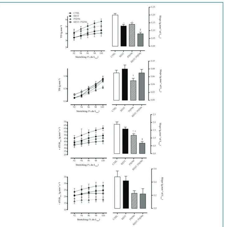
Figure 2 – Effect of resistance training and paradoxical sleep deprivation on papillary muscle contractile parameters in terms of length-tension relationship. One-way ANOVA with Duncan post-hoc test. Data expressed in mean ± standard error, with significance set at p = 0.05. DT: maximum developed tension; RT: isometric resting tension; +dT/dt: temporal variation of contraction force; -dT/dt: temporal variation of relaxation force;
CTRL: control group; REST: resistance training group; PSD96: paradoxical sleep deprivation for 96 hours; REST/PSD96: resistance training followed by 96-hour paradoxical sleep deprivation. *significantly different from CTRL; †different from REST group; ‡ different from all groups
in REST/PSD96 (p = 0.01), (F(3,21) = 15.741, p < 0.01). No difference in -dT/dt slopes was found between the groups (F(3,20) = 2.4210, p > 0.05).
Also, TTP was not different between the groups. The PSD96 group had higher RT50% for all stretching percentages tested as compared with the other groups (F(3,25) = 5.1010, p < 0.05) (Figure 3).
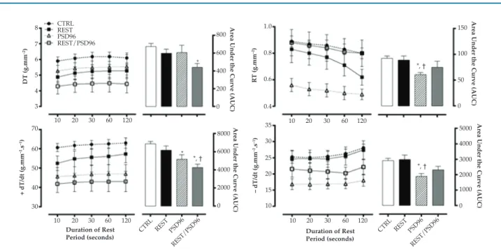
Contractile mechanics of papillary muscle in post-rest potentiation
250
200
150
100
TPP (ms)
250
200
150
100
R
T50% (ms)
92 94 96 98 100
Stretching (% do Lmax)
92 94 96 98 100
Stretching (% do Lmax)
CTRL
REST
PSD96
REST/PSD96
Figure 3 – Effect of resistance training and paradoxical sleep deprivation on papillary muscle temporal parameters in terms of length-tension relationship. Two-way ANOVA with Duncan post-hoc test. Data expressed in mean ± standard error, with significance set at p = 0.05. TTP: time to developed tension peak; RT50%: time required for maximum developed tension to decrease by 50%; CTRL: control group; REST: resistance training group; PSD96: paradoxical sleep deprivation for 96 hours; REST/PSD96: resistance training followed by 96-hour paradoxical sleep deprivation; Lmax: maximum papillary muscle length. ‡ different from all groups in each stretching percentage tested
PSD96 (p = 0.02) and REST/PSD96 (p = 0.002) than in CTRL, and significantly different between RST/PSD96 and REST (p = 0.01) (F(3,23) = 4.6147, p < 0.05) (Figure 4).
No difference in time parameters was observed between the groups (F(12,96) = 0.7959, p < 0,05), except for RT50% at 60s in comparison with other rest times – 10 (p = 0.00004), 20 (p = 0.001), 30 (p = 0.004) and 120 seconds (p = 0.0006), (F(4,88) = 3.8313, p < 0.05) in REST/PSD96 (Figure 5).
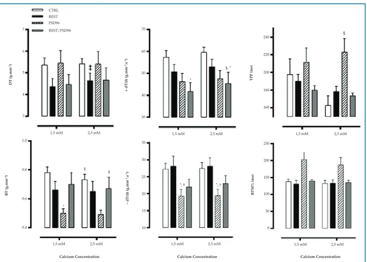
Contractile mechanics of papillary muscle in response to extracellular calcium
There was an increase in DT in the REST group (F(1,18) = 5.1523, p = 0.02), and hence greater response to calcium-induced inotropy. RT was lower at Ca2+ 2,5 mM as compared with Ca2+ 1.5 mM levels in CTRL (p = 0.001) and REST/PSD96 (p = 0.04) (F(1,18) = 14,0730, p < 0,01), and lower at both Ca2+ 1.5 mM (p=0.009) and 2.5 mM (p = 0.02) in the PSD96 group than in CTRL group (F(3,18) = 3.2331, p < 0.05) (Figure 6).
Regarding +dT/dt, this variable was higher at Ca2+ 2.5 mM than Ca2+ 1.5 mM (p = 0.03) in the REST/ PSD96 group, however, at both concentrations (Ca2+ 1.5 mM, p = 0.01 and Ca2+ 2.5mM, p=0.03), +dT/dt was lower in this group as compared with CTRL (F(1,18) = 9.354531, p < 0.01).
Similarly, reduced–dT/dt was observed at Ca2+ 1.5 mM and Ca2+ 2.5 mM in the PSD96 group as compared with CTRL (p = 0.03) and REST (p = 0.02), (F(3,20) = 3.6516, p < 0.05).
TTP increased at Ca2+ 2.5mM in PSD96 as compared with CTRL (p = 0.003), REST (p = 0.02) and REST/PSD96 (p = 0.01) (F(3,19) = 5.538, p < 0.05). No difference in RT50% was found between the groups (F(3,19) = 0.69, p < 0.05).
Discussion
Our results demonstrated the deleterious effect of PSD on myocardial contractility and the role of resistance training on partially attenuating such effect. Resistance training and PSD cause myocardial hypertrophy associated with reduced Lmax. This may be due to an increase in cardiac collagen content18 and reduced papillary muscle elongation resulting from an increase in corticosterone levels induced by PSD.5,19
250
200
150
10 20 30 60 120 10 20 30 60 120
Duration of Rest Period (seconds) Duration of Rest Period (seconds)
250
200
150
TPP (ms) RT50% (ms)
CTRL REST PSD96
REST/PSD96
Figure 5
Effect of resistance training and paradoxical sleep deprivation on papillary muscle temporal parameters in terms of post-rest potentiation. Two-way ANOVA with Duncan post-hoc test. Data expressed in mean ± standard error, with significance set at p = 0.05. TTP: time to developed tension peak; RT50%: time required for maximum developed tension to decrease by 50%; CTRL: control group; REST: resistance training group; PSD96: paradoxical sleep deprivation for 96 hours; REST/PSD96: resistance training followed by 96-hour paradoxical sleep deprivation; *different from CTRL; † different from the REST group; ‡different from all other rest periods within the same group
8
7
6
5
4
3
DT (g.mm
–2)
R
T (g.mm
–2)
CTRL REST PSD96 REST/PSD96
10 20 30 60 120
800
600
400
200
0
Area Under the Curve (AUC)
Area Under the Curve (AUC)
Area Under the Curve (AUC)
Area Under the Curve (AUC)
1.0
0.8
0.6
0.4
10 20 30 60 120
10 20 30 60 120
150
100
50
0
10 20 30 60 120
CTRL REST PSD96
REST/PSD96
CTRL REST PSD96
REST/PSD96 Duration of Rest
Period (seconds) Duration of RestPeriod (seconds)
8000
6000
4000
2000
0 70
60
50
40
30
+ dT/dt (g.mm
–2.s –1)
– dT/dt (g.mm
–2.s –1)
35
30
25
20
15
10
5000
4000
3000
2000
1000
0 *
* *, †
*, †
*, †
CTRL REST PSD96 REST/PSD96
DT (g.mm
–2)
+ dT/dt (g.mm
–2.s –1)
– dT/dt (g.mm
–2.s –1)
R
T (g.mm
–2)
R
T50% (ms)
TPP (ms)
Calcium Concentration Calcium Concentration Calcium Concentration
7
6
5
4
3
1,5 mM 2,5 mM 1,5 mM 2,5 mM
1,5 mM 2,5 mM
1,5 mM 2,5 mM 1,5 mM 2,5 mM
1,5 mM 2,5 mM 70
60
50
40
30
240
220
200
180
160
1.0
0.8
0.6
0.4
35
30
25
20
15
10
250
200
150
100
50
0 *
*, † *, † *
‡, * ‡
‡ ‡
§
Figure 6 – Effect of resistance training and paradoxical sleep deprivation on papillary muscle contractile mechanics in response to extracellular Ca2+.
Two-way ANOVA with Duncan post-hoc test. Data expressed in mean ± standard error, with significance set at p = 0.05. DT: maximum isometric developed tension; RT: isometric resting tension; +dT/dt: temporal variation of contraction force; -dT/dt: temporal variation of relaxation force; TTP: time to developed tension peak; RT50%: time required for maximum developed tension to decrease by 50%; CTRL: control group; REST: resistance training group; PSD96: paradoxical sleep deprivation for 96 hours; REST/PSD96: resistance training followed by 96-hour paradoxical sleep deprivation; *different from CTRL at same Ca2+ concentration; † different from the REST group at same Ca2+ concentration; ‡different from Ca2+ at 1.5 mM within the same
group; § different from all groups at same Ca2+ concentration
Temporal parameter of cardiac relaxation was increased in PSD96, possibly due to cardiac muscle myosin composition22 and calcium transient duration,23 both influenced by calcium signaling dysfunction.24 On the other hand, resistance training prevented the slowing down of relaxation time of papillary muscle, although it was not effective in preventing DT decrease.
The decrease in DT in the REST/PSD96 group may be explained by the high intensity of the resistance training. Nevertheless, although it promotes beneficial changes, high-intensity resistance training combined with PSD resulted in papillary muscle contractile function depression. No difference in DT between CTRL and REST was observed, suggesting preservation of contractile function of papillary muscle. In contrast, resistance training at pre-determined
loads (60-70% of 1RM) has been shown to improve myosin ATPase activity and papillary muscle contractility.8 Regarding aerobic training, previous studies have shown higher DT in trained animals than in controls.16,25
filaments, and consequent binding of Ca2+ to troponin. Therefore, it is plausible to say that the distance between the filaments is a determining factor for cardiac muscle physiological behavior.26,27
The decrease in RT corroborates the lower contractile response in the PSD96 group in relation to the Frank-Starling mechanism, indicating stiffness and reduced contractility and relaxation capacity, as observed in infarcted animals.20 The lower the RT, the higher the muscle distension capacity, which is associated with increased collagen type III: collagen type I ratio.28 Another hypothesis is an impaired muscle oxygenation.29-31 Besides, similarly to baseline analysis, there was an increase in the relaxation time in the PSD96.
In our study, there was no increase in contractile parameters in the REST group. However, data on the literature have shown that regular exercise has an impact on length-tension relationship of left ventricular cardiomyocytes, thereby increasing contraction force.29-31 On the other hand, length-DT relationship was decreased in animals subjected to aerobic exercise, suggesting no effect of this type of exercise on attenuating this parameter.25
Post-rest potentiation of electrical stimulations allows to indirectly determine the balance between the processes of Ca+2 reuptake, storage and release, controlled by sarcoplasmic reticulum, and intracellular Ca+2 release through the sarcolemma. A pause in electrical stimulation potentiates contraction, due to Ca+2 accumulation in sarcoplasmic reticulum during such pause, increased Ca2+released by sarcoplasmic reticulum, and reduced Na+/Ca+2 exchange activity in removing Ca+2 from the cells.32,33 In infarcted rats, post-rest potentiation was decreased in papillary muscle.20,25 These processes could be evidenced in our study when temporal variations of contraction (+dT/dt) and relaxation force (-dT/dt) were assessed in the PSD96 group, suggesting a possible kinetic dysfunction of Ca2+ associated with increased Na+/Ca+2 exchanger activity.
Therefore, our findings suggest that PSD can affect cardiac function, and such effect is not prevented by high-intensity resistance training, since a +dT/dt depression was found in animals subjected to the exercise. It is possible that protocols of moderate- or low-intensity resistance training or even
aerobic exercise would be more effective in improving contractile function of papillary muscle. Experimental data have shown an increased post-rest potentiation in infarcted rats submitted to aerobic training, which highlights the preventive role of this type of exercise.
Finally, we evaluated the contractile response of papillary muscle to increasing concentrations of Ca2+ in bathing solution. This approach aims to evaluate whether there is an improvement in contraction-relaxation behavior in response to increasing availability of Ca2+.34 Our results have shown that, in response to increasing Ca2+ concentrations, DT increased in the REST group and both RT and –dT/dt decreased in PSD96. The variable +dT/dt decreased in the REST/PSD96 group and temporal variation of cardiac contraction increased in the PSD96 group, suggesting an increase in the time to systole.
Altogether, these results suggest that both contractile and temporal response was negatively affected by PSD. In response to Ca2+, regardless of the ion concentration, there was an impaired response of papillary muscle of animals submitted to PSD. These findings are in accordance with those reported in infarcted animals.35
Conclusion
High-intensity resistance training was effective in improving temporal but not contractile parameters. Less intense exercise protocols may be more effective for myocardial protection, represented by the papillary muscle.
Study limitations
The present study aimed to investigate the effects of sleep deprivation on myocardial contractility in the absence of associated comorbidities, which may represent confounding factors in clinical trials. For this reason, caution is needed in extrapolating these results to the clinical setting.
Acknowledgements
1. Gottlieb DJ, Redline S, Nieto FJ, Baldwin CM, Newman AB, Resnick HE, et al. Association of usual sleep duration with hypertension: the Sleep Heart Health Study. Sleep. 2006;29(8):1009-14.
2. Mullington JM, Haack M, Toth M, Serrador JM, Meier-Ewert HK. Cardiovascular, inflammatory, and metabolic consequences of sleep deprivation. Prog Cardiovasc Dis. 2009;51(4):294-302.
3. Joo EY, Yoon CW, Koo DL, Kim D, Hong SB. Adverse effects of 24 hours of sleep deprivation on cognition and stress hormones. J Clin Neurol. 2012;8(2):146-50.
4. Joukar S, Ghorbani-Shahrbabaki S, Hajali V, Sheibani V, Naghsh N. Susceptibility to life-threatening ventricular arrhythmias in an animal model of paradoxical sleep deprivation. Sleep Med. 2013;14(12):1277-82.
5. Andersen ML, Martins PJ, D'Almeida V, Bignotto M, Tufik S. Endocrinological and catecholaminergic alterations during sleep deprivation and recovery in male rats. J Sleep Res. 2005;14(1):83-90.
6. Perry JC, Bergamaschi CT, Campos RR, Andersen ML, Montano N, Casarini DE, et al. Sympathetic and angiotensinergic responses mediated by paradoxical sleep loss in rats. Journal of the renin-angiotensin-aldosterone system. J Renin Angiotensin Aldosterone Syst. 2011;12(3):146-52.
7. De P, Roy SG, Kar D, Bandyopadhyay A. Excess of glucocorticoid induces myocardial remodeling and alteration of calcium signaling in cardiomyocytes. J Endocrinol. 2011;209(1):105-14.
8. de Cassia Cypriano Ervati Pinter R, Padilha AS, de Oliveira EM, Vassallo DV, de Fucio Lizardo JH. Cardiovascular adaptive responses in rats submitted to moderate resistance training. Eur J Appl Physiol. 2008;103(5):605-13.
9. Fernandes T, Soci UP, Oliveira EM. Eccentric and concentric cardiac hypertrophy induced by exercise training: microRNAs and molecular determinants. Braz J Med Biol Res. 2011;44(9):836-47.
10. Mole PA. Increased contractile potential of papillary muscles from exercise-trained rat hearts. Am J Physiol. 1978;234(4):H421-5.
11. Monico-Neto M, Antunes HK, Dattilo M, Medeiros A, Souza HS, Lee KS, et al. Resistance exercise: a non-pharmacological strategy to minimize or reverse sleep deprivation-induced muscle atrophy. Med Hypotheses. 2013;80(6):701-5.
12. Tobaldini E, Cogliati C, Fiorelli EM, Nunziata V, Wu MA, Prado M, et al. One night on-call: sleep deprivation affects cardiac autonomic control and inflammation in physicians. Eur J Intern Med. 2013;24(7):664-70.
13. Monico-Neto M, Antunes HK, Lee KS, Phillips SM, Giampa SQ, Souza Hde S, et al. Resistance training minimizes catabolic effects induced by sleep deprivation in rats. Appl Physiol Nutr Metab. 2015;40(11):1143-50.
14. Suchecki D, Tufik S. Social stability attenuates the stress in the modified multiple platform method for paradoxical sleep deprivation in the rat. Physiol Behav. 2000;68(3):309-16.
15. Machado RB, Hipolide DC, Benedito-Silva AA, Tufik S. Sleep deprivation induced by the modified multiple platform technique: quantification of sleep loss and recovery. Brain research. 2004;1004(1-2):45-51.
16. Andrews Portes L, Magalhaes Saraiva R, Alberta Dos Santos A, Tucci PJ. Swimming training attenuates remodeling, contractile dysfunction and congestive heart failure in rats with moderate and large myocardial infarctions. Clin Exper Pharmacol Physiol. 2009;36(4):394-9.
17. Cicogna AC, Padovani CR, Georgette JC, Aragon FF, Okoshi MP. Effects of protein-calorie restriction on mechanical function of hypertrophied cardiac muscle. Arq Bras Cardiol. 1999;72(4):431-40.
18. Kamphuis PJ, de Vries WB, Bakker JM, Kavelaars A, van Dijk JE, Schipper ME, et al. Reduced life expectancy in rats after neonatal dexamethasone treatment. Pediatr Res. 2007;61(1):72-6.
19. Dattilo M, Antunes HK, Medeiros A, Monico-Neto M, Souza Hde S, Lee KS, et al. Paradoxical sleep deprivation induces muscle atrophy. Muscle Nerve. 2012;45(3):431-3.
20. Bocalini DS, dos Santos L, Antonio EL, Santos AA, Davel AP, Rossoni LV, et al. Myocardial remodeling after large infarcts in rat converts post rest-potentiation in force decay. Arq Bras Cardiol. 2012;98(3):243-51.
21. Luiz Rda S, Silva KA, Rampaso RR, Antonio EL, Montemor J, Bocalini DS, et al. Exercise attenuates renal dysfunction with preservation of myocardial function in chronic kidney disease. PloS One. 2013;8(2):e55363.
22. Sugiura S, Kobayakawa N, Fujita H, Yamashita H, Momomura S, Chaen S, et al. Comparison of unitary displacements and forces between 2 cardiac myosin isoforms by the optical trap technique: molecular basis for cardiac adaptation. Circ Res. 1998;82(10):1029-34.
23. Morgan JP, Chesebro JH, Pluth JR, Puga FJ, Schaff HV. Intracellular calcium transients in human working myocardium as detected with aequorin. J Am Coll Cardiol. 1984 Feb;3(2 Pt 1):410-8.
24. Cicogna AC, Padovani CR, Okoshi K, Aragon FF, Okoshi MP. Myocardial function during chronic food restriction in isolated hypertrophied cardiac muscle. Am J Med Sci. 2000;320(4):244-8.
References
Author contributions
Conception and design of the research: Mônico-Neto M, Antunes HKM. Acquisition of data: Giampá SQC, Mônico-Neto M, Souza HS, Portes LA, Serra AJ, Antunes HKM. Analysis and interpretation of the data: Giampá SQC, Mônico-Neto M, Portes LA, Serra AJ, Tucci PJF, Antunes HKM. Statistical analysis: Giampá SQC. Obtaining financing: Mello MT, Tufik S, Tucci PJF, Antunes HKM. Writing of the manuscript: Giampá SQC, Mônico-Neto M, Portes LA, Antunes HKM. Critical revision of the manuscript for intellectual content: Mônico-Neto M, Souza HS, Mello MT, Tufik S, Portes LA, Serra AJ, Tucci PJF, Antunes HKM.
Potential Conflict of Interest
No potential conflict of interest relevant to this article was reported.
Sources of Funding
This study was funded by FAPESP (2013/00152-5, 2011/15962-7).
Study Association
25. Veiga EC, Portes LA, Bocalini DS, Antonio EL, Santos AA, Santos MH, et al. Cardiac implications after myocardial infarction in rats previously undergoing physical exercise. Arq Bras Cardiol. 2013;100(1):37-43.
26. Endoh M. Force-frequency relationship in intact mammalian ventricular myocardium: physiological and pathophysiological relevance. Eur J Pharmacol. 2004;500(1-3):73-86.
27. Yagi N, Okuyama H, Toyota H, Araki J, Shimizu J, Iribe G, et al. Sarcomere-length dependence of lattice volume and radial mass transfer of myosin cross-bridges in rat papillary muscle. Pflugers Arch. 2004;448(2):153-60.
28. Jugdutt BI. Remodeling of the myocardium and potential targets in the collagen degradation and synthesis pathways. Curr Drug Targets Cardiovasc Haematol Disord. 2003;3(1):1-30.
29. Diffee GM, Nagle DF. Exercise training alters length dependence of contractile properties in rat myocardium. J App Physiol. 2003;94(3):1137-44.
30. Diffee GM, Nagle DF. Regional differences in effects of exercise training on contractile and biochemical properties of rat cardiac myocytes. J Appl Physiol. 2003;95(1):35-42.
31. Natali AJ. Effects of chronic exercise on cardiac myocytes: a review about mechanical adaptations. Rev Bras Ci e Mov. 2004;12(1):91-6.
32. Abreu GR, Vassallo DV, Mill JG. The Na+-Ca2+ exchange mechanism as a regulator of post rest contractions in cardiac muscle. Braz J Med Biol Res. 1987;20(6):817-20.
33. Mill JG, Vassallo DV, Leite CM. Mechanisms underlying the genesis of post-rest contractions in cardiac muscle. Braz J Med Biol Res. 1992;25(4):399-408.
34. Wiegner AW, Bing SH. Isometric relaxation of rat myocardium at end-systolic fiber length. Circ Res. 1978;43(6):865-9.