SPONTANEOUSCAROTIDDISSECTION
REV ASSOC MED BRAS 2017; 63(5):397-400 397
IMAGE IN MEDICINE
Spontaneous carotid dissection
CAROLINA DUTRA QUEIROZ FLUMIGNAN1, RONALD LUIZ GOMES FLUMIGNAN2*, LUIS CARLOS UTA NAKANO2, JORGE EDUARDODE AMORIM2
1MD, Research Physician, Escola Paulista de Medicina da Universidade Federal de São Paulo (EPM-Unifesp), São Paulo, SP, Brazil 2MD, PhD, Adjunct Professor, EPM-Unifesp, São Paulo, SP, Brazil
S
UMMARYStudy conducted at Division of Vascular and Endovascular Surgery, Department of Surgery, EPM-Unifesp, São Paulo, SP, Brazil
Article received: 8/29/2016
Accepted for publication: 10/19/2016
*Correspondence:
Address: Rua Borges Lagoa, 754 São Paulo, SP – Brazil Postal code: 04038-001 [email protected]
http://dx.doi.org/10.1590/1806-9282.63.05.397
Carotid dissection is a rare occurrence but it is the main cause of stroke in indi-viduals aged less than 45 years, and can be the etiology in up to 25% of strokes in young adults. We report a case with classic image of ying yang on vascular ultrasound, which was treated according to the best available medical evidence, yielding a favorable outcome.
Keywords: carotid artery internal dissection, evidence-based medicine, platelet aggregation inhibitors, stroke, Doppler ultrasonography.
I
NTRODUCTIONCarotid dissection (CD) accounts for only 1-2% of all isch-emic strokes. In young individuals and middle-aged adults, however, this etiology accounts for 10-25% of these events.1
Population incidence is around 1.7 to 3/100,000 per year, but it is the main cause of stroke in people aged less than 45 years.2 Etiopathogenesis is still controversial but it is
believed that an association of genetic predisposition (Ehler-Danlos syndrome, Marfan, fibromuscular dysplasia, osteogenesis imperfecta, etc.), environmental factors (recent infection, trauma or cervical manipulation) and risk fac-tors (hypertension, migraine, low cholesterol levels, and body mass index) may lead to the development of CD.1-9
Clinical presentation varies according to the artery involved. Ipsilateral headache and focal symptoms are often associ-ated with the area of cerebral or retinal ischemia. After clinical suspicion, additional diagnostic tests are essential for diagnostic confirmation. Despite the good accuracy of Doppler ultrasonography, confirmation with mag-netic resonance imaging (MRI) or computed tomography (CT scan) is still routine. Endovascular angiography, as a resource in the diagnostic stage, is used with caution due to the possibility of iatrogenic worsening.1
C
ASE REPORTA 52-year-old female patient, caucasian, homemaker, com-plaining of left hemicranial headache and speech diffi-culty upon awakening three hours earlier. She denied previous episodes and other complaints such as paresis
FLUMIGNAN CDQ ETAL.
398 REV ASSOC MED BRAS 2017; 63(5):397-400
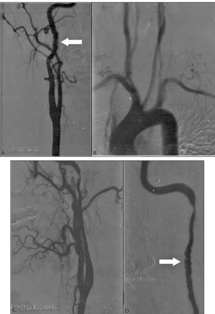
FIGURE 1 Digital luoroscopy angiography. A. Right carotid, oblique view. B. Aortic arch, oblique view. C. Left carotid, lateral view (proile). D. Left internal carotid artery, oblique view. Arrows: “stacked coins” appearance.
FIGURE 2 Gadolinium-enhanced T1-weighted cranial magnetic resonance imaging, six months after initial event. Arrows: area of ischemia.
A B
SPONTANEOUSCAROTIDDISSECTION
REV ASSOC MED BRAS 2017; 63(5):397-400 399
similar (true lumen: 37 cm/s, and false lumen: 31 cm/s). This evidenced the high flow character in the false light, but without causing significant stenosis in the true light. No subintimal carotid hematoma was identified. The evalu-ation of renal arteries on CDUS did not confirm a typical pattern of fibromuscular dysplasia with a succession of dilatations and stenoses (“string of beads”). Then, clopi-dogrel was suspended, while ASA was maintained at a dose of 100 mg/day. The patient remained with no recur-rence of stroke after one year of conservative treatment. She had complete remission of dysphasia and showed, on CDUS, a reduction in the extent of dissection to 1 cm, without hematoma or significant stenosis.
D
ISCUSSIONThe case corroborates the epidemiology of CD, having occurred in a young adult and affected the internal ca-rotid artery hereafter the bifurcation as seen in up to 2.5% of all cases of stroke.1 The only probable risk factor was
the suggestive pattern of fibromuscular dysplasia on ca-rotid endovascular angiography. The situation illustrates multifactorial etiopathogenesis and the difficulty in es-tablishing a specific etiology. Currently, the main scien-tific evidence for clinical decision making is a systematic
review of randomized clinical trials.10,11 A Cochrane
sys-tematic review of randomized clinical trials on antithrom-botic drugs for carotid artery dissection conducted in 2010 yielded no evidence in this regard.2 In 2015,
how-ever, new evidence was produced. A randomized clinical trial found no significant difference between treatment with anticoagulants or antiplatelet agents to prevent death and stroke in symptomatic dissection of carotid or ver-tebral arteries.12 In this clinical trial, recurrent events were
much rarer than in previous observational studies, and the initial diagnosis of dissection was quite misleading after a thorough review of the images. This shows that radiographic criteria are not always correctly applied in clinical practice. Lower levels of evidence report that en-dovascular treatments, such as angioplasty with stenting or thrombolysis, may be superior to conservative treatment in selected populations.13-16 The case presented is in line
with the best scientific evidence available at the time. The patient underwent conservative treatment with antiplate-let agents and did not present recurrence of stroke or death within one year of follow-up.
Although rare, CD should always permeate the diag-nostic suspicion in cases of stroke, especially in young patients and/or those with risk factors for developing the
FIGURE 3 Doppler vascular ultrasound, six months after initial event. A. Color mode with ying yang image. B. Spectral mode, true lumen. C. Spectral mode, false lumen. LICA: left internal carotid artery; LECA: left external carotid artery.
A
B C
LECA
LICA
FLUMIGNAN CDQ ETAL.
400 REV ASSOC MED BRAS 2017; 63(5):397-400
disease. Diagnostic confirmation and follow-up should include non-invasive ancillary methods, with thorough collection and evaluation of these images. The best level of available evidence still recommends conservative treat-ment as a general measure, either with anticoagulation or anti-platelet aggregation.
A
CKNOWLEDGMENTSWe thank the patient for authorizing her case report.
R
ESUMODissecção espontânea de carótida
A dissecção de carótida é entidade rara, mas é a principal causa de acidentes vasculares cerebrais isquêmicos em me-nores de 45 anos e pode ser a etiologia de até 25% dos aci-dentes vasculares cerebrais em adultos jovens. Apresenta-se um caso com imagem clássica de ying yang à ultrassonogra-fia vascular, que foi tratado de acordo com as melhores evidências médicas disponíveis e apresentou boa evolução.
Palavras-chave: dissecção da artéria carótida interna, medi-cina baseada em evidências, inibidores da agregação de pla-quetas, acidente vascular cerebral, ultrassonografia Doppler.
R
EFERENCES1. Blum CA, Yaghi S. Cervical artery dissection: a review of the epidemiology, pathophysiology, treatment, and outcome. Arch Neurosci. 2015; 2(4):e26670. 2. Lyrer P, Engelter S. Antithrombotic drugs for carotid artery dissection.
Cochrane Database Syst Rev. 2010; (10):CD000255.
3. Rusu O, Vasile M, Bajenaru O, Antochi F. Evolution of internal carotid artery occlusion in non-traumatic carotid dissection. Mædica (Buchar). 2014; 9(2):194-7.
4. Nasser M, Vega MB, Pivetta LGA, Nasser AI, Melo DG. Internal carotid artery dissection in a patient with Ehlers-Danlos syndrome type IV: diagnosis and management. J Vasc Bras. 2013; 12(2):174-9.
5. Campos CR, Bassi TG, Pinto F, Abrahão DKP. [Internal carotid artery dissection in a patient with recent respiratory infection: case report of a possible link]. Arq Neuropsiquiatr. 2005; 63(2B):523-6.
6. Campos-Herrera CR, Scaff M, Yamamoto FI, Conforto AB. Spontaneous cervical artery dissection: an update on clinical and diagnostic aspects. Arq Neuropsiquiatr. 2008; 66(4):922-7.
7. Pieri A, Spitz M, Valiente RA, Avelar WM, Silva GS, Massaro AR. Dissecção espontânea das artérias carótidas e vertebrais em uma população multiétnica. Arq Neuropsiquiatr. 2007; 65(4A):1050-5.
8. Marin LF, Bichuetti DB, Felício AC, Santos WAC, Oliveira FF, Morita ME, et al. Hypoglossal ner ve palsy as the sole manifestation of spontaneous internal carotid artery dissection. Arq Neuropsiquiatr. 2009; 67(1):107-8.
9. Silvariño R, Mérola V, Firpo M, Pino A, Fraga L, Tafuri J, et al. Disección espontánea de carótida interna como causa de accidente cerebrovascular isquémico en el joven. Arch Med Int. 2009; 31(1):37-9.
10. Howick J, Chalmers I, Glasziou P, Greenhalgh T, Heneghan C, Liberati A, et al. Explanation of the 2011 Oxford Centre for Evidence-Based Medicine (OCEBM) Levels of Evidence (Background Document); 2011 [cited 2016 Mar 3]. Available from: http://www.cebm.net/index.aspx?o=5653. 11. OCEBM Levels of Evidence Working Group. The Oxford 2011 Levels of
Evidence; 2011 [cited 2016 Mar 3]. Available from: http://www.cebm.net/ index.aspx?o=5653.
12. CADISS trial investigators, Markus HS, Hayter E, Levi C, Feldman A, Venables G, et al. Antiplatelet treatment compared with anticoagulation treatment for cervical artery dissection (CADISS): a randomised trial. Lancet Neurol. 2015; 14(4):361-7.
13. Huang R, Niu L, Wang Y, Jia G, Jia L, Wang Y, et al. Endovascular versus non-interventional therapy for cervicocranial artery dissection in east Asian and non-east Asian patients: a systematic review and meta-analysis. Sci Rep. 2015; 5:10474.
14. Tsivgoulis G, Zand R, Katsanos AH, Sharma VK, Goyal N, Krogias C, et al. Safety and outcomes of intravenous thrombolysis in dissection-related ischemic stroke: an international multicenter study and comprehensive meta-analysis of reported case series. J Neurol. 2015; 262(9):2135-43. 15. Haussen DC, Jadhav A, Jovin T, Grossberg JA, Grigoryan M, Nahab F, et al.
Endovascular management vs intravenous thrombolysis for acute stroke secondary to carotid artery dissection: local experience and systematic review. Neurosurgery. 2016; 78(5):709-16.