Cardiac remodeling indicators in adolescent athletes
JOAQUIM CASTANHEIRA1,2, JOÃO VALENTE-DOS-SANTOS2,4,5,6, DANIELA COSTA2,3, DIOGO MARTINHO2,3,7, JORGE FERNANDES3,8,
JOÃO DUARTE2,3,9, NUNO SOUSA2,10, VASCO VAZ2,3, LUIS RAMA2,3, ANTÓNIO FIGUEIREDO2,3, MANUEL COELHO-E-SILVA2,3,4,7,9*
1Department of Clinical Physiology, School of Health and Technology, Instituto Politécnico de Coimbra, Coimbra, Portugal 2CIDAF (uid/dtp/04213/2016), Universidade de Coimbra, Coimbra, Portugal
3Faculty of Sport Science and Physical Education, Universidade de Coimbra, Coimbra, Portugal 4Fundação para a Ciência e a Tecnologia (SFRH/BPD/100470/2014), Lisboa, Portugal
5Institute for Biomedical Imaging and Life Sciences (IBILI), Faculty of Medicine, Universidade de Coimbra, Coimbra, Portugal 6Faculty of Physical Education and Sport, Universidade Lusófona de Humanidades e Tecnologias, Lisboa, Portugal 7Fundação para a Ciência e a Tecnologia (SFRH/BD/121441/2016), Lisboa, Portugal
8Federação Portuguesa de Judô, Lisboa, Portugal
9Fundação para a Ciência e a Tecnologia (SFRH/BD/101083/2014), Lisboa, Portugal 10Faculdade Estácio de Sá de Vitória, Vitória, ES, Brazil
SUMMARY
Study conducted by the UID/DTP/ 04213/2016, Faculty of Sport Sciences
and Physical Education, Universidade de Coimbra, and by the Department of
Clinical Physiology, School of Health and Technology, Instituto Politécnico de
Coimbra, Coimbra, Portugal
Article received: 10/26/2016
Accepted for publication: 12/1/2016
*Correspondence:
Faculdade de Ciências do Desporto e Educação Física, Universidade de Coimbra
Address: Estádio Universitário de Coimbra, Pavilhão III,
Santa Clara, Portugal Postal code: 3040-156
http://dx.doi.org/10.1590/1806-9282.63.05.427
Objective: The idea that different sports and physical training type results in different cardiac adaptations has been widely accepted. However, this remodelling process among different sport modalities is still not fully understood. Thus, the current study aims to investigate the heart morphology variation associated with a set of different modalities characterized by distinct models of preparation and different methods and demands of training and completion.
Method: The sample comprises 42 basketball players, 73 roller hockey players, 28 judo athletes and 21 swimmers. Anthropometry was assessed by a single and experienced anthropometrist and the same technician performed the echocar-diographic exams. Analysis of variance was used to study age, body size and echocardiograph parameters as well as different sport athlete’s comparison. Results: Basketball players are taller (F=23.448; p<0.001; ES-r=0.553), heavier (F=6.702; p<0.001; ES-r=0.334) and have a greater body surface area (F=11.896; p<0.001; ES-r=0.427). Basketball and hockey players have larger left auricle diameters compared with judo athletes (F=3.865; p=0.011; ES-r=0.316). Interventricular end-diastolic septal thickness (F=7.287; p<0.001; ES-r=0.347) and left ventricular posterior wall thickness (F=8.038; p<0.001; ES-r=0.362) of the judokas are smaller compared to the mean values of other sports participants. In addition, relative left parietal ventricular wall thickness is lower among swimmers compared with judokas (F=4.127; p=0.008; ES-r=0.268).
Conclusion: The major contributors to changes in heart morphology are for the most part associated with sport-specific training and competition and the specific dynamics and adaptive mechanisms imposed by each sport.
Keywords: sports modalities, young athletes, echocardiography, left ventricle mass, body size, growth.
I
NTRODUCTIONThe practice of sports offers a field of application to un-derstand left ventricular (LV) cardiac remodeling, and is addressed in several studies1-3 pointing to the occurrence of concentric or eccentric hypertrophy depending on the modality practiced. However, athletes of the same modal-ity, exposed to apparently similar training methodologies, evidence different cardiac remodeling processes,4 which implies the need to systematize knowledge across the vast
spectrum of sports modalities and, within each of them, sex and age group by competitive level. Complementa-rily, no evidence of dichotomous cardiac adaptation was found among athletes of modalities with predominance of strength in the performance structure.5 The level of demand for the upper limbs is considered relevant in the process of cardiac remodeling.6
is characterized by the existence of multiple and repeat-ed episodes of high intensity and short duration exertion,7 and therefore is naturally regarded as a modality in which short-term energy demands predominate. Roller hockey, like most general sports games, has a complex structure, and it has been estimated that 71% of playing time (n=14, 20-32 years) includes intermittent high intensity activi-ties, with an average heart rate of 163.5 bpm and a refer-ence lactate concentration of 4.20±0.95 mmol/L.8 Despite some similarity between collective sports games in rela-tion to the intermittent nature of the effort, roller hock-ey players are characterized by a predominance of mus-culoskeletal structures,9 as opposed to basketball, which attracts and promotes players of high stature. Modalities such as basketball, roller hockey and swimming were classified as sports with a low or moderate static compo-nent and high dynamic compocompo-nent, unlike judo,10 a mo-dality in which repeated application of force and resis-tance is fundamental.11
Cardiac dimensions in children and young people were assumed to be directly proportional to height.12 In adolescent athletes, the available studies describe LV mor-phology and function as well as other cardiac parameters,1,13 but it is not always the interpretation of results relating to the relationships between sporting modalities, cavity dimensions and wall thickness that consider the addi-tional effect attributable to body size, proporaddi-tionality between trunk and limbs, and components of body com-position. Our study examines the variation in the morphol-ogy of the heart associated to a set of sports modalities that are characterized by distinct long-term preparation models, different training and competition methodologies, and unique processes of selection and promotion of the most talented athletes. Our objective is to complement the studies that only proceed to the comparative analysis of athlete and non-athlete adolescents.
M
ETHODStudy design and population
Our study has a cross-sectional design and comprises young male athletes of various modalities (n=164, 15.28±0.76 years): 42 basketball players (including 24 international players with participation in European championship games in their respective age groups), 73 hockey players (including 12 international and European champions in their respective age groups), 28 judokas (recruited to train with the national team in their respec-tive age groups) and 21 swimmers (classified among the best swimmers of the Central Region of Portugal in their respective age groups). Several groups were formed
ac-cording to age: 14.0-14.9 years (n=41); 15.0-15.9 years (n=100) and 16.0-16.9 years (n=23). All athletes were in-volved in federated sports for at least five years. None of the participants had evidence of organic disease or were under the effect of any active substance (medication or ergogenic supplementation). The study was approved by the Research Ethics Council and the Scientific Council of the FCDEF, University of Coimbra, and later by the Portuguese Foundation for Science and Technology (SFRH/PROTEC, process no. 67972/2010), in accordance with the principles of the Declaration of Helsinki. Par-ticipants and their representatives signed free and in-formed consent terms, ensuring the voluntary nature of the participation, as well as the confidentiality of the data.
Whole body anthropometric measures
Stature, body mass, and triceps subcutaneous and sub-scapular fat folds were evaluated by a single experienced anthropometrist, following the procedures described by Lohman et al.14 Stature was measured using a Harpenden stadiometer (model 98.603, Holtain Ltd, Crosswell, UK) and body mass using a SECA scale (model 770, Hanover, MD, United States). The folds of subcutaneous fat were measured using a Lange Skinfold Caliper adipometer (Beta Technology, Ann Arbor, MI, United States). Technical er-rors of measurement for height, body mass and folds of subcutaneous fat were 0.27 cm, 0.47 kg, and 0.47-0.72 mm, respectively. Percentage of fat mass was estimated ac-cording to the protocol by Slaughter et al.15 Subsequently, the absolute values for fat and lean mass were determined, in kilograms.
Echocardiographic evaluation
measure-ments were obtained: aortic root (AoR), left auricle (LA), left ventricular end-diastolic and end-systolic (LVED and LVES, respectively) diameters, and interventricular septal wall (IVSW) and left ventricular posterior wall (LVPW) thicknesses. Based on the previous dimensions, we calcu-lated LV mass (LVM) using the ASE cube formula modi-fied by Devereux et al.19 and measures of the predictors (IVSW, LVED and LVPW) expressed in millimeters:
LVM (g) = 0.8 × [1.04 × ((IVSW + LVED + LVPW)3 - LVED3)] + 0.6
(Equation 1)
LVM was adjusted for body surface area (BSA, equation 2), allowing the calculation of the LV mass index (g/m2). Sub-sequently, left ventricular relative wall thickness (LVRPT) was determined as described by equation 3 to distinguish an LV concentric (≥0.44) or eccentric (<0.44) profile:1,13
BSA (m2) = 0.007184 × [body mass (kg)]0.425× [height (cm)]0.725
(Equation 2)
LVRTP = [(IVSW + LVPW) / LVED]
(Equation 3)
LV systolic function is translated by calculating the LV shortening (LVSF) and ejection fraction (LVEF). The short-ening fraction uses the diastolic (LVED) and end-systolic (LVES) diameters, while the ejection fraction uses the end-diastolic (LVEDV) and the end-systolic (LVESV) volumes, respectively, according to equations 4 and 5:18
LVSF = [(LVED – LVES) / LVED] × 100
(Equation 4)
LVEF = [(LVEDV – LVESV) / LVEDV] × 100
(Equation 5)
Data quality was assessed by determining the intraob-server variability using a random subgroup of 20 indi-viduals (basketball players, n=5; hockey players, n=5; non-athletes, n=10) who were evaluated and reevaluated after one week by the same investigator, using the same equip-ment and following the procedures described above. The difference and percentage of difference between the two measurements were determined considering a 95% con-fidence interval for the dimensions of the cavities and cardiac thicknesses (0.02-0.17 mm, 95CI -1.95–2,28 mm). The parameters resulting from the evaluation of the pre-vious measurements had a percentage of intraobserver variability ranging from 0.3 to 0.8% (95CI -4.1–8.1%).
Statistical analysis
Minimum, maximum, mean and standard deviation values were calculated for all variables. The absolute frequencies of adolescents divided by age group were calculated, con-sidering also their classification by sport modality. We then used variance analysis (ANOVA) to study the age-related variation in body size measurements and parameters result-ing from the echocardiographic evaluation, considerresult-ing the entire sample. Finally, we used ANOVA once again to obtain a new comparison between athletes in different sports modalities, and covariance analysis (ANCOVA) adjusted for the additional effect of age, height and body mass. For the two analyzes of variance, we calculated the magnitude of the correlation effect (ES-r) estimated by the square root of the ratio of the t value squared and the
dif-ference between t value squared and the degrees of freedom.
The level of significance was maintained at 5%, as estab-lished for social and behavioral sciences. For statistical analysis of the data, we use Statistical Program for Social Sciences – SPSS software, version 19.0 for Windows.
R
ESULTSThe characteristics of the study’s total sample are shown in Table 1. The dispersion measures given by the range of variation are quite high, that is, 52 cm for height and 72.9 kg for body mass. The central tendency values found for the echocardiographic parameters show a slight in-crease in left ventricular cavity diameter (54.5 mm) for these age groups, suggesting a markedly eccentric profile (LVRPT=0.28).
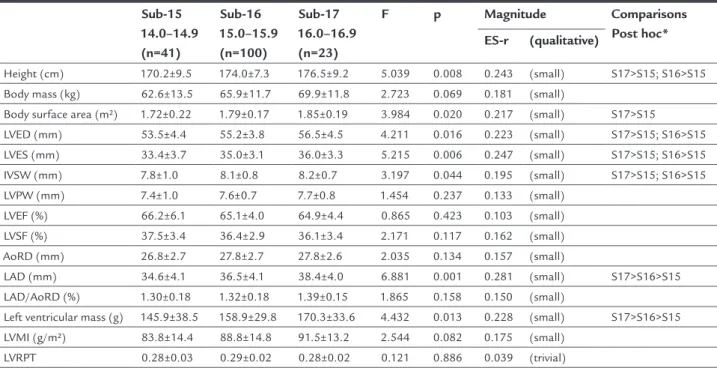
The age-related variation for the entire sample of athletes is shown in Table 2. Understandably, the groups of athletes aged 15 and 16 years had higher mean values for height (F=5.039; p<0.01; ES-r=0.243) and BSA (F=3.984; p<0.05; ES-r=0.217). Although this trend also occurs in relation to body mass, the values are not statistically sig-nificant. Additionally, in relation to the echocardiograph-ic parameters, those in the groups of 15 and 16 year-old athletes, compared to their 14-year-old peers, had higher means in left ventricular end-diastolic (F=4.211; p<0.05; ES-r=0.223) and end-systolic (F=5.215; p<0.01; ES-r=0.247) diameters, as well as IVSW thickness (F=3.197; p<0.05; r=0.195). Also, LA diameter (F=6.881; p<0.01; ES-r=0.281) and LVM (F=4.432; p<0.05; ES-r=0.228) were higher in the 16 year-old group.
TABLE 1 Characteristics of the total study sample (n=164).
Variable Unit
measured
Abbreviation Amplitude Mean Standard deviation Minimum Maximum Value (95CI)
Age years 13.31 16.66 15.28 (15.19-15.39) 0.76
Height cm 143.6 195.6 172.7 (172.1-174.7) 8.4
Body mass kg 38.4 111.3 65.2 (63.7-67.5) 12.6
Body surface m² BSA 1.24 2.32 1.77 (1.75-1.81) 0.19
LV end-diastolic diameter mm LVED 44.2 65.0 54.9 (54.3-55.6) 4.2 LV end-systolic diameter mm LVES 24.9 45.5 34.7 (34.2-35.3) 3.4
Interventricular end-diastolic septal wall thickness mm IVSW 5.8 10.6 8.1 (7.9-8.2) 0.8 LV end-diastolic posterior wall thickness mm LVPW 5.5 10.0 7.6 (7.5-7.7) 0.8
LV ejection fraction % LVEF 42.0 76.0 65.4 (64.7-66.1) 4.5
LV shortening fraction % LVSF 29.8 45.3 36.7 (36.2-37.1) 3.1
Aortic root diameter mm AoRD 19.6 34.0 27.5 (27.1-27.9) 2.7
LA diameter mm LAD 20.0 47.9 36.2 (35.6-36.9) 4.2
LA diameter:aortic root ratio mm/mm LAD/AoRD 0.77 2.00 1.33 (1.30-1.35) 0.17
Left ventricular mass g LVM 83.1 258.1 157.3 (152.1-162.4) 33.6
Left ventricular mass index g/m² LVMI 54.6 136.1 87.9 (85.7-90.2) 14.9
LV relative parietal thickness LVRPT 0.22 0.36 0.29 (0.28-0.30) 0.03
Note: 95CI: 95% conidence interval; LV: left ventricle; LA: left auricle.
TABLE 2 Descriptive statistics (mean ± standard deviation) and age-related variation for the entire sample of athletes.
Sub-15 14.0–14.9 (n=41)
Sub-16 15.0–15.9 (n=100)
Sub-17 16.0–16.9 (n=23)
F p Magnitude Comparisons Post hoc* ES-r (qualitative)
Height (cm) 170.2±9.5 174.0±7.3 176.5±9.2 5.039 0.008 0.243 (small) S17>S15; S16>S15 Body mass (kg) 62.6±13.5 65.9±11.7 69.9±11.8 2.723 0.069 0.181 (small)
Body surface area (m²) 1.72±0.22 1.79±0.17 1.85±0.19 3.984 0.020 0.217 (small) S17>S15
LVED (mm) 53.5±4.4 55.2±3.8 56.5±4.5 4.211 0.016 0.223 (small) S17>S15; S16>S15 LVES (mm) 33.4±3.7 35.0±3.1 36.0±3.3 5.215 0.006 0.247 (small) S17>S15; S16>S15
IVSW (mm) 7.8±1.0 8.1±0.8 8.2±0.7 3.197 0.044 0.195 (small) S17>S15; S16>S15 LVPW (mm) 7.4±1.0 7.6±0.7 7.7±0.8 1.454 0.237 0.133 (small)
LVEF (%) 66.2±6.1 65.1±4.0 64.9±4.4 0.865 0.423 0.103 (small) LVSF (%) 37.5±3.4 36.4±2.9 36.1±3.4 2.171 0.117 0.162 (small)
AoRD (mm) 26.8±2.7 27.8±2.7 27.8±2.6 2.035 0.134 0.157 (small)
LAD (mm) 34.6±4.1 36.5±4.1 38.4±4.0 6.881 0.001 0.281 (small) S17>S16>S15 LAD/AoRD (%) 1.30±0.18 1.32±0.18 1.39±0.15 1.865 0.158 0.150 (small)
Left ventricular mass (g) 145.9±38.5 158.9±29.8 170.3±33.6 4.432 0.013 0.228 (small) S17>S16>S15 LVMI (g/m²) 83.8±14.4 88.8±14.8 91.5±13.2 2.544 0.082 0.175 (small)
LVRPT 0.28±0.03 0.29±0.02 0.28±0.02 0.121 0.886 0.039 (trivial)
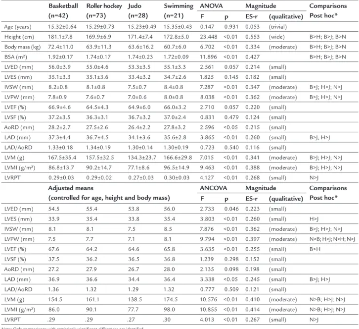
hockey players, judokas and swimmers. The variation associated with sports modality for echocardiographic parameters was only significant for some variables of car-diac morphology. Basketball players, hockey players and swimmers had higher values for IVSW thickness (F=7.287; p<0.001; ES-r=0.347) and LVPW thickness (F=8.038; p<0.001; ES-r=0.362) compared to the judokas. The same happens in the test of comparison of means having as dependent variables the LVM (F=7.015; p<0.001; ES-r=0.341) and left ventricular mass index (F=9.463; p<0.001; ES-r=0.388). Basketball players and hockey players have a larger left atrial cavity, compared to judokas (F=3.865; p=0.011; ES-r=0.316). Finally, the data points to a higher LVRPT in swimmers compared to judokas (F=4.127; p=0.008; ES-r=0.268), being swimmers characterized by a markedly eccentric left ventricular profile (Table 3). Table 3 also shows the results of covariance analysis. Co-variates included: chronological age, stature and body mass, allowing to verify that, in general, the differences between the size of the cavities and thickness of the heart tissue structures, as well as composite measures, confirm the main conclusions previously described. In fact, it suggests that the results found are mainly due to chron-ic adaptations to the effects of training and competition and not so much to the sport selection factor that associ-ates particular morphological archetypes to each of the modalities, particularly, tall adolescents to basketball. The results presented in Table 3, adjusted for mean values
of age equal to 15.30 years, height equal to 173.4 cm and body mass equal to 65.6 kg, allow us to conclude that judo athletes have less increase in IVSW (F=7.876; p<0.01; ES-r=0.362) and LVPW (F=9.794; p<0.01; ES-r=0.397) thicknesses. As for the latter variable, after adjustment for age and body size, it is still possible to distinguish swimmers, who present significantly higher values com-pared to basketball players and hockey players. The small-er dimensional development of a judoka’s heart is also noted with respect to the left atrial cavity (F=3.338; p<0.05; ES-r=0.245). Finally, in agreement with the previous points and after controlling a number of additional effects, the comparison between modalities, with respect to the mean values of the left ventricular mass, presents the swimmers positively and the judokas negatively.
D
ISCUSSIONRecognizing the complexity derived from difficulties in distinguishing the effects of sports training, growth and biological maturation, our study analyzed the effects of age and sports modality on the remodeling process of the heart cavities of young athletes in their late teens.
Manolas et al.20 refer to this age group as the period of greatest increase in cardiac chambers. In our study, the mean LV end-diastolic diameter was 1.7 mm greater in 15-year-old athletes compared to the mean value deter-mined for athletes at an immediately younger age, i.e., 14 years. Likewise, a mean difference of 1.3 mm was found between the 15 and 16 year olds, with the highest values among the older athletes. This direct variation between age and measures of LV end-diastolic diameter coincides with the increase in height and body surface of younger adolescents compared to the older ones.
Recently, a longitudinal study21 with healthy adoles-cents aged 11 to 15 years determined that sports partici-pation for 3.1±1.2 hours per week plus one match per week was not associated with increased LV growth, after statistical adjustment for individual differences in growth and biological maturation. However, it has been widely accepted that different modalities of physical training result in diverse patterns of cardiac adaptation in athletes.17 The association established between anthropometry of the whole body and the generalities of the echocardio-graphic parameters should also be recognized.21-23 It is true that higher values for body size translate into high-er values for the dimensions of aortic root, left atrium, ventricular cavities and LV wall thickness.16 Recently, a study with male hockey players aged 14.5 to 16.5 years determined correlations between left ventricular mass and several indicators: 0.36 with chronological age, 0.50 with skeletal age, 0.52 with height, 0.61 with seated height, 0.56 with lean mass.22
After organizing the analysis of the means by sports modality, we found that the basketball players were tall-er, heavier and consequently presented greater body sur-face compared to their peers in the remaining three sports modalities. In fact, basketball players are recognized and consensually among the groups of athletes with larger body size and are also those with higher values for LA diameter, IVSW and LVPW thickness, LVM and left ven-tricular mass index.24 In this particular aspect, the results of our study are similar to those of Madeira et al.,1 which also included 15-16 year-old participants. Variations of heart dimensions cannot be attributed solely to the adap-tive effect to training.
but higher values for IVSW and LVPW thickness compared to young hockey players. Although we have also found a trend towards slightly higher mean values in our study, the differences are not statistically significant.
Swimmers, compared to control groups, often present higher values for cardiac cavity diameters and LV wall thickness.25 Future studies should have a mixed longitu-dinal design with samples derived from a single modal-ity or, desirably, sub-samples extracted from more than
one modality, associating measures capable of describing the volume and weekly intensity of training and compe-titions to the longitudinal predictors. Regarding LVRPT, all modalities studied have a markedly eccentric profile, although in the judokas this profile is significantly more pronounced than in swimmers. While some authors men-tion that the enlargement of the left atrium is also a com-ponent of the athlete’s heart,26 we did not find significant changes in the other echocardiographic variables between
TABLE 3 Descriptive statistics (mean ± standard deviation) for the groups of athletes organized by sport modality and
ANOVA to test the variation associated to the modality practiced, followed by ANCOVA with adjustment for age (centered in 15.30 years), height (centered in 173.4 cm) and body mass (centered at 65.6 kg).
Basketball (n=42)
Roller hockey (n=73)
Judo (n=28)
Swimming (n=21)
ANOVA Magnitude Comparisons Post hoc* F p ES-r (qualitative)
Age (years) 15.32±0.64 15.29±0.73 15.23±0.49 15.35±0.43 0.147 0.931 0.053 (trivial)
Height (cm) 181.1±7.8 169.9±6.9 171.4±7.4 172.8±5.0 23.448 <0.01 0.553 (wide) B>H; B>J; B>N
Body mass (kg) 72.4±11.0 63.9±11.3 63.6±16.2 60.7±6.0 6.702 <0.01 0.334 (moderate) B>H; B>J; B>N BSA (m²) 1.92±0.17 1.74±0.17 1.74±0.23 1.72±0.09 11.896 <0.01 0.427 B>H; B>J; B>N LVED (mm) 56.0±3.9 55.0±4.6 53.3±3.5 55.1±3.3 2.561 0.057 0.214 (small)
LVES (mm) 35.1±3.3 35.1±3.6 33.4±3.2 34.7±2.6 1.825 0.145 0.182 (small)
IVSW (mm) 8.2±0.8 8.1±0.8 7.5±0.7 8.4±0.8 7.287 <0.01 0.347 (moderate) B>J; H>J; N>J LVPW (mm) 7.8±0.9 7.6±0.7 7.0±0.6 8.0±0.8 8.038 <0.01 0.362 (moderate) B>J; H>J; N>J
LVEF (%) 66.9±4.6 64.5±4.3 64.9±6.0 66.0±3.2 2.710 0.057 0.220 (small) LVSF (%) 37.2±3.5 36.3±3.1 36.7±3.2 37.0±2.4 0.831 0.479 0.124 (small) AoRD (mm) 28.2±2.7 27.5±2.6 26.4±2.2 27.8±3.2 2.596 <0.05 0.215 (small)
LAD (mm) 37.3±4.4 36.7±4.5 34.1±3.6 35.6±2.8 3.865 <0.01 0.260 (small) B>J; H>J
LAD/AoRD 1.33±0.18 1.34±0.19 1.30±0.14 1.30±0.19 0.723 0.540 0.116 (small)
LVM (g) 167.5±35.4 157.5±32.5 134.3±23.7 166.6±29.8 7.015 <0.01 0.341 (moderate) B>J; H>J; N>J LVMI (g/m²) 86.8±13.7 90.2±14.7 77.1±8.6 96.5±14.9 9.463 <0.01 0.388 (moderate) B>J; H>J; N>J
LVRPT 0.29±0.03 0.29±0.02 0.27±0.03 0.30±0.03 4.127 <0.01 0.268 (small) N>J Adjusted means
(controlled for age, height and body mass)
ANCOVA Magnitude Comparisons Post hoc* F p ES-r (qualitative)
LVED (mm) 54.5 55.4 53.8 56.0 2.733 0.046 0.223 (small)
LVES (mm) 33.9 35.4 33.8 35.4 3.803 <0.01 0.260 (small) H>J IVSW (mm) 8.1 8.1 7.5 8.5 7.876 <0.01 0.362 (moderate) B>J; H>J; N>J
LVPW (mm) 7.5 7.7 7.1 8.1 9.794 <0.01 0.397 (moderate) N>B; H>J; N>H; N>J LVEF (%) 67.6 64.2 64.6 65.8 3.635 <0.01 0.255 (small) B>H
LVSF (%) 37.5 36.2 36.5 36.8 1.239 0.298 0.152 (small) AoRD (mm) 27.2 27.9 26.7 28.0 2.135 0.098 0.198 (small)
LAD (mm) 36.9 36.6 34.4 36.4 3.338 <0.05 0.245 (small) B>J; H>J LAD/AoRD 1.36 1.32 1.29 1.32 0.777 0.509 0.121 (small)
LVM (g) 154.5 161.1 138.5 174.5 10.576 <0.01 0.410 (moderate) N>B; H>J; N>J
LVMI (g/m²) 86.0 90.1 77.7 98.0 10.855 <0.01 0.414 (moderate) N>B; H>J; N>J
LVRPT .29 .29 .27 .30 4.013 <0.01 0.267 (small) N>J
Note: Only comparisons with statistically signiicant differences are identiied.
the groups, so it is possible that there are more differ-ences between athletes and non-athletes than between athletes of different modalities.
The study of systolic function, performed by calculat-ing the shortencalculat-ing and ejection fractions of the left ven-tricle, confirms the results reported in the literature,27 that is, no significant differences were found between groups. This fact demonstrates that the increase of the cavities and the thickness of the walls of the LV represents for the athletes an increase in cardiac performance, with-out any impairment at functional level, like other (path-ological) situations that can progress to increased car-diac dimensions. In this case, the increase in dimensions is due to the sports training and is performed in a har-monic way, in accordance with the proportion with the body weight.28
Limitations
Four sporting modalities have been taken into account, making it difficult to obtain statistically significant results with so many groups. Therefore, it is possible that the choice of only two clearly contrasting modalities would result in greater association. Magnetic resonance imaging is the most appropriate method to determine LVM, al-though echocardiographic examination is still the most used methodology, particularly in the context of clinical practice and, especially, in the child and adolescent popu-lation. Future studies should consider female athletes and other age groups coinciding with periods of greater sport-ing specialization, i.e., 17-19 years. Variability of measures resulting from echocardiographic examination with mea-sures of body composition should also be considered.
C
ONCLUSIONBriefly, modalities such as basketball and roller hockey tend to attract, respectively, tall and short adolescents, due to different aspects of performance structure, name-ly airspace dispute in basketball and skills related to fast movements with the ball next to the ground in roller hockey. It seems that differences in cardiac morphology between athletes of different modalities should also be understood in the light of adapting to the chronic effects of exertion. This is supported by the analyzes in our study, both in relation to the treatment of absolute val-ues and the control of additional effects related to age, stature and body mass. The differences found between judokas and swimmers corroborate the effect of the sports modalities on the process of cardiac remodeling. While the judokas practice an acyclic modality, with great peripheral resistance and less development of ventricular
mass, cavities and walls of the ventricle, swimmers are exposed to cyclical movements facilitating the ejection fraction and cardiovascular return. Swimmers training and competition loads are also characterized by less intermittent heart rate regimens, which contributes to the existence of higher mean values both for cardiac mass and cavities, and thickness of the walls limiting them.
A
CKNOWLEDGMENTSThe authors thank the coaches in charge of the national and district teams, national training centers, athletes, and those responsible for the athletes’ education, for their willingness to participate in our study. In addition, spe-cial thanks are due to the young researcher Filipe Simões (deceased on 02/22/2013), who participated in the col-lection of data and, although he did not write the article, he was mainly concerned with the data of the echocar-diographic evaluation. Three authors of our study received grants from the Foundation for Science and Technology [JC: SFRH/PROTEC/67972/2010; JVS: SFRH/ BPD/100470/2014; JPMD: SFRH/BD/101083/2014]. Data collection from the basketball athletes was par-tially supported by the Foundation for Science and Tech-nology [PTDC/DES/70918/2006]. CIDAF has the FCT register uid/dtp/04213/2016.
C
ONFLICT OF INTERESTThe authors declare no conflict of interest.
R
ESUMOIndicadores de remodelagem cardíaca em atletas adolescentes
Objetivo: Os efeitos decorrentes da prática de diferentes modalidades desportivas resultam em padrões divergentes de adaptação cardíaca. A presente pesquisa procura estu-dar a variação da morfologia do coração associada a um conjunto de modalidades desportivas distintas quanto à natureza do esforço e aos modelos de preparação, incluin-do metoincluin-dologias de treino e sistemas de competição. Método: Foram estudados 42 basquetebolistas, 73 hoquis-tas, 28 judocas e 21 nadadores. A antropometria foi avalia-da por um único e experiente antropometrista e os exames ecocardiográficos foram realizados pelo mesmo operador. Recorreu-se à análise da variância para estudar a variação associada a idade, medidas de tamanho corporal e parâme-tros ecocardiográficos, bem como para a comparação entre os atletas de diferentes modalidades desportivas.
p<0,001; ES-r=0,334), com maior superfície corporal (F=11,896; p<0,001; ES-r=0,427) e, com os hoquistas, apre-sentam um diâmetro da aurícula esquerda superior ao dos judocas (F=3,865; p=0,011; ES-r=0,316). A espessura telediastólica do septo interventricular (F=7,287; p<0,001; ES-r=0,347) e da parede posterior do ventrículo esquerdo (F=8,038; p<0,001; ES-r=0,362) dos judocas é inferior à dos outros atletas, mesmo quando controlado para o tamanho corporal. Os nadadores apresentam uma espes-sura parietal relativa do ventrículo esquerdo superior à dos judocas (F=4,127; p=0,008; ES-r=0,268).
Conclusão: As diferentes fontes de variação da morfolo-gia cardíaca prendem-se com as dinâmicas do processo de treino, competição e correspondentemente com os mecanismos adaptativos, sobrepondo-se ao processo de formação desportiva a longo prazo.
Palavras-chave: modalidades desportivas, jovens atletas, ecocardiografia, massa do ventrículo esquerdo, tamanho corporal, crescimento.
R
EFERENCES1. Madeira RB, Trabulo M, Alves F, Pereira JG. Effects of chronic exercise training on left ventricular dimensions and function in young athletes. Rev Port Cardiol. 2008; 27(7-8):909-22.
2. Naylor LH, George K, O’Driscoll G, Green DJ. The athlete’s heart: a contemporary appraisal of the ‘Morganroth hypothesis’. Sports Med. 2008; 38(1):69-90.
3. Sagiv M, Sagiv M, Ben-Sira D. Weight lifting training and left ventricu-lar function in adolescent subjects. J Sports Med Phys Fitness. 2007; 47(3):329-34.
4. Morganroth J, Maron BJ, Henry WL, Epstein SE. Comparative left ventricular dimensions in trained athletes. Ann Intern Med. 1975; 82(4):521-4. 5. Wernstedt P, Sjöstedt C, Ekman I, Du H, Thuomas KA, Areskog NH, et al.
Adaptation of cardiac morphology and function to endurance and strength training. A comparative study using MR imaging and echocardiography in males and females. Scand J Med Sci Sports. 2002; 12(1):17-25.
6. Gates PE, George KP, Campbell IG. Concentric adaptation of the left ventricle in response to controlled upper body exercise training. J Appl Physiol. 2003; 94:549-54.
7. Ben Abdelkrim N, El Fazaa S, El Ati J. Time-motion analysis and physiological data of elite under-19-year-old basketball players during competition. Br J Sports Med. 2007; 41(2):69-75.
8. Bonafonte LF, Pérez JR, Marrero RM. Physiological characteristics of roller-hockey: Study in the sports field. Arch Med Dep. 1994; 11(44):345-50. 9. Coelho-e-Silva MJ, Vaz V, Simoes F, Carvalho HM, Valente-Dos-Santos J,
Figueiredo AJ, et al. Sport selection in under-17 male roller hockey. J Sports Sci. 2012; 30(16):1793-802.
10. Mitchell JH, Haskell W, Snell P, Van Camp SP. Task Force 8: classification of sports. J Am Coll Cardiol. 2005; 45(8):1364-7.
11. Ullrich B, Pelzer T, Oliveira S, Pfeiffer M. Neuromuscular responses to short-term resistance training with traditional and daily undulating periodization in adolescent elite judoka. J Strength Cond Res. 2016; 30(8):2083-99. 12. Epstein ML, Goldberg SJ, Allen HD, Konecke L, Wood J. Great vessel,
car-diac chamber, and wall growth patterns in normal children. Circulation. 1975; 51(6):1124-9.
13. Zdravkovic M, Perunicic J, Krotin M, Ristic M, Vukomanovic V, Soldatovic I, et al. Echocardiographic study of early left ventricular remodeling in highly trained preadolescent footballers. J Sci Med Sport. 2010; 13(6):602-6. 14. Lohman TG, Roche AF, Martorell R. Anthropometric standardization
reference manual. Champaign: Human Kinetics; 1988.
15. Slaughter MH, Lohman TG, Boileau RA, Horswill CA, Stillman RJ, Van Loan MD, et al. Skinfold equations for estimation of body fatness in children and youth. Hum Biol. 1988; 60(5):709-23.
16. Pelliccia A, Di Paolo FM, Maron BJ. The athlete’s heart: remodeling, electro-cardiogram and preparticipation screening. Cardiol Rev. 2002; 10(2):85-90. 17. Basavarajaiah S, Boraita A, Whyte G, Wilson M, Carby L, Shah A, et al. Ethnic
differences in left ventricular remodeling in highly-trained athletes relevance to differentiating physiologic left ventricular hypertrophy from hypertrophic cardiomyopathy. J Am Coll Cardiol. 2008; 51(23):2256-62.
18. Lang RM, Bierig M, Devereux RB, Flachskampf FA, Foster E, Pellikka PA, et al. Recommendations for chamber quantification. Eur J Echocardiogr. 2006; 7(2):79-108.
19. Devereux RB, Alonso DR, Lutas EM, Gottlieb GJ, Campo E, Sachs I, et al. Echocardiographic assessment of left ventricular hypertrophy: comparison to necropsy findings. Am J Cardiol. 1986; 57(6):450-8.
20. Manolas VM, Pavlik G, Bánhegyi A, Faludi J, Sidó Z, Olexó Z. Echocardiographic changes in the development of the athlete’ s heart in 9 to 20-year-old male subjects. Acta Physiol Hung. 2001; 88(3-4):259-70. 21. Valente-Dos-Santos J, Coelho-e-Silva MJ, Castanheira J, Machado-Rodrigues
AM, Cyrino ES, Sherar LB, et al. The effects of sports participation on the development of left ventricular mass in adolescent boys. Am J Hum Biol. 2015; 27(4):530-7.
22. Valente-Dos-Santos J, Coelho-e-Silva MJ, Vaz V, Figueiredo AJ, Castanheira J, Leite N, et al. Ventricular mass in relation to body size, composition, and skeletal age in adolescent athletes. Clin J Sport Med. 2013; 23(4):293-9. 23. Valente-Dos-Santos J, Coelho-e-Silva MJ, Ferraz A, Castanheira J, Ronque
ER, Sherar LB, et al. Scaling left ventricular mass in adolescent boys aged 11-15 years. Ann Hum Biol. 2014; 41(5):465-8.
24. Corrado D, Pelliccia A, Bjørnstad HH, Vanhees L, Biffi A, Borjesson M, et al. Cardiovascular pre-participation screening of young competitive athletes for prevention of sudden death: proposal for a common European protocol. Consensus Statement of the Study Group of Sport Cardiology of the Working Group of Cardiac Rehabilitation and Exercise Physiology and the Working Group of Myocardial and Pericardial Diseases of the European Society of Cardiology. Eur Heart J. 2005; 26(5):516-24.
25. Triposkiadis F, Ghiokas S, Skoularigis I, Kotsakis A, Giannakoulis I, Thanopoulos V. Cardiac adaptation to intensive training in prepubertal swimmers. Eur J Clin Invest. 2002; 32(1):16-23.
26. D’Ascenzi F, Cameli M, Lisi M, Zacà V, Natali B, Malandrino A, et al. Left atrial remodelling in competitive adolescent soccer players. Int J Sports Med. 2012; 33(10):795-801.
27. Hoogsteen J, Hoogeveen A, Schaffers H, Wijn PF, van Hemel NM, van der Wall EE. Myocardial adaptation in different endurance sports: an echocardiographic study. Int J Cardiovasc Imaging. 2004; 20(1):19-26. 28. Dickhuth HH, Roecker K, Niess A, Hipp A, Heitkamp HC. The