AUDIOLOGIC PROFILE OF OLDER ADULTS SUBJECTED
TO VESTIBULAR REHABILITATION THERAPY
Perfil audiológico de idosos submetidos à reabilitação vestibular
Sara Alois de Abreu Martins (1), Iara Bassi (2), Patrícia Cotta Mancini (3)
(1) Universidade Federal de Minas Gerais, UFMG, Belo Hori-zonte, MG, Brasil.
(2) Faculdade de Medicina da Universidade Federal de Minas Gerais, UFMG, Belo Horizonte, MG, Brasil.
(3) Departamento de Fonoaudiologia da Faculdade de Medi-cina da Universidade Federal de Minas Gerais, UFMG, Belo Horizonte, MG, Brasil.
Support: Pró-Reitoria de Pesquisa, Universidade Federal de Minas Gerais
Conlict of interest: non-existent
Aging is directly associated with the presence of
otoneurological symptoms4. As the auditory system
exhibits anatomical-physiological continuity with the labyrinth, aging is associated with physiological changes in both the cochlea, which is responsible for hearing, and the vestibular system, which is responsible for balance5. Both are located in the
temporal bone, speciically in the labyrinth4,6, and
communicate through the ductus reuniens, which
connects the saccule to the cochlear duct4. The
organ of Corti is part of the membranous labyrinth, being contained in the same compartment as the
utricular macula, saccular macula, and crista
ampul-laris. Therefore, otological complaints related to the
inner ear are common among older adults.
The body balance is maintained by the vestibular,
visual, and proprioceptive systems, and when those
skills are affected, the response of those systems
decreases, resulting in vertigo or dizziness7,8. The
risk factors associated with dizziness include cardio
-vascular, cerebro-vascular, neurological, sensory, and metabolic diseases9.
The prevalence of presbycusis, i.e., age-related
auditory loss, is high among older adults; this
INTRODUCTION
Older adults, according to the World Health Organization (WHO), are individuals older than 65 years old; however, in Brazil, old age is considered
to begin at 601,2. Aging is associated with progressive
and dynamic physiological changes, resulting in
increased vulnerability and higher incidence of
pathologies1,3. According to the WHO, by 2025,
Brazil will rank sixth in the number of older adults worldwide, with an estimated elderly population of 32
million people2,3. The ongoing increase in life expec
-tancy requires establishing care and adjustment to meet the needs of the elderly3.
ABSTRACT
Purpose: to characterize the auditory proile of older adults with dizziness undergoing Vestibular Rehabilitation and compare the results obtained in the auditory evaluation of elderly without dizziness. Methods: a cross-sectional observational study of 87 seniors, including 35 in the group with dizziness
and 52 in the group without dizziness. History, pure tone audiometry and speech audiometry were
conducted. For statistical analysis, Statistical Package for Social Sciences version 17.0, with a signiicance level of 5% was used in all analyzes. Results: sensorineural hearing loss from mild to moderate degree was present in 72.4% of the sample, with worsening of hearing thresholds in frequencies above 4000Hz in both groups. Tinnitus was the most frequent symptom observed in the
sample. Conclusion: the hearing proile of elderly patients with dizziness does not differ from that found in older adults without dizziness, being observed more frequently bilateral mild sensorineural hearing loss with downward sloping coniguration.
a public hospital from August to December 2013
who met the inclusion criteria and voluntarily agreed to participate in the study. The older adults with
dizziness were referred from otorhinolaryngolo
-gists at primary care facilities to perform vestibular rehabilitation therapy, while the ones without dizziness were referred by geriatrists for auditory
testing.
Individuals aged 60 years old or older from both genders were considered eligible to participate in the
study. Individuals with physical, cognitive, or sensory
limitations that could hinder the performance of the auditory tests and individuals unable to understand and respond to simple verbal commands were excluded from the study.
The participants were informed as to the voluntary nature of participation, as well as on the
study procedures, stressing that they would not pose any health risk. The participants responded to a questionnaire that collected data on hearing
complaints, family history of hearing loss, history of acoustic trauma, tinnitus, and balance-related
complaints. Based on the questionnaire data, the participants were allocated into two groups: Group 1 – individuals with dizziness; and Group 2 – individuals without dizziness.
The participants were then subjected to inspection of the external auditory meatus to establish whether its condition was adequate for auditory assessment. Next, pure–tone threshold audiometry was performed in a sound-isolated
room using an audiometer Interacoustics® model
AVS-500. The air conduction audibility thresholds
were measured using TDH-39 earphones at
the frequencies 250, 500, 1,000, 2,000, 3,000, 4,000, 6,000 and 8,000 Hz. The bone-conduction thresholds were measured using vibrator B71 at the frequencies 500, 1,000, 2,000, 3,000 and 4,000 Hz bilaterally. Speech audiometry was performed by a single examiner using a loudspeaker, the intensity of which was controlled by the audiometer speech
level indicator. The parameters assessed were the
Speech Discrimination Score of 100% (SDS) and Speech Reception Threshold (SRT) of both ears. The results of pure-tone audiometry were analyzed according to the BIAP (1997) classiication, which is based on the average air conduction audibility
threshold at 500, 1,000, 2,000 and 4,000 Hz and
considers values up to 20 dBal as normal21. The
classiication of the hearing loss type (sensorineural, conductive, or mixed) and audiometric conigu
-ration followed the criteria formulated by Silman
and Silverman (1997)21. The results of speech
audiometry were classiied according to Jerger,
Speaks & Trammell (1968)22.
condition impairs the ability to understand
speech4,10. From the audiological perspective, it is
characterized by sensorineural hearing loss, with the auditory threshold for high frequencies affected. As a function of age, the mitotic capacity of some
cells decreases, intracellular pigments accumulate,
and the intercellular luid undergoes changes11.
Thus, the entire auditory organ (outer, middle, and
inner ear, central auditory pathways) is affected by aging12,13. As a rule, the hearing loss is due to
degeneration of the cochlea, affecting the basal portion of this organ, where the high-frequency sounds are identiied. Therefore, the appearance of those structural changes throughout the system is expected to cause hearing decline with age6,13,14.
Presbycusis is considered to be the main cause of hearing deiciency among older adults, with a preva
-lence of 30% among individuals older than 65 years
old15,16.
Among older adults, hearing loss can be associated with dificulties in oral communication and
in understanding the spoken language, especially in noisy environments, with relevant consequences
for the elderly within their social and familial environment. Among sensory deicits, hearing loss is the one that exerts the greatest impact on the life of individuals because it reduces the ability to share
ideas and thoughts through the oral language16-18.
The main dificulties exhibited by older adults with hearing loss concern the ability to talk on the phone, understand some words, hear alarms and doorbells, locate the source of sounds, and talk with a large
audience19,20.
As a function of the aforementioned facts, the degree of hearing loss might be greater in older
adults with dizziness compared to older adults
without this complaint: the affection of the vestibular
system might imply concomitant cochlear disorders,
as both systems are anatomically located within the
same organ.
The aim of this study was to characterize the auditory proile of older adults with dizziness subjected to vestibular rehabilitation therapy and to compare the results with the proiles of older adults
without dizziness.
METHODS
This cross-sectional, observational and analytical study was approved by the research ethics committee of Federal University of Minas
Gerais, ruling no. 0551/11. All the participants were
informed as to the study procedures and voluntarily signed an informed consent form.
sample was non-probabilistic, and more individuals without dizziness were referred for auditory testing
than individuals with dizziness. Nevertheless, the
average age was similar in both groups, 75.7 years
old in Group 1 and 76.9 years old in Group 2; that
difference was not statistically signiicant (p=0.661). The number of women with dizziness was larger; however, the difference in the number of women between the groups was not signiicant (p=1). Audiometry detected a predominance of bilateral hearing loss, as shown in Table 1.
The data collected in the interviews indicated
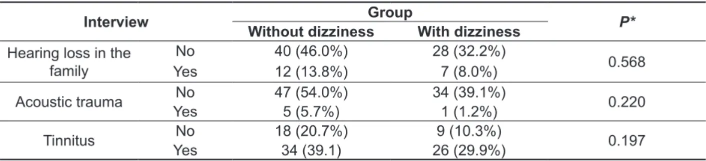
that tinnitus was the most frequent complaint in either group. No statistically signiicant difference was detected regarding history of acoustic trauma, tinnitus, or family history of hearing loss between the groups (Table 2).
The database was discussed by the investi
-gators and built using the software Excel. Statistical analysis was performed using the software SPSS (Statistical Package for the Social Sciences) version 17.0. The data were irst subjected to descriptive analysis, including measures of proportion, central
tendency, and dispersion, and then, the chi-square
test was used to assess the categorical variables
and the non-parametric Mann-Whitney test was
used to assess the continuous variables. The signii
-cance level was set to 5% in all analyses.
RESULTS
The sample comprised 87 older adults, 35 of whom complained of dizziness (Group), while 52 did not (Group 2). That difference was because the
Table 1 – Descriptive data relative to the age, gender, and audiometry results of the sample
Variable
Group With dizziness
(N=35)
Without dizziness (N=52)
Age
Mean (SD) 75.7 (7.4) 76.9 (7.7)
Minimum 60 62
Maximum 93 97
Gender Male N (%) 9 (25.7%) 12 (42.3%)
Female N (%) 26 (74.3%) 30 (57.7%)
Audiometry result
Normal hearing 2 (5.7%) 5 (9.6%)
Unilateral HL 4 (11.4%) 7 (13.5%)
Bilateral HL 29 (82.9%) 40 (76.9%)
Caption: N: absolute number; HL: hearing loss; SD: standard deviation; %: percentage
Table 2 – Data on the presence of trauma, tinnitus, and family history in both groups
Interview Group P*
Without dizziness With dizziness
Hearing loss in the
family
No 40 (46.0%) 28 (32.2%)
0.568
Yes 12 (13.8%) 7 (8.0%)
Acoustic trauma No 47 (54.0%) 34 (39.1%) 0.220
Yes 5 (5.7%) 1 (1.2%)
Tinnitus No 18 (20.7%) 9 (10.3%) 0.197
Yes 34 (39.1) 26 (29.9%)
Audiometry revealed a predominance of senso
-rineural hearing loss, 79.3%, followed by normal hearing, 14.4%, and mixed hearing loss, 6.3%. Mild
sensorineural hearing loss prevailed in the study
sample. The audiometry indings corresponding to both groups are described in Table 4.
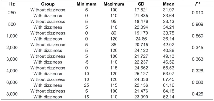
Analysis of the audibility thresholds found a predominance of sensorineural hearing loss. A loss of 5-10 dB per octave was detected in the average
air conduction auditory thresholds toward the high
frequencies, thus deining a mild downward sloping coniguration (Table 3).
Table 3 – Data on air conduction thresholds of audibility in groups with or without dizziness
Hz Group Minimum Maximum SD Mean P*
250 Without dizziness 5 100 17.521 31.97 0.910
With dizziness 0 110 21.835 33.64
500 Without dizziness 5 95 18.476 33.13 0.909
With dizziness 5 110 22.094 34.21
1,000 Without dizziness 0 80 19.179 33.75 0.869
With dizziness 0 120 24.66 36.14
2,000 Without dizziness 5 85 20.745 42.02 0.345
With dizziness 5 120 24.122 40.86
3,000 Without dizziness 5 100 21.727 49.13 0.363
With dizziness -5 110 22.237 46.52
4,000 Without dizziness 0 115 24.662 55.53 0.328
With dizziness 10 120 25.127 53.07
6,000 Without dizziness 10 120 24.336 67.45 0.088
With dizziness 25 115 22.136 61.16
8,000 Without dizziness 5 100 21.476 64.18 0.425
With dizziness 15 110 23.399 62.14
*p signiicance value (Mann-Whitney test) Caption: SD: standard deviation
Table 4 – Descriptive statistics of audiometry indings in the right and left ears
Classiication
Group 1 (N=70 ears) Group 2 (N=104 ears) Total
RE LE RE LE
N %
N % N % N % N %
Normal hearing 3 4.3 5 7.2 8 7.7 9 8.7 25 14.4
Mild SNHL 9 12.9 16 22.9 15 14.4 15 14.4 55 31.6
Moderate I SNHL 13 18.5 8 11.4 18 17.4 14 13.5 53 30.5
Moderate II SNHL 3 4.3 2 2.9 4 3.8 9 8.7 18 10.3
Severe I SNHL 2 2.9 1 1.4 1 1.0 0 0 4 2.3
Severe II SNHL 1 1.4 0 0 2 1.9 2 1.9 5 2.8
Profound I SNHL 0 0 1 1.4 0 0 0 0 1 0.6
Profound II SNHL 1 1.4 1 1.4 0 0 0 0 2 1.2
Mixed HL 3 4.3 1 1.4 4 3.8 3 2.8 11 6.3
Total 35 50 35 50 52 50 52 50 174 100
consequent impact on the mental domain, inducing
irritation, anxiety, depression, and insomnia27. Some
studies have found bilateral tinnitus in more than 50% of individuals with presbycusis26,28. In this study,
tinnitus was the symptom most frequently reported by the participants in both groups, with prevalences similar to the values reported by the abovemen -tioned studies.
From the audiological perspective, presbycusis is characterized by sensorineural hearing loss, with poorer auditory thresholds in the high frequencies1,16.
It is considered to be the sensory deiciency most
commonly associated with aging, with a prevalence
of up to 35% among individuals aged 60 to 70 years
old 26. In this study, more than 70% of the sample
exhibited mild-to-moderate hearing loss (Table 4), with a mild downward sloping coniguration in both groups (Table 3). These indings corroborate the results of other studies that investigated hearing in
older adults10,23,24,29. As presbycusis is associated
with aging, being caused by auditory degeneration of the basal portion of the cochlea12,24, the
predomi-nance of sensorineural hearing loss attended by gradual affection of the audibility thresholds was expected, as the high frequencies are the most severely affected in presbycusis.
That fact might be explained on physiological grounds, as the loss of hair cells occurs in the basal portion of the cochlea, where high-frequency sounds stimulate the local nerve ibers12,13,16.
Understanding speech is an essential
requirement for eficacious communication; for that reason, the performance of speech audiometry is
DISCUSSION
The sample of this study comprised 87 partici
-pants, and women predominated in both groups
(with or without dizziness). According to the Brazilian
Institute of Geography and Statistics, the number of
women in the Brazilian population is larger than the
number of men, as the life expectancy of the former
is longer compared to the latter23. Those facts
account for the larger percentage of women among
older adults1,9. In this study, dizziness predominated
among the women, which corroborates reports in the literature indicating that the frequency of dizziness is signiicantly higher among women 1,13,20.
Presbycusis is a form of sensorineural hearing loss, and it is considered to be the main cause of hearing deiciency among older adults, its preva
-lence varying from 30 to 66.4% among individuals
older than 65 years old15,16. In this study, bilateral
hearing loss predominated in both groups, with a much higher proportion than has been reported in
other studies15,16. Moreover, other factors might also
affect hearing in older adults11,24, which makes it
dificult to establish whether the high proportion of bilateral hearing loss found in this study might be exclusively attributed to aging of the inner ear.
The prevalence of hearing loss and tinnitus
increases with age25. Tinnitus is one of the main
symptoms associated with hearing loss and is
sometimes more disturbing than deafness itself26.
According to some studies, tinnitus might interfere with leisure activities, rest, sleep, social life, activ
-ities of daily living, and professional activ-ities, with
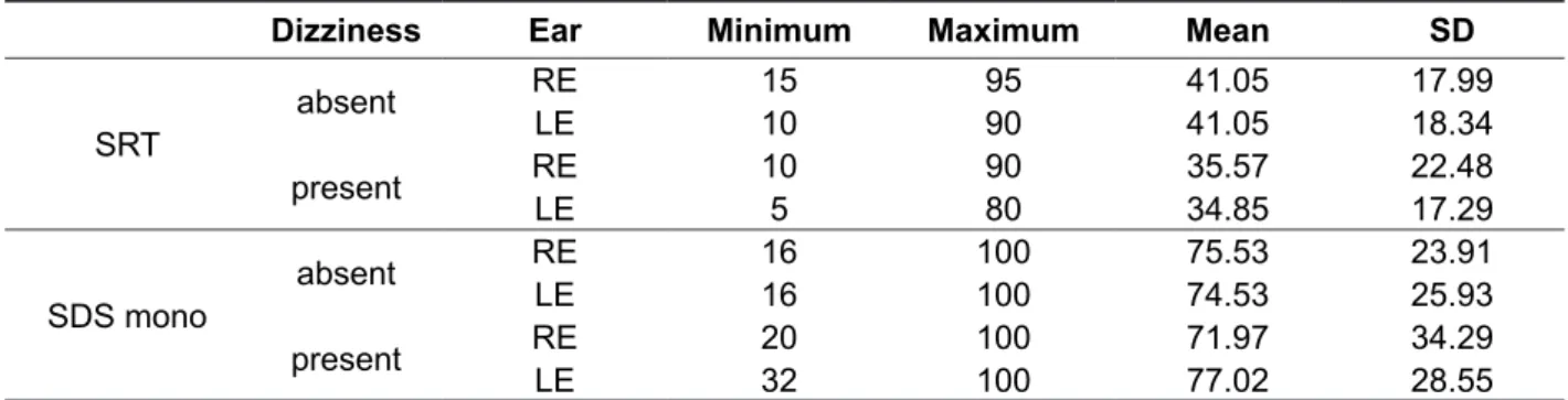
Table 5 – Speech audiometry indings in groups with or without dizziness
Dizziness Ear Minimum Maximum Mean SD
SRT
absent RE 15 95 41.05 17.99
LE 10 90 41.05 18.34
present RE 10 90 35.57 22.48
LE 5 80 34.85 17.29
SDS mono
absent RE 16 100 75.53 23.91
LE 16 100 74.53 25.93
present RE 20 100 71.97 34.29
LE 32 100 77.02 28.55
Caption: RE: right ear; LE: left ear; SD: standard deviation, SRT: Speech Reception Threshold; SDS mono: Speech Discrimination Score for monosyllabic words.
In speech audiometry, the SDS values found indicated dificulty with speech at a normal conver -sational level21. That inding is compatible with the
predominance of participants with mild to moderate
of participants with dizziness exhibited profound sensorineural hearing loss (Table 4), no difference was found in the auditory proile between the groups. Among the limitations of this study, the difference in the size of the groups stands out. As this study was merely observational, further studies with larger numbers of participants with severe and profound
sensorineural hearing loss are needed to elucidate
the possible association.
CONCLUSION
Although the vestibular and auditory systems are
anatomically located in the same organ,
character-istics indicative of differences in the auditory proile between individuals with or without dizziness were not found. The highest proportion of cases corre
-sponded to mild bilateral sensorineural hearing loss, attended by mild downward sloping toward the high frequencies.
indispensable in the assessment of hearing30. In this
study, the average SDS for monosyllabic words was approximately 70% in both ears, which indicates moderate dificulty in understanding speech22.
Sensorineural hearing loss is characterized by reduced speech discrimination, resulting in difi -culties in understanding the spoken language and in oral communication, especially in noisy
environ-ments, with consequent reduction of the social and family activities of older adults16,17,19. Hearing loss
is a serious limitation for the elderly, eventually resulting in social isolation as a function of the difi -culty in communicating with their environment28.
Body balance is maintained by the vestibular,
visual, and proprioceptive systems. Older adults
with hearing loss might exhibit greater dificulty in maintaining body balance due to the reduction in auditory feedback, in addition to reduction of the ability to locate sounds and of auditory discrimi -nation7,8. Although in this study, only the group
RESUMO
Objetivo: caracterizar o peril auditivo de idosos com tontura submetidos à Reabilitação Vestibular e comparar os resultados obtidos nas avaliações auditivas de idosos sem tontura. Métodos: estudo
observacional analítico transversal com 87 idosos, sendo 35 no grupo com tontura e 52 no grupo sem tontura. Foram realizadas anamnese, audiometria tonal limiar e vocal. Para a análise estatística foi utilizado o programa estatístico Statistical Package for the Social Sciences versão 17.0, com nível de
signiicância de 5% nas análises. Resultados: a perda auditiva neurossensorial de grau leve e
mode-rada esteve presente em 72,4% da amostra, com piora dos limiares de audibilidade por via aérea a partir de 4000Hz em ambos os grupos. O zumbido foi a queixa mais frequente observada na amostra. Conclusão: o peril auditivo de idosos com tontura não se diferencia daquele encontrado em idosos sem tontura, sendo observada com maior frequência a perda auditiva neurossensorial leve bilateral de coniguração descendente.
DESCRITORES: Fonoaudiologia; Audição; Idoso; Presbiacusia; Vestíbulo do Labirinto
REFERENCES
1. Guerra TM, Estevanovic LP, Cavalcante MAM,
Silva RCL, Miranda ICC, Quintas VG. Peril dos limiares audiométricos e curvas timpanométricas
de idosos. Brazilian J Otorhinolaryngol. 2010;76(5):663-6.
2. Esteves CC, Brandão FN, Siqueira, CGA, Carvalho SAS. Audição, zumbido e qualidade de vida: um
estudo piloto. Rev CEFAC. 2012;14(5):836-43.
3. Veras RP, Ramos LR, Kalache A. Crescimento da população idosa no Brasil: transformações
e consequências na sociedade. Rev Saúde Públ.1987;21: 225-33.
4. Ganança MM, Albernaz PLM, Fukuda Y, Munhoz MSL, Caovilla HH. Neuroanatomoisiologia do Sistema Vestibular – Correlações Clínicas. In: Filho
OL, Campos CAH. Tratado de Otorrinolaringologia.
São Paulo: Roca; 1994. P. 814-25.
5. Santos EM, Gazzola JM, Ganança CF, Caovilla HH, Ganança FF. Impacto da tontura na qualidade de vida de idosos com vestibulopatia crônica.
Pró-Fono R Atual Cient. 2010;22(4):427-32.
(estudo SABE, 2006). Cad. Saúde Pública.
2012;28(8):1479-92.
21. Sistemas de Conselhos Federal e Regionais
de Fonoaudiologia. Manual de procedimentos em
audiometria tonal limiar, logoaudiometria e medidas
de imitância acústica. Fevereiro, 2013. Disponível em: http://www.fonoaudiologia.org.br/cffa/index.
php/guias-e-manuais/
22. Jerger J, Speaks C, Trammell J. A new
approach to speech audiometry. J Speech Hear
Disord.1968;33:318.
23. Instituto Brasileiro de Geograia e Estatística 2000 [acessado em 2012]. Disponível em: http:// www.ibge.gov.br
24. Huang Q, Tang J. Age-related hearing loss or
presbycusis. Eur Arch Otorhinolaryngol (2010)
267:1179-91.
25. Pinto PCL, Sanchez TG, Tomita S. Avaliação da relação entre severidade do zumbido e perda auditiva, sexo e idade do paciente. Braz J
Otorhinolaryngol. 2010;76(1):18-24.
26. Sogebi OA, Olusoga-Peters OO, Oluwapelumi O. Clinical and audiometric features of presbycusis in Nigerians. African Health Sciences. 2013;
13(4):886-92.
27. Humes LE, Dubno JR, Gordon-Salant S, Lister JJ, Cacace AT, Cruickshanks KJ et al. Central Presbycusis: a review and evaluation of the
evidence. J Am Acad Audiol 2012;23:635-66.
28. Mondelli MFCG, Souza PES. Qualidade de vida
em idosos antes e após a adaptação do AASI. Braz
J Otorhinolaryngol. 2012;78(3):49-56.
29. Meneses C, Mário MP, Marchori LLM, Melo
JJ, Freitas ERFS. Prevalência de perda auditiva e fatores associados na população idosa de
Londrina, Paraná: estudo preliminar. Rev CEFAC.
2010;12(3):384-92.
30. Menegotto IH. Logoaudiometria básica. In:
Bevilacqua MC, Martinez MAN, Balen SA, Pupo AC,
Reis ACMB, Frota S. Tratado de Fonoaudiologia.
São Paulo: Santos Editora; 2012. P. 81-99. de Otorrinolaringologia. São Paulo: Roca; 1994. P.
481-509.
7. Patatas OHG, Ganança CF, Ganaça FF. Qualidade de vida de indivíduos submetidos à reabilitação vestibular. Brazilian J Otorhinolaryngol.
2009;75(3):387-94.
8. Ricci NA, Gazzola, JM, Coimbra, IB. Sistemas sensoriais no equilíbrio corporal de idosos. Arq Bras Ciên Saúde. 2009;34(2):94-100.
9. Silva MC. O processo de envelhecimento no
Brasil: desaios e perspectivas. Textos sobre
Envelhecimento [Internet] 2005 [citado 2010 Jul 17];
8(1). Disponível em: http://www.unati.uerj.br.
10. Kim S, Lim EJ, Kim HS, Park JH, Jarng SS, Lee SH. Sex differences in a cross sectional study of age-related hearing loss in Korean. Clin Exp
Otorhinolaryngol. 2010;3(1):27-31.
11. Gonçalves CGO, Mota PHM, Marques JM. Ruído e idade: análise da inluência na audição em indivíduos com 50-70 anos. Pró-Fono Rev Atual
Cient. 2009;21(4):57-62.
12. Ribeiro LCC, Alves PB, Meira EP. Percepção dos idosos sobre as alterações isiológicas do envelhecimento. Revista Ciência, Cuidado e Saúde.
2009;8(2):220-7.
13. Raynor LA, Pankow JS, Miller MB, Huang GH,
Dalton D, Klein R et al. Familial aggregation of
age-related hearing loss in an epidemiological study
of older adults. Am J Audiol. 2009;18:114-8.
14. Liporaci FD, Frota SMMC. Resolução temporal
auditiva em idosos. Rev Soc Bras Fonoaudiol. 2010;15(4):533-9.
15. Kano CE, Mezzena LH, Guida HL. Estudo comparativo da classiicação do grau de perda
auditiva em idosos institucionalizados. Rev CEFAC. 2009;11(3):473-7.
16. Teixeira AR, Freiras CLR, Milão LF, Gonçalves AK, Junior BB, Santos AMPV, et. al. Relação entre a queixa e a presença de perdas auditivas
entre idosos. Arq. Int. Otorrinolaringol. / Intl. Arch. Otorhinolaryngol. 2009;13(1):78-82.
17. Souza MGC, Russo ICP. Audição e percepção
da perda auditiva em idosos. Rev Soc Bras Fonoaudiol. 2009;14(2):241-6.
18. Tenório JP, Guimarães JATL, Flores NGC, Iório MCM. Comparação entre critérios de classiicação dos achados audiométricos em idosos. J Soc Bras
Fonoaudiol. 2011;23(2):114-8.
19. Azzolini VC, Ferreira MID. Processamento
Auditivo Temporal em Idosos. Arq. Int. Otorrinolaringol.2010;14(1):95-102.
20. Cruz MS, Lima MCP, Santos JLF, Duarte YAO,
Received on: July 03, 2014
Accepted on: November 03, 2014
Mailing address:
Patrícia Cotta Mancini
Universidade Federal de Minas Gerais – Faculdade de Medicina
Av. Professor Alfredo Balena, 190 / 251, Santa Eigênia
Belo Horizonte – MG – Brasil CEP: 30130-100