LATERALITY OF ACTIVITY OF MEDIAL OLIVOCOCHLEAR
EFFERENT SYSTEM: PRELIMINARY STUDY
Lateralidade da atividade do sistema olivococlear medial eferente:
estudo preliminar
Tatiana Rocha Silva(1), Fernanda Abalen Martins Dias(2)
(1) Pontifícia Universidade Católica de Minas Gerais – PUC
Minas – Belo Horizonte (MG), Brasil.
(2) Departamento de Fonoaudiologia, Pontifícia Universidade
Católica de Minas Gerais – PUC Minas – Belo Horizonte (MG), Brasil.
Study developed at Centro Clínico de Fonoaudiologia (Clinical Center for Speech – Language Pathology and Audiology), Ponti-fícia Universidade Católica de Minas Gerais – PUC Minas – Belo Horizonte (MG), Brazil.
Conlict of interest: non-existent
understood. It is believed that the medial
olivo-cochlear eferent tractfunctions as a modulator.
The medial olivocochlear eferent system adjusts
active cochlear process through the slow
contrac-tions of the external hair cells, attenuates the rapid contractions and providing, speciically, a protective
mechanism of the inner ear structures, by acoustic stimulation3-5.
Researchers described diferences in patterns
suppression of evoked otoacoustic emissions between the right and left ear. Found interaural asymmetry, relative to the size of the amplitude of evoked otoacoustic emissions, It is greater right, and
also as the action of the eferent auditory system, also more efectively to right6-9. The activity of the medial olivocochlear eferent tract, in both ears of 44
individuals between 19 and 29 years, with normal hearing, It was investigated to compare the inhibition of the bilateral tract. The evoked otoacoustic emissions by transient stimulus were researched with intensity of the click stimulus varying of 59-71 dB NPS, with contralateral acoustic stimulation of white noise to 30 dB the loudness level. It was
observed higher activity of the eferent system on the
INTRODUCTION
Otoacoustic emissions are sounds produced in
the cochlea and detected in the external acoustic meatus, it is speciically the registry of mobility
and of mechanical skill of external hair cells1. The
contralateral noise exerts inhibitory efect on the functioning of the external hair cells, the result is the
reduction of the amplitude of otoacoustic emissions.
This phenomenon, known as suppression efect of
otoacoustic emissions, demonstrates the integrity of the medial system olivocochlear, since that
innervate the external hair cells2-4.
The mechanism by which the suppression
efect of otoacoustic emissions occurs is not fully
ABSTRACT
Purpose: to evaluate the diference in the suppression of otoacoustic emissions between the right and left ears Methods: participated in this study 36 individuals, being 18 right-handed and left-handed 18, all without hearing complaints and hearing within normal limits. Data collection was performed by: transient otoacoustic emissions and suppression of the otoacoustic emissions. The analysis of the
presence / absence of the suppression efect was made based on the value obtained in response.
Results: there was no diference between the right and left ears for the response values of otoacoustic
emissions with contralateral noise for right and left handed individuals. In the male, there was
a diference between the right and left ears for the response values of otoacoustic emissions with
contralateral noise. Conclusion: the evaluation through the suppression of otoacoustic emissions
showed no diferences between the right and left ears in the groups studied.
KEYWORDS: Auditory Perception; Suppression; Eferent Pathways; Otoacoustic Emissions,
METHODS
The procedures in this study were approved by the Research Ethics Committee of Pontifícia Universidade Católica de Minas Gerais (PUC
Minas), under protocol number 0342.0.213.000-10
(Resolution 196/96 National Health Council – CONEP).
This research characterized by a pilot study, of descriptive typology, of qualitative and quantitative analysis. Were invited to participate of the study
60 subjects. These, ten refused to participate of the research, six not attend for the evaluation and the other 8 were excluded of the study by present
hearing loss, hearing complaints and/or previous ear disease. The sample, so, was composed by 36
subjects (18 right-handed and left-handed 18), of
both genders, in the age range of 18-25 years. The research participants were selected in the
undergraduate courses ofered by the Instituto
de Educação Continuada – IEC da Pontifícia Universidade Católica de Minas (PUC Minas) and in the social environment of the Researchers. The
subjects of this study were selected by means of the
non-random sampling technique, of the type conve-nience sampling.
The participants of the research were
commu-nicated personally about the objectives of the
research, the absence of damage to your health,
the guarantee of conidentiality of their identities or
any other characteristics which could identify them, and on the roadmap of the research. Being properly informed, all signed the Consent Term.
The data collection was carried in the Clinical Center for Speech – Language Pathology and Audiology of PUC Minas. All the individuals were submitted to basic audiologic evaluation. This evalu-ation consisted of: anamnesis, otoscopy, pure tone audiometry, logoaudiometry, tympanometry and
research of acoustic relexes.
In the anamnesis the individual provided infor-mation as personal data, audiological history, health-related aspects and domain of manual motor ability (right or left handed). The anamnesis was conducted with the same protocol used in the Clinical Center for Speech – Language Pathology and Audiology of PUC Minas.
To perform the visual inspection of the external
auditory canal (otoscopy) was used otoscope, of the brand TK®, model 22. The Pure tone audiometry and logoaudiometry were performed in acoustically treated booth and with two-channel audiometer, model Midimate 622, of the brand Madsen Electronics®, using phone TDH-39 and bone vibrator B-71. The tympanometry and the research
of acoustic relexes were performed through the right side, without signiicant diference according to
gender9.
In a study that compared the presence of the
suppression efect of otoacoustic emissions and its
amplitude in normal hearing adults individuals the authors observed no side dominance. There was no
diference between the results of the right ear and
left ear as to asymmetry of the amplitude size and
as to presence of suppression efect10.
Some authors veriied the occurrence and the magnitude of the suppression efect of otoacoustic
emissions by transient stimulus, in preterm infants, with risk factors for hearing loss and observed greater responses in the right ear11. Values
greater of the amplitude suppression of transient otoacoustic emissions in the right ear using a noise in the ear contralateral and changes in amplitude of these emissions during tasks of auditory and visual attention indicate that the medial olivocochlear tract may be involved in maintaining this asymmetric
peripheral standard and through him, the cortex can
modulate cochlear function8,9,12.
The laterality of the central nervous system or the predominance of one brain hemisphere above the other it is a topic much studied since dominant areas for language in the left hemisphere have been described. However, only with the advent of
functional imaging exams it was possible better
understand this asymmetric functioning and analyze whether the absence of this predominance can collaborate with the appearance of conditions such
as dyslexia, aphasia, schizophrenia and autism13-15.
On the other hand, determine this asymmetry is not easy task. Beyond the various levels of hemispheric
dominance, the asymmetry may be diferent in
the same individual, for the various members and sensory organs. Thus, there can be crossed
later-ality for diferent organs and functions12.
However, there is evidence that the central and peripheral auditory system also function in lateral way. More amplitude of wave III in brainstem audiometry and of the transient otoacoustic emissions in the right ear n counterpart with most presence of tinnitus and of temporary hearing loss
after exposure to noise in the left ear suggest, in
addition to an asymmetry between the ears, that the left ear is more vulnerable to hearing alterations8.
Therefore, this research was justiied by the
possibility of understanding the performance of the
medial olivocochlear eferent system as it relates to the hearing dominance. Thus, the objective of the study was to evaluate the diference in the
model ILO version 6, of the brand Otodynamics®.
To investigate the efect of suppression of transient
otoacoustic emissions was used broadband noise transmitted by the two-channel audiometer, model Midimate 622, of the brand Madsen Electronics®, by means of the phone TDH-39, in the intensity of 60 dB NPS.
The transient evoked otoacoustic emissions were found to be present when the amplitude of
the frequencies of 1 e 1,4, 2, 2,8 e 4 kHz were
greater or equal to 3 dB in at least three consecutive frequencies reproducibility having values greater
than 50% and values of stability of the adjustment
of the probe greater than 70%. Only individuals who had transient evoked otoacoustic emissions present were included in the study.
For the efect of suppression observed the
variation of the response of amplitude in the presence of noise, in relative the response of amplitude in the absence of noise. The value of the suppression referring to the action of the olivocochlear system is
given by the diference of the values obtained in the
conditions with and without contralateral stimulation, in each ear, and that value determines whether or
not suppression. The value of this diference repre
-sents quantitatively the magnitude of the reduction / suppression. So, if the value is positive, there suppression and if it is negative or zero, there is no suppression in the emission amplitudes10. In
this study we chose to by considering a minimum variation of 0.5 dB NPS and the analysis of the
presence / absence of efect reduction / suppression
was made based on the value obtained in response,
excluding the interference of background noise.
Then the data collected were tabulated and
subjected to statistical analysis. The statistical
analysis was performed by of the software Statistical Package for Social Sciences (SPSS) version 20.0. Initially was performed the descriptive analysis, that understood measures of central tendency (mean and median), of dispersion (standard deviation) and
of position (maximum and minimum).
Besides descriptive statistics was performed the inferential statistics by of the test t of Student
paired. It was adopted the level of signiicance of 5% (p≤0.05). It was considered as trend to statistical signiicance the results signiicant at level of 10% (p≤0.10).
middle ear analyzer, model AZ7, of the brand Interacoustics®.
Were used as inclusion criteria to constitute the study group, individuals without hearing complaints and/or previous ear disease and with audiometry assessment within the normal range. It was considered individuals with hearing evaluation within normal standards those with pure tone thresholds by air conduction to 25 dBNA, in the frequencies of 250 Hz to 8 kHz, and pure tone thresholds by bone conduction to 15 dBNA, in the frequencies of de 500
Hz to 4 kHz, with diference between the thresholds
of air conduction and bone conduction less or equal to 10 dB, tympanometric curve type A and presence
of acoustic relexes in the frequencies of 500 Hz, 1, 2 and 4 kHz.
Were excluded from the study neurological
disorders individuals, cancer, ear infections, tympanic membrane perforation, he with a history of head trauma and otologic surgery, the with tinnitus,
with exposure history of noise, in use of ototoxic
drugs, with hearing complaints and hearing loss. Then, the individuals were submitted to research of transient evoked otoacoustic emissions and the suppression of transient otoacoustic emissions. Each participant submitted himself only an assessment of transient otoacoustic emissions and suppression of transient otoacoustic emissions. First, was held a record of transient otoacoustic emissions in one ear and, then, a record in the other ear. Posteriorly, was held a suppression recording of transient otoacoustic emissions in one ear and, then, a record in the other ear. Therefore, the collection was held alternately between the ears.
The transient otoacoustic emissions were performed in comfortable and quiet environment with linear stimulus, type click, with intensities from 80 e 85 dB NPS. The number of stimuli used during registration of transient otoacoustic emissions
did not sufer variation (260 series), wherein the procedure presented in the maximum duration
of 75 seconds in each ear. Was considered noise level below 16dB NPS. The transient otoacoustic emissions were taken initially without contralateral noise, and then, with contralateral noise with the order not to change the placing the probe during the two test cases.
mean age was of 22,3 years (standard deviation 2,08) and in the group of left-handed individuals, the mean age was of 21,7 years (standard deviation 2,81).
The descriptive analysis, considering the whole sample, can be visualized in the Table 1. It can be observed that, the mean of response values of otoacoustic emissions with and without contralateral noise were higher for the right ear.
RESULTS
The mean age of the study population was
22,0 years (standard deviation 2,46). Being that 18 subjects were female, mean age of 22,8 years (standard deviation 1,84) and 18 subjects were
male, mean age of 21,2 years (standard deviation 2,75). In the group of right-handed individuals, the
Table 1 – Measures of central tendency, dispersion and position for the results otoacoustic emissions and otoacoustic emissions suppression (n=36)
Parameters Mean Median DP Maximum Minimum
Response SR
OD 18,97 18,65 5,74 27,1 8,2 OE 17,77 18,25 4,30 25,7 10,8 Response CR
OD 17,36 16,75 6,00 26 5,6
OE 15,95 16,85 4,90 24,7 7,5 Magnitude reduction
OD 1,60 1,55 0,97 5,5 0,5
OE 1,82 1,2 1,73 8,4 0,5
Legend: DP = standard deviation; SR = without contralateral noise; CR = with contralateral noise; OD = right ear; OE = left ear
The Table 2 shows the comparison between right-handed and left-handed individuals for the measures of central tendency. In the Table 3 shows the comparison between the female and male genders for the measures of central tendency.
In the inferential statistical analysis, consid-ering the whole sample, was found that there was
no diference between the response values of
otoacoustic emissions without contralateral noise between the right and left ears (p=0,155) and that
also there was no diference between the response
values of otoacoustic emissions with contralateral
noise between the right and left ears (p=0,104).
In relation to magnitude of the reduction, it was
observed that there was no diference between the right and left ears (p=0,420).
In the group of right-handed individuals was
found that there was no diference between the
response values of otoacoustic emissions without contralateral noise between the right and left ears
(p=0,523) and that also there was no diference
between the response values of otoacoustic emissions with contralateral noise between the right and left ears (p=0,360). In relation to magnitude of the reduction, it was observed that there was no
diference between the right and left ears (p=0,380).
In the group of left-handed individuals, was
found that there was no diference between the
response values of otoacoustic emissions without contralateral noise between the right and left ears
(p=0,151) and that also there was no diference
between the response values of otoacoustic emissions with contralateral noise between the right and left ears (p=0,166). In relation to magnitude of the reduction, it was observed that there was no
diference between the right and left ears (p=0,891).
In female gender, was found that there was
no diference between the response values of
otoacoustic emissions without contralateral noise between the right and left ears (p=0,808) and that
also there was no diference between the response
values of otoacoustic emissions with contralateral noise between the right and left ears(p=0,637). In relation to magnitude of the reduction, it was
observed that there was no diference between the right and left ears (p=0,457).
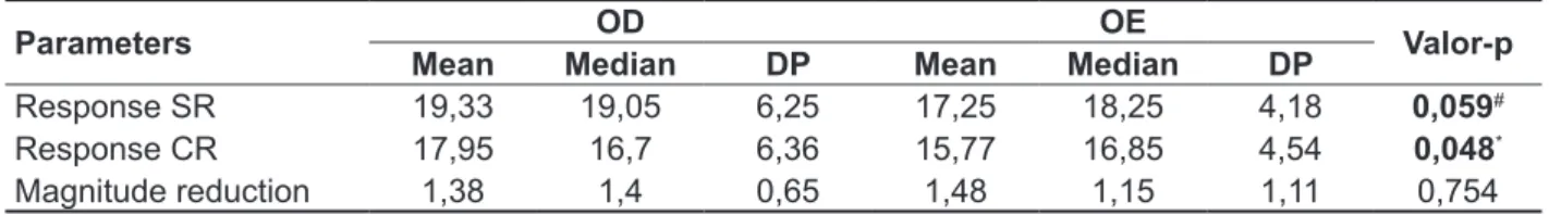
The Table 4 allows observing that, in the male gender, there was trend to diference between the
right ear and left ear for the response values of otoacoustic emissions without contralateral noise
and that there was diference between the right
Table 2 – Measures of central tendency, dispersion and position for the results otoacoustic emissions and otoacoustic emissions suppression for right-handed and left-handed individuals
Parameters Mean Median DP Maximum Minimum
Right-handed
Response SR
OD 18,1 18,4 5,88 27,1 8,2
OE 17,23 18,25 4,46 25,7 10,8 Response CR
OD 16,45 17 6,23 25,8 5,6
OE 15,2 16,85 5,29 24,7 7,5
Magnitude reduction
OD 1,64 1,3 1,21 5,5 0,5
OE 2,03 1,1 2,10 8,4 0,8
Left-handed
Response SR
OD 19,85 19 5,63 27 9,1
OE 18,31 19,2 4,19 24,6 11,4 Response CR
OD 18,27 16,45 5,80 26 7,3
OE 16,7 17,75 4,49 22,5 9,2
Magnitude reduction
OD 1,57 1,65 0,69 3,1 0,5
OE 1,61 1,4 1,29 6,4 0,5
Legend: DP = standard deviation; SR = without contralateral noise; CR = with contralateral noise; OD = right ear; OE = left ear
Table 3 – Measures of central tendency, dispersion and position for the results otoacoustic emissions and otoacoustic emissions suppression for male and female gender
Parameters Mean Median DP Maximum Minimum
Female gender
Response SR
OD 18,61 18,65 5,34 26,5 9
OE 18,3 18,05 4,48 25,7 12,1 Response CR
OD 16,78 16,75 5,74 25,5 5,6 OE 16,12 16,85 5,36 24,7 8,7 Magnitude reduction
OD 1,83 1,8 1,19 5,5 0,5
OE 2,17 1,4 2,16 8,4 0,9
Male gender
Response SR
OD 19,33 19,05 6,25 27,1 8,2 OE 17,25 18,25 4,18 22,6 10,8 Response CR
OD 17,95 16,7 6,36 26 6,6
OE 15,77 16,85 4,54 21,3 7,5 Magnitude reduction
OD 1,38 1,4 0,65 2,8 0,5
OE 1,48 1,15 1,11 5,5 0,5
asymmetric activity between the ears and, conse-quently, a magnitude of greater suppression on the right ear3,11. Such indings provide new arguments
in favor of the peripheral auditory lateralization,
especially in regard to the medial eferent system.
Some authors argue that there is a predominance of the right ear on the left ear the that indicates a
probable inluence of olivocochlear tract. Thus, believe that a better function of the eferent medial
system on the right would lead to greater protection
of the external hair cells that, consequently, would generate greater suppression efect. Besides, would trigger eferent relexes more efective on this side of, relecting a balance between the function of the external hair cells and of the medial eferent
system12.
It is emphasized that the studies about suppression of otoacoustic emissions that the
objective of investigating diferences between the laterality patterns are scarce. Existing studies show
great methodological variability, the that undertakes the comparison. Besides, the same level of stimulus presented in otoacoustic emissions can produce
diferent answers in diferent individuals, and this inter-subject variability will be relected, conse -quently, in level of suppression2.
Therefore, it is necessary that other studies be developed to propitiate new comparisons and
discov-eries. The existing studies have deined various
parameters for get the best catchment records of the
otoacoustic emissions and of the suppression efect otoacoustic emissions, for example, the analysis
interval of the amplitude response, the intensity of contralateral noise versus suppression amplitude, the bilateral mode of catchment in comparison to the ipsilateral or contralateral and time intervals1,8.
However, there is still a gap how much the gender factor and the lateralization factor for right and left ears.
In the present study, the participants were also
divided according to gender, but no diferences
were found between right and left ears for the suppression of otoacoustic emissions in the female
DISCUSSION
In similar studies no diferences were found for the presence of suppression efect of evoked
otoacoustic emissions as regards right and left dominance in adults with hearing within normal limits6,10.
However, in another study, the researchers have
documented diferences in patterns of suppression
of evoked otoacoustic emissions as the right and left ears. The researchers showed interaural asymmetry, in relation to evoked otoacoustic emissions, being
greater right, and, as the action of the eferent auditory system, also with more efectively right. However, these researchers did not explain the motives of their indings, highlighting the need for further investigations to explain the asymmetry
found9.
In the present study the mean of the response values of otoacoustic emissions without contra-lateral noise and with contracontra-lateral noise was higher to right. However, this result was not observed for the magnitude of the reduction of otoacoustic emissions. The mean of the magnitude values of reduction of otoacoustic emissions it was higher to left.
In research conducted with right-handed with hearing within normal limits, the suppression was
signiicantly greater in the right ear. As most of the
study participants was right-handed, the probable
explanation for this inding is the lateralization of the
medial olivocochlear system7,4,12.
In the present study, the participants were divided into 2 groups, a group of right-handed individuals and a group of left-handed individuals. However, the mean of the magnitude values of reduction of otoacoustic emissions it was higher to left both for right-handed individuals as for left-handed individuals.
In other studies statistically signiicant difer -ences between the side of the ear in nursing infants were observed wherein the right side had higher responses2. The authors suggested that there is an
Table 4 – Comparison between right and left ears for the results otoacoustic emissions and otoacoustic emissions suppression for male gender
Parameters OD OE Valor-p
Mean Median DP Mean Median DP
Response SR 19,33 19,05 6,25 17,25 18,25 4,18 0,059#
Response CR 17,95 16,7 6,36 15,77 16,85 4,54 0,048*
Magnitude reduction 1,38 1,4 0,65 1,48 1,15 1,11 0,754
Legend: DP = standard deviation; SR = without contralateral noise; CR = with contralateral noise; OD = right ear; OE = left ear
* Signiicant values (p≤0,05) - Test t paired
listening they should be realized at the includion time of participants for determining of the real hearing predominance9-12.
CONCLUSION
The results of this study were not conclusive. The assessment carried out by means of otoacoustic
emissions suppression showed no diferences
between the right and left ears for all groups
studied. Although without statistically signiicant diference, was observed right ear advantage for
the response values otoacoustic emissions with and without contralateral noise for all groups studied.
The diference between the ears in the male gender
should be considered with caution, once there is data not consistent the literature studied. Therefore,
to airm that there is acting of the medial olivoco
-chlear eferent system as it relates to the hearing
dominance, other studies must be performed with larger casuistry and having criteria of inclusion
diferent, as, for example, the determining auditory
predominance.
gender. This fact, probably, could be justiied, also, by the absence of diference between right and
left ears for the response values of the transient otoacoustic emissions without contralateral noise.
However, the male gender diferences were found
between right and left ears for the suppression of otoacoustic emissions and for the response values of the transient otoacoustic emissions without contralateral noise.
The physiology and anatomy of the eferent
pathway, particularly of the tract medial olivocochlear
eferent, are still unknown. It is emphasized, then,
the need for more detailed knowledge of the impli-cations of the occurrence of a lateral dominance for the otoacoustic emissions mechanism suppression.
It is emphasized yet that a rigorous sample selection is necessary in studies of laterality and functional predominance. It is possible to have
cross-laterality for diferent organs and functions,
therefore the selection of right-handed individuals does not prove that these too have predominant right auditory systems. For this proof, electrophysi-ological tests with stimulus verbal, tonal and dichotic
RESUMO
Objetivo: avaliar a diferença na supressão das emissões otoacústicas entre as orelhas direita e esquerda. Métodos: participaram da pesquisa 36 indivíduos, sendo 18 destros e 18 canhotos, todos
sem queixa auditiva e com audição dentro dos padrões de normalidade. A coleta de dados foi reali
-zada por meio das emissões otoacústicas transientes e pela supressão das emissões otoacústicas. A análise da presença/ausência do efeito de supressão foi realizada baseada no valor obtido em response. Resultados: não houve diferença entre a orelha direita e esquerda para os valores de response das emissões otoacústicas com ruído contralateral para os indivíduos destros e canhotos. No gênero masculino houve diferença entre a orelha direita e esquerda para os valores de response das emissões otoacústicas com ruído contralateral. Conclusão: a avaliação realizada por meio da supressão das emissões otoacústicas não evidenciou diferenças entre as orelhas direita e esquerda nos grupos estudados.
DESCRITORES: Percepção Auditiva; Supressão; Vias Eferentes; Emissões Otoacústicas
9. Khalfa S, Collet L. Functional asymmetry of medial olivocochlear system in humans. Towards a peripheral auditory lateralization. Neuroreport. 1996;7:993-6.
10. Oliveira JRM, Fernandes CF, Filho OAC. Estudo da supressão da amplitude das emissões otoacústicas: dominância lateral. Braz J
Otorhinolaryngol. 2011;77(5):547-54.
11. Amorim AM, Lewis DR, Rodrigues GRI, Fiorini AC, Azevedo MF. Efeito de supressão das emissões otoacústicas evocadas por estímulo transiente em lactentes de risco para perda auditiva nascidos
pré-termo. Rev CEFAC. 2010;12(5):749-55.
12. Fávero ML, Sanchez TG, Bento RF, Nascimento
AF. Atividade coclear assimétrica: inluência do SNC? Arquivos Int Otorrinolaringol. 2005;9(4):300-4.
13. Foundas AL. Is language laterality established
by 5 years os age? Neurology. 2003;60:1573-4. 14. Binder JR, Frost JA, Hammeke TA, Rao SM, Cox
RW. Function of the left planum temporale in auditory
and linguistic processing. Brain. 1996;119:1239-47. 15. Veuillet E, Georgief N, Philibert B, Dalery J,
Marie-Cardine M, Collet L. Abnormal peripheral auditory asymmetry in schizophrenia. J neurol
neurosurg psychiatry. 2001;70:88-94.
REFERENCES
1. Kemp DT. Otoacoustic emissions: basic facts and
applications. Audiol Practice. 1989;3(1):1-4.
2. Hill JC, Prasher DK, Luxon LM. Evidence eferent efects on auditory aferent activity and their functional relevance. Clin Otolaryngol. 1997;22:394-402.
3. Burguetti FAC, Carvalho RMM. Sistema auditivo eferente: efeito no processamento auditivo. Rev
Bras Otorrinolaringol. 2008;74(5):737-45.
4. Leme VN, Carvallo RMM. Efeito da estimulação
acústica contralateral nas medidas temporais das emissões otoacústicas. Rev CEFAC. 2009;11(Suppl
1):24-30.
5. Perez AP, Kós MI, Frota S. A supressão das emissões otoacústicas transitórias em mulheres com
audição normal. Rev CEFAC. 2006;8(3):368-74.
6. Urnau D, Tochetto TM. Ocorrência e efeito de supressão das emissões otoacústicas em adultos normo-ouvintes com zumbido e hiperacusia. Braz J
Otorhinolaryngol. 2012;78(1):87-94.
7. Breuel MLF, Sanchez TG, Bento RF. Vias auditivas eferentes e seu papel no sistema auditivo. Arquivos Int. Otorrinolaringol. 2001;5(2):62-7. 8. Khalfa S, Morlet T, Micheyl C, Morgon A. Evidence of peripheral hearing asymmetry in humans: clinical implications. Acta otolaryngol. 1997;117:192-6.
http://dx.doi.org/10.1590/1982-021620151768615
Received on: June 18, 2015 Accepted on: August 12, 2015
Mailing address: Tatiana Rocha Silva R. Boninas, 1070, Pompeia Belo Horizonte – MG – Brasil CEP: 30280-220