DOI: 10.1590/0004-282X20130108
VIEW AND REVIEW
ABSTRACT
Myasthenia gravis (MG) is an autoimmune disorder affecting neuromuscular transmission leading to generalized or localized muscle weak-ness due most frequently to the presence of autoantibodies against acetylcholine receptors in the postsynaptic motor end-plate. Myasthe-nic crisis (MC) is a complication of MG characterized by worsening muscle weakness, resulting in respiratory failure that requires intubation and mechanical ventilation. It also includes postsurgical patients, in whom exacerbation of muscle weakness from MG causes a delay in extubation. MC is a very important, serious, and reversible neurological emergency that affects 20–30% of the myasthenic patients, usually within the first year of illness and maybe the debut form of the disease. Most patients have a predisposing factor that triggers the crisis, generally an infection of the respiratory tract. Immunoglobulins, plasma exchange, and steroids are the cornerstones of immunotherapy. Today with the modern neurocritical care, mortality rate of MC is less than 5%.
Key words: myasthenic crisis, myasthenia gravis, respiratory failure, immunosupressive therapy, thymectomy.
RESUMO
Miastenia grave (MG) é um distúbio autoimune que afeta principalmente a transmissão neuromuscular, levando a fraqueza muscu-lar generalizada ou localizada. É devida mais frequentemente à presença de auto-anticorpos anti-receptores de acetilcolina na fenda pós-sináptica da placa motora. A crise miastênica (CM) é uma complicação da MG caracterizada por piora da fraqueza muscular, resultando en falência respiratória, o que requer entubação endotraqueal e ventilação mecânica.Isto ocorre também em pacientes pós-cirúrgicos, em que há piora da fraqueza muscular devido à MG, causando um atraso na extubação. MC é uma emergência neurológica importante, séria e reversível que afeta 20–30% dos pacientes miastênicos, usualmente duranteo primeiro ano de enfermidade, podendo a crise miastênica ser a manifestação inicial da MG. A maioria dos pacientes tem fatores predisponentes que desencadeiam a crise, geralmente uma infecção do trato respiratório. Imunoglobulina, plasmaférese e esteróides são a pedra angular da imunoterapia. Hoje, dentro da terapia neurocrítica, a taxa de mortalidade na CM é menor que 5%.
Palavras-Chave: crise miatênica, miastenia gravis, falência respiratória, terapia imunosupressiva, timectomia.
The myasthenic patient in crisis: an update of
the management in Neurointensive Care Unit
Pacientes miastênicos em crise: uma melhora de conduta na unidade de terapia intensiva
Daniel Agustin Godoy1,2, Leonardo Jardim Vaz de Mello3,4, Luca Masotti5, Mario Di Napoli6,7
1Intensive Care Unit, Hospital San Juan Bautista, Catamarca, Argentina; 2Neurointensive Care Unit, Sanatorio Pasteur, Catamarca, Argentina;
3Neurology Service, Santa Casa de São João del Rei, São João del Rei MG, Brazil; 4Hospital Nossa Senhora das Merces, São João del Rei MG, Brazil;
5Internal Medicine, Cecina Hospital, Cecina, Italy;
6Neurological Service, San Camillo de’ Lellis General Hospital, Rieti, Italy;
7Neurological Section, SMDN – Center for Cardiovascular Medicine and Cerebrovascular Disease Prevention, Sulmona, L’Aquila, Italy.
Correspondence: Daniel Agustín Godoy; Unidad de Cuidados Neurointensivos Sanatorio Pasteur; Chacabuco 675; 4700 Catamarca - Argentina; E-mail: [email protected]
Conlict of Interest: There is no conflict of interest to declare.
Received 1 March 2013; Received in final form 3 April 2013; Accepted 10 April 2013. Myasthenic crisis (MC) is an uncommon life-threatening neurological emergency1-3. It may occur in patients who have
previously diagnosed myasthenia gravis (MG) or may be the onset of the disease, generally during the irst year after diag-nosis4-6. he hallmark of MC is the bulbar or respiratory
fail-ure1,2,5,7-12. he management of these patients is challenging
due to the luctuating nature of the disease4,5,8-12. Prevention
and treatment of MC requires admission to intensive care unit
(ICU) — preferably a neuroscience ICU — close observation, and, when necessary, intubation for ventilatory and feed-ing support2,8-13. In addition, acute care should be focused on
reducing circulating antibody titers with immunologic ther-apy such as plasmapheresis (PE), immunoglobulin (IVIg), and corticosteroids2,8-13. Despite the growing interest and newer
should be considered in MC2,4-12,18. Although there is no
uni-versally accepted deinition, MC should be considered a true neurological emergency characterized by “Severe weak-ness of the bulbar (innervated by cranial nerves) and/or respiratory muscles, enough to cause inability to maintain adequate ventilation and/or permeability of upper airways, causing respiratory failure that requires artiicial airway or ventilatory support”2,6. Postoperative myasthenic patients
in whom extubation has been delayed more than 24 hours also should be considered crisis7. Generally, patients with
MC correspond to class 3 or 4 in Osserman and Genkins classiication14 or class V according to Myasthenia Gravis
Foundation14 (Table 1).
PREDISPOSING FACTORS
Patients who develop MC in their great majority have a precipitating factor, although, in 30–40% of cases, none is found2,5-7,9-12,19,21. Respiratory infection (40%), emotional
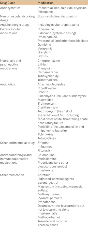
stresses, microaspirations (10%), changes in medication regi-men (8%), surgery, or trauma are among the most common predisposing factors2,5-7,9-12,16,18. Many drugs exacerbate MG
and may determine MC19. hey should be avoided or used
with caution. Some examples have been listed in Table 2. It is important to note that telithromycin, a macrolide, is absolutely contraindicated in MG14,19-21. Initial treatment
with prednisone led to exacerbation of MG in almost half of of MG. his paper reviews the available evidence in the
detec-tion and treatment of the MC from a multidisciplinary per-spective, with the intention to help to correct management.
EPIDEMIOLOGICAL DATA
he annual incidence of MG is 1–2/100,00014, with an
estimated prevalence of 5–15/100,0003,14; 21% of patients had
onset after 60 years15 and 30% of them will develop some
degree of bulbar or respiratory muscle weakness15. About
15–20% of MG patients will develop MC, usually within the irst year of illness4,6-11. MC may be the initial presentation
of MG in about 20% of patients and one-third of surviving may experience another crisis4,6-11. Overall, women are twice
as likely as men to be afected3,14,. he average age of
admis-sion with crisis is 59 years16. he occurrence of MG shows a
bimodal distribution, with the following male:female ratios: 3:7 if aged <40 years, 1:1 if aged 40–49 years, 3:2 if aged >50 years14,17. he outcome has improved signiicantly, and
today the reported mortality rate is around 5%2,7,9,10,16.
HOW WE DEFINED MYASTHENIC CRISIS?
By deinition, all MG patients with acquired (neona-tal) or autoimmune form showing a respiratory failure due to muscle weakness and requiring ventilatory assistance
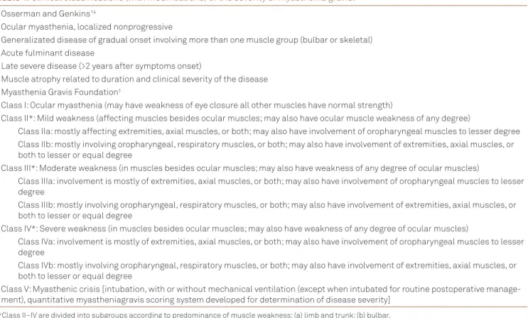
Table 1. Clinical classifications (with modifications) of the severity of myasthenia gravis.
Osserman and Genkins14
Ocular myasthenia, localized nonprogressive
Generalizated disease of gradual onset involving more than one muscle group (bulbar or skeletal) Acute fulminant disease
Late severe disease (>2 years after symptoms onset)
Muscle atrophy related to duration and clinical severity of the disease Myasthenia Gravis Foundation†
Class I: Ocular myasthenia (may have weakness of eye closure all other muscles have normal strength)
Class II*: Mild weakness (affecting muscles besides ocular muscles; may also have ocular muscle weakness of any degree)
Class IIa: mostly affecting extremities, axial muscles, or both; may also have involvement of oropharyngeal muscles to lesser degree Class IIb: mostly involving oropharyngeal, respiratory muscles, or both; may also have involvement of extremities, axial muscles, or
both to lesser or equal degree
Class III*: Moderate weakness (in muscles besides ocular muscles; may also have weakness of any degree of ocular muscles)
Class IIIa: involvement is mostly of extremities, axial muscles, or both; may also have involvement of oropharyngeal muscles to lesser degree
Class IIIb: mostly involving oropharyngeal, respiratory muscles, or both; may also have involvement of extremities, axial muscles, or both to lesser or equal degree
Class IV*: Severe weakness (in muscles besides ocular muscles; may also have weakness of any degree of ocular muscles)
Class IVa: involvement is mostly of extremities, axial muscles, or both; may also have involvement of oropharyngeal muscles to lesser degree
Class IVb: mostly involving oropharyngeal, respiratory muscles, or both; may also have involvement of extremities, axial muscles, or both to lesser or equal degree
Class V: Myasthenic crisis [intubation, with or without mechanical ventilation (except when intubated for routine postoperative manage-ment), quantitative myastheniagravis scoring system developed for determination of disease severity]
*Class II–IV are divided into subgroups according to predominance of muscle weakness: (a) limb and trunk; (b) bulbar.
†Myasthenia Gravis Foundation of America (MGFA) clinical classification based on neurologic examination limitations of clinical classification fluctuating
MG patients receiving immunosuppression2,7,19. Contrast
agents20 and electrolyte alterations (hypokalemia,
hypophos-phatemia) may exacerbate muscle weakness2,7. hyroid
dis-ease, which can coexist with MG, can exacerbate or unmask MG weakness when untreated, while over-replacement with levothyroxine may also cause MC2,7,19. If a MG patient requires
general anaesthesia, neuromuscular-blocking agents should be used cautiously since they are particularly sensitive to non-depolarizing agents and the response to non-depolarizing drugs is variable2,7,21. he association of MG with thymic
pathol-ogy is well known. MC is almost as twice more frequent in patients with thymoma2,7,14,23-25. Pregnancy aggravates MG in
33% of the cases, and MC in pregnancy carries high perinatal mortality2,7,26.
PATHOPHYSIOLOGY OF MYASTHENIC GRAVIS AND MYASTHENIC CRISIS
MG is an autoimmune disorder resulting from antibody-complement-mediated and T-cell-dependent immunologic attack on the postsynaptic membrane of the neuromuscular junction, mainly against acetylcholine receptor (AchR)14,27,28
(Fig 1). he antibodies that bind to epitopes of the skeletal muscle end-plate region result in abnormal neuromuscular transmission and clinical weakness14,27,28. here are
difer-ent antibodies directed at the neuromuscular junction and detectable in the plasma (Table 4)14,27,28. AChR antibodies bind
to the main immunogenic region of alpha subunit of AChR of postsynaptic membrane resulting in decreased numbers and density of AChR14,27,28. hey are present in 70–90% of patients
with generalized MG and between 30 and 70% of patients with ocular form14,27,28.
About 10% of patients show antibodies to muscle-speciic tyrosine kinase (Anti MuSK)14,27,28. Patients with MuSK
typi-cally are female and have characteristical weakness pattern involving principally bulbar, neck, shoulder, and respiratory muscles14,27,28. MuSK is a protein located at the postsynaptic
membrane, which is responsible for clustering the AChR at the muscle membrane surface during development, but the function in mature skeletal muscle and its role in pathophysi-ology of MG is unknown14,27,28. Other muscle autoantibodies
reacting with striated muscle titin and ryanodin receptor (RyR) antigens are found in up to 95% of MG patients with thymoma and in 50% of late-onset MG patients29 (>50 years).
hymomas are present in <2% of patients without antistri-ated antibodies30. Following thymectomy, rise in antistriated
muscle antibody titer may be a sign of recurrent tumor30.
hese antibodies are usually associated with more severe MG30. Titin is a protein, providing a direct link between
mechanical muscle strain and muscle gene activation14,28,28.
Antititin antibodies may also be detected in 50% of patients with late-onset generalized MG without thymoma28,29. he
RyR is the calcium channel of the sarcoplasmic reticulum
Table 2. Medication and drugs that may provoke myasthenia crisis.
Drug Class Medication
Antipsychotics Phenothiazines, sulpiride, atypicals (clozapine)
Neuromuscular-blocking drugs
Succinylcholine, Vecuronium
Anticholinergic drugs Including ocular proparacaine Cardiovascular
medications
Cibenzoline
Lidocaine (systemic dosing) Procainamide
Propranolol (and other beta blockers) Quinidine
Verapamil Bretylium Statins Neurologic and
psychoactive medications
Chlorpromazine Lithium Phenytoin Carbamazepin Trihexyphenidyl Trimethadione
Antibiotics All aminoglycosides
Ciprofloxacin Colistin
Lincomycins (includes clindamycin) Macrolides
Erythromycin Clarithromycin
Telithromycin (has risk of exacerbation of MG, including rapid onset of life-threatening acute aespiratory failure
Penicillins (include ampicillin and imipenem-cilastatin)
Polymyxins Tetracyclines Other antimicrobial drugs Emetine
Imiquimod Ritonavir Antirheumatologic and
immunosuppressive medications
Chloroquine Penicillamine Prednisone (and other glucocorticosteroids) Interferons
Other medication Aprotinin
Iodinated-contrast agents Levonorgestrel
Magnesium (including magnesium sulfate)
Methoxyflurane Pyrantel pamoate Propafenone
Dextro carnitine-levocarnitine but not levocarnitine alone
Interferon alfa Methocarbamol Transdermal nicotine Acetazolamide
patients, whereas 9–18% of them develop MC19,22. herefore,
initiation of corticosteroids should always occur in a hospi-tal setting, where respiratory function can be monitored19,22.
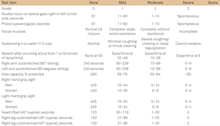
Table 3. Quantitative myasthenia gravis score for disease severity.
Test item None Mild Moderate Severe Score
Grade 0 1 2 3
Double vision on lateral gaze right or left (circle
one), seconds 61 11–60 1–10 Spontaneous
Ptosis (upward gaze), seconds 61 11–60 1–10 Spontaneous
Facial muscles Normal lid
closure
Complete, weak, some resistance
Complete, without
resistance Incomplete
Swallowing 4 oz water (1/2 cup) Normal Minimal coughing
or throat clearing
Severe coughing/ choking or nasal regurgitation
Cannot swallow
Speech after counting aloud from 1 to 50 (onset
of dysarthria) None at 50
Dysarthria at 30–49
Dysarthria at
10–29 Dysarthria at 9
Right arm outstretched (90° sitting), 240 seconds 90–239 10–89 0–9
Left arm outstretched (90 degrees sitting), 240 seconds 90–239 10–89 0–9
Vital capacity, % predicted ≥80 65–79 50–64 <50
Right-hand grip, kgW
Men ≥45 15–44 5–14 0–4
Women ≥30 10–29 5–9 0–4
Light-hand grip, kgW
Men ≥35 15–34 5–14 0–4
Women ≥25 10–24 5–9 0–4
Head lifted (45° supine), seconds 120 30–119 1–29 0
Right leg outstretched (45° supine), seconds 100 31–99 1–30 0
Right leg outstretched (45° supine), seconds 100 31–99 1–30 0
Fig 1. Normal neuromuscular junction and pathophysiology of myasthenia gravis. In the normal neuromuscular junction, acetylcholine (Ach) released from the nerve terminal following a nerve action potential binds to the acetylcholine receptor (AChRs) on the postsynaptic muscle, triggering a muscle action potential propagated by the voltage-gated sodium channel. Acetylcholinesterase scavenges and breaks down unbound ACh. In a separate pathway, neural agrin binds muscle-specific tyrosine kinase (MuSK) initiating clustering of phosphorylated rapsyn and AChRs, stabilizing the postsynaptic structure opposite the nerve. MuSK initiates clustering of the cytoplasmic protein rapsyn and AChRs and is believed to maintain normal postsynaptic architecture.
In myasthenia gravis caused by antibodies to the AChRs, there is blockade of the binding site for ACh, cross-linking of the AChR with subsequent internalization and reduction in its surface expression, and initiation of complement and cellular inflammatory cascades with damage to the post- and presynaptic structures. The molecular physiology of myasthenia gravis mediated by antibodies to MuSK has not been established.
Nerve terminal acetylcholine
agrin
acetylcholine in a presynaptic vesicle
nicotinic acetylcholine receptor
muscle specific tyrosine kinase rapsyn
voltage gated sodium channel acetylcholine binding site
acetylcholinesterase
Ta e 4. Clinical subtypes and the occurrence of the various muscle autoantibodies in the different subgroups of
myasthenia gravis.
Muscle autoantibodies (percentage of patients)
MG subgroups Age of
onset
Thymic histology
HLA associations
AChR MuSK Titin RyR Clinical findings
Early-onset non-MuSK nonthymoma
<40 Hyperplasia DR3-B8
DR9 (in Asians)
+ (100%) – (100%) + (10%) – (100%) These patients are more often female. In addition to anti-AChR antibodies, other organ-specific autoantibodies might be present, and patients might be affected by other autoimmune dis-eases, most commonly autoimmune thyroid disease. Antibodies to non-AChR muscle components are not typically seen in early-onset MG Late-onset
non-MuSK non-thymoma
>40 Normal/
thymic atrophy
DR2-B7 + (100%) – (100%) + (58%) + (14%) These patients are more often male
and usually have normal thymic his-tology or thymic atrophy. They can present with ocular or generalized weakness, but typically have a more severe disease course compared with early-onset MG, and spontane-ous remissions are rare. The pres-ence of anti-ryanodine receptor an-tibodies has been associated with more severe, generalized, or predom-inantly oropharyngeal weakness, and frequent myasthenic crises MuSK positive
(regardless of onset age)
<40 (most patients)
Normal DR14-DQ5 – (100%) + (100%) NA NA Whereas patients with anti-MuSK
antibodies can have presentations similar to anti-AChR-positive MG, they commonly have atypical clini-cal features, such as selective facial, bulbar, neck, and respiratory mus-cle weakness and marked musmus-cle atrophy, occasionally with relative sparing of ocular muscles. Respira-tory crises are more common than in generalized anti-AChR–positive dis-ease. Weakness can involve muscles that are not usually symptomatic in MG, such as paraspinal and up-per esophageal muscles. Enhanced sensitivity, nonresponsiveness, and even clinical worsening in response to anticholinesterase agents have also been reported. Disease onset in patients with anti-MuSK MG tends to be earlier, and patients are pre-dominantly female
Seronegative (regardless of onset age)
– (100%) – (100%) – (100%) – (100%) Patients with MG who lack both anti-AChR and anti-MuSK antibodies (so-called seronegative MG) are clini-cally heterogeneous and can have purely ocular, mild generalized, or severe generalized disease. The true prevalence of seronegative MG might be quite low, because some patients might have low-affinity anti-AChR antibodies that are not detected with currently available assays. Not sur-prisingly, these patients are essen-tially indistinguishable from patients with anti-AChR–positive MG in terms of clinical features, pharmacological treatment response, and even thym-ic abnormalities in some cases
are considered seronegative14,27. Clinically they are similar to
patients with AChR antibodies.
During MC, the respiratory failure can be hypoxemic, hypercapnic, or both and result from poor airway protection, inadequate secretions clearance, and hypoventilation. Bulbar (oropharyngeal) muscle dysfunction may be the predominant feature in some patients31. In MuSK-MG, bulbar weakness
always precedes respiratory failure7. he dysfunction of
bul-bar muscles alters cough, swallowing relexes, as well as sigh mechanisms2,5,7,9-11,31,32. Signs of bulbar weakness include
dys-phagia, nasal regurgitation, nasal and staccato speech, jaw and tongue weakness, and bifacial paresis32. It is diicult to handle
secretions that accumulate in the oropharynx. Upper airway patent is lost2,5,7,9-11,31,32. hese alterations increase the
likeli-hood of microaspiration, atelectasis, upper airway resistance, dead space, and work of breathing2,5,7,9-11,31,32. Muscle weakness
in AchR-MG tends to initially afect intercostals and accessory muscles and then the diaphragm7. he recruitment of
acces-sory muscles indicates signiicant inspiratory weakness32. Weak
involved in excitation-contraction coupling of striated mus-cle14,28-30. It is found in 50% of patients with MG and
thy-moma14,28-30. Higher RyR antibody levels are associated with
severity14,28-30. Patients with RyR antibodies are
character-ized by frequent involvement of bulbar, respiratory, and neck muscles14,28-30. Neck weakness at onset is a distinctive feature
of patients with RyR antibodies, while respiratory symptoms are found in patients with titin antibodies with and without RyR antibodies14,28-30. Limb involvement with few or no bulbar
signs is typical in RyR-antibody-negative MG28-30. Since many
thymoma patients have RyR antibodies, neck weakness and nonlimb bulbar distribution of symptoms are initial char-acteristic features. Such symptom distribution should raise the suspicion of thymoma28-30. hymoma and late-onset MG
share similar serological proile with high prevalence of titin and RyR antibodies and lower AChR antibody concentra-tions compared with early-onset MG29,30.
Finally, there is a remaining group of patients who do not have either AChR or MuSK antibodies and they actually
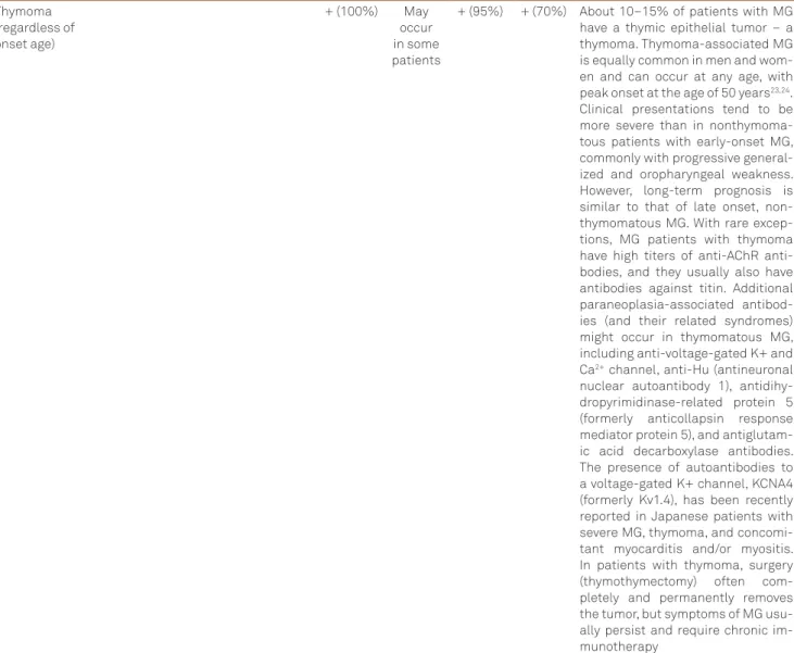
Table 4. Continuation
Thymoma (regardless of onset age)
+ (100%) May
occur in some patients
+ (95%) + (70%) About 10–15% of patients with MG have a thymic epithelial tumor – a thymoma. Thymoma-associated MG is equally common in men and wom-en and can occur at any age, with peak onset at the age of 50 years23,24.
Clinical presentations tend to be more severe than in nonthymoma-tous patients with early-onset MG, commonly with progressive general-ized and oropharyngeal weakness. However, long-term prognosis is similar to that of late onset, non-thymomatous MG. With rare excep-tions, MG patients with thymoma have high titers of AChR anti-bodies, and they usually also have antibodies against titin. Additional paraneoplasia-associated antibod-ies (and their related syndromes) might occur in thymomatous MG, including anti-voltage-gated K+ and Ca2+ channel, anti-Hu (antineuronal
nuclear autoantibody 1), antidihy-dropyrimidinase-related protein 5 (formerly anticollapsin response mediator protein 5), and antiglutam-ic acid decarboxylase antibodies. The presence of autoantibodies to a voltage-gated K+ channel, KCNA4 (formerly Kv1.4), has been recently reported in Japanese patients with severe MG, thymoma, and concomi-tant myocarditis and/or myositis. In patients with thymoma, surgery (thymothymectomy) often com-pletely and permanently removes the tumor, but symptoms of MG usu-ally persist and require chronic im-munotherapy
deterioration of bulbar and respiratory muscles. For these reasons, a strict monitoring of respiratory status with regular bedside pulmonary function testing is appropriate2,5,7,9-11,31-33.
Patients with previous diagnosed Myasthenia gravis
h e presence or worsening of clinical features, such as progressive muscle weakness (arms, limbs), palpebral pto-sis, bulbar muscle envolvement, and disphagia together with the presence of respiratory distress (dyspnea, shortness of breath, tachypnea, use of accessory muscles) may help to identify patients at risk for MC2,5,7,9-11,31-33.
Patients without a previous diagnosis of Myasthenia gravis
If MC is the i rst presentation of the disease, the specii c clin-ical features of the myasthenic state cannot be evident. h ese patients quite suddenly show a severe respiratory distress, facial weakness, airway collapse, and muscle failure. Initially, oxygena-tion is preserved32. A suspected clinical diagnosis should be
con-i rmed uscon-ing electrophyscon-iologcon-ical, pharmacologcon-ical, and labora-tory testing2,7,14,17,36, usually not available on an emergent basis36.
Electrophysiological testing
In MG, repetitive nerve stimulation (RNS) shows a sig-nii cant decremental response (>9%) between the i rst and fourth or i fth compound muscle action potential (CMAP) at low rates (2–5 Hz)14,17,36. RNS depletes Ach stores at
neuromus-cular junction, reducing the safety factor and the probability of successful neuromuscular transmission CMAP becomes reduced in amplitude and area14,17,36. In patients with
respira-tory involvement, phrenic and long thoracic nerves should also be tested37. h e results of repetitive long thoracic nerve
and phrenic stimulation show a good correlation with res-piratory symptoms and management requirements37.
Single-i ber electromyography (SFEMG)38 is the most sensitive test
for abnormal neuromuscular transmission detection; how-ever, it is time consuming and requires special expertise38.
Pharmacologic testing: edrophonium (Tensilon) h e Tensilon (edrophonium) test is useful in diagnosing MG and in distinguishing MC from cholinergic crisis2,7,14,17.
Edrophonium is a acetylcholinesterase inhibitor with rapid onset (30 seconds), and ef ects lasting 5 minutes reported a sensitivity of 86% for ocular MG and 95% for generalized MG14,17. Edrophonium temporarily improves the safety factor
of neuromuscular transmission and may elicit improved mus-cle strength2,7,14,17. Once airway and ventilation are secured,
give an initial dose of 1–2 mg and watch for 1 minute, then give 3 mg, and another 3 mg if neccesary14,17. Typical side ef ects
of sweating, tearing, fasciculations, and abdominal cramping may indicate peak edrophonium ef ect. Observe for possible serious adverse ef ects such as hypotension or arrhythmias and have always atropine available as antidote14,17.
cough or dii culty in counting notes weakness of expiratory muscles32. Anxiety, accompanied by tachycardia and
tachyp-nea, may be the i rst sign of air hunger32. Respiratory muscles
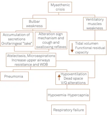
are unable to maintain adequate tidal volume. Ventilation becomes rapid and shallow, decreasing pulmonary functional residual capacity, resulting in atelectasis, closing a vicious circle that increases work of breathing with exacerbation of muscle weakness that culminates altering the ventilation/perfusion relationship causing hypoxia and hypercapnia2,5,7,9-11,31,32 (Fig 2).
h e signs of MC should be sought in all patients with MG, even when they do not complain weakness because central ventilatory drive usually remains intact during crisis; so, even when minute ventilation response to CO2 is poor, the
gener-alized weakness can mask the usual signs of respiratory dis-tress. Respiratory muscles may suddenly fatigue, producing precipitous respiratory collapse32. In addition, some patients
may present with respiratory insui ciency out of proportion to limb or bulbar weakness2,5,7,9-11,31-33. In rare cases of MC,
ven-tilatory failure is the only clinically overt manifestation34,35.
HOW TO MAKE A CORRECT DIAGNOSIS?
MC is an acute respiratory failure due to worsening MG, characterized by forced vital capacity (FVC) below 1 L, nega-tive inspiratory force (NIF) of 20 cm H2O or less, and the need for ventilatory support2,5,7,9-11,31-33. Arterial blood gas analysis
commonly shows hypercarbia before hypoxia. h ere should be a low threshold for endotracheal intubation due to rapid
Fig 2. Pathophysiology of myasthenic crisis. Myasthenic
crisis
Bulbar weakness
Accumulation of secretions Orofaringeal “lake”
Alteration sign mechanism and
cough and
swallowing reflexes Tidal volumenFunctional residual capacity
Atelectasis, Microaspirations Increase upper airways
resistance and WOB
Pneumonia Hypoventilation Dead space V/Q alterations
Hypoxemia-Hypercapnia
Respiratory failure
Ventilatory muscles weakness
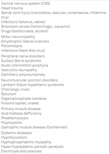
A number of disorders that cause respiratory failure due to muscle weakness should be considered in the diferential diagnosis (Table 5).
ACUTE MANAGEMENT
General evaluation
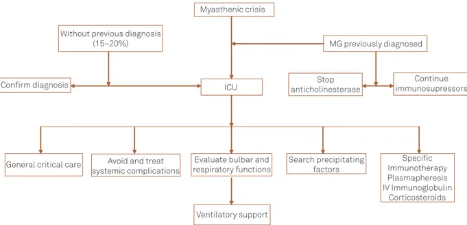
he management of MC should follow a step by step, sequential, and multidisciplinary protocol (Fig 3), based on guidelines of the European Federation of Neurological Socie-ties40. Prompt recognition of impending respiratory paralysis
is the key to successful management31-33. he evolution of
res-piratory muscle weakness in AChR-MG often follows a pat-tern where the intercostals and accessory muscles weaken irst, followed by the diaphragm31-33. In MuSK-MG, bulbar
weakness always precedes respiratory failure7.
Trigger detection and assessment of the respiratory and bulbar functions
It is essential to evaluate bulbar and respiratory func-tions together with ensure life support and detecting trigger conditions (precipiting factors)1,2,5,7-13,16,33. Habitually, bulbar
and respiratory dysfunction occurs simultaneously with If muscle strength improves within 1 minute of any dose
increment, test is positive and no further edrophonium needs to be administered14,17. Edrophonium test is not
rec-ommended in patient in crisis because of likelihood of false- positive or false-negative results, and the risk of worsening muscle weakness above all in patients with anticholinest-erase overdose2,7,14,17. Patients with a cholinergic crisis may
respond to edrophonium challenge by increasing salivation and bronchopulmonary secretions, diaphoresis, and gastric motility2,5,7,14,17. hese changes should be managed expectantly,
as the half-life of edrophonium is short (10 min). In addition, worsening of bulbar and respiratory symptoms in MuSK-MG after anticholinesterase administration is known and could confound the clinical diagnosis17. If the patient requires
ven-tilatory support there is no need to distinguish the two crisis entities17. False-positives have been also reported in lower
motor neuron diseases and brainstem tumors17.
Serological testing
If MG is suspected, the patients should be tested for AChR antibodies14,17,39. If these are negative, MuSK
antibod-ies should also be tested14,17,39. Antibodies should be sent for
analysis before the institution of any immunotherapy. Anti-AChR antibodies are elevated in 85–90% of patients with gen-eralized MG14,17,28,39. MuSK-related autoimmune-acquired MG
presents with slightly diferent phenotype 14,17,28,39.
Other testing
Chest computerized tomography (CT) should be per-formed in patients with MG to exclude thymoma14,17. Chest
CT is more sensitive than plain chest radiographs for delin-eating anterior mediastinal masses. MRI does not improve diagnostic sensitivity. Iodinated contrast agents may pre-cipitate worsening of myasthenic weakness20. Although
this is an uncommon phenomenon, we do not routinely use iodinated contrast agents during chest CT to assess for thymoma. hese examination should be made in a stable patient. Since MG often coexists with other autoimmune disorders, particularly thyroid disease, patients should undergo thyroid and other autoimmune testing when clini-cally appropriate14,17,39.
DIFFERENTIAL DIAGNOSIS
Diferential diagnosis includes other disorders of the neu-romuscular junction including Lambert–Eaton syndrome, botulism, congenital myasthenic syndromes, and tick paraly-sis2,5,7,9-11,14,17. In addition, acute inlammatory demyelinating
polyradiculoneuropathy (AIDP) and variants, particularly those featuring external ophthalmoplegia and ptosis, may simulate MG,2,5,7,9-11,14,17. Motor neuron disease and brainstem
ischemia involving oropharyngeal weakness may appear in MG14,17.
Table 5. Neurologic and systemic causes of muscle/respiratory weakness/failure.
Central nervous system (CNS) Head trauma
Spinal cord Injury (traumatica, vascular, compressive, inflamma-tory)
Infections (tetanus, rabies)
Brainstem stroke (hemorrhagic, isquemic) Drugs (barbiturates, alcohol)
Motor neuronopathy Amyotrophic lateral sclerosis Poliomielytis
Infections (West Nile virus) Peripheral nerve disorders Guillain Barrè syndrome Acute intermittent porphyria Vasculitis neuropathy Diphtheric polyneurophaty Neuromuscular junction disorders Lambert-Eaton myasthenic syndrome Cholinergic crisis
Botulism
Organophosphate overdose Poisons (spider, snake) Primary muscle disease Acid maltase defficiency Rhabdomyolysis Polymyositis
Dystrophic muscle disease (Duchenne’s) Systemic diseases
Hypothyroidism
Fig 3. Algorithm for myasthenic crisis management.
generalized muscle weakness characteristic of myasthenic patients. Typical clinical features include shortness of breath, tachypnea, orthopnea, discomfort, tachycardia, sweating, use of accessory muscles of respiration, or paradoxical ven-tilation31-33. he collapse of the airway is marked by coughing
and swallowing disability, leading to accumulation of secre-tions in the pharynx31-33. Patients are unable to swallow 5 cc
of water or count until 20 in a single respiratory cycle7,31-33.
Intubation and mechanical ventilation
he decision of the mode of ventilatory support should be based on clinical judgement. Careful observation and bedside measurements (vital capacity, peak low measure-ment, pulse rate, and blood pressure) are more important than repeated monitoring of blood gases1,2,5,7-13,16,33.
Never-theless, certain ventilatory tests can be performed, includ-ing forced vital capacity (FVC), negative inspiratory pres-sure (NIP), positive expiratory prespres-sure (PEP), and arterial blood gases1,2,5,7-13,16,33. he 20/30/40 rule (FCV<20 mL/kg;
NIP<30 cmH2O; and PEP<40 cmH2O) is probably the most helpful guide to decide intubation (Table 6). FVC<30 mL/kg is associated with inefective cough, poor handling of secretions, atelectasis, and hypoxemia. NIP<20 cmH2O
sig-nals marked weakness of the inspiratory muscles and dia-phragm, while PEP<40 cmH2O indicates involvement of
expiratory muscle function closely linked with the ability to cough and clear secretions1,2,5,7-13,16,33. hese
determina-tions require training, depending on the patients’ efort, and require proper closure of the mouth — all conditions are diicult to obtain during the crisis. Furthermore, these threshold values have not been established through pro-spective studies. In addition, muscle weakness often
luc-tuates, and patients can develop apnea suddenly, or may precipitously fatigue with the rapid development of respira-tory failure before a downward trend in these parameters is noted1,2,5,7-13,16,31-33. Moreover, none of them have been shown to
be reliable predictors of the need for mechanical ventilation. Life-threatening hypoxemia (PaO2<60 mmHg) occurs late in neuromuscular respiratory failure and generally improves with supplemental oxygen1,2,5,7-13,16,31-33. In this situation,
we can attempt the use of bilevel positive airway pressure (BiPAP), since the application of positive pressure helps to endure increased resistance of the upper airways in addition to preventing alveolar collapse and atelectasis2,5,7,31-33. Severe
hypercapnia (PaCO2>50 mmHg) predicts BiPAP failure and
indicates that muscle fatigue is imminent36. he absolute
indications for intubation may include cardiac or respira-tory arrest, impaired consciousness, shock, life-threatening arrhythmias, severe blood–gas alterations, and bulbar dys-function with conirmed aspiration1,2,5,7-13,16,50. Much more
diicult is the decision to intubate when such strict criteria are not met. If doubt exists, it is recommended to intubate and ventilate inmediately2,7-13,33. Endotracheal intubation Table 6. Ventilatory test in patients with myasthenic crisis.
Normal Intubation criteria
Weaning criteria
Extubation criteria FVC
(mL/kg) >60 <20 >15 >25
NIP (cmH2O)
>-70 <-30 >20 >40
PEP (cmH2O)
>100 <40 >40 >50
FVC: indicates forced vital capacity; NIP: negative inspiratory pressure; PEP: positive expiratory pressures.
Myasthenic crisis
MG previously diagnosed Without previous diagnosis
(15–20%)
ICU Confirm diagnosis
Search precipitaing factors General criical care Evaluate bulbar and
respiratory funcions
Specific Immunotherapy Plasmapheresis IV Immunoglobulin
Coricosteroids Coninue immunosupressors Stop
anicholinesterase drugs
Avoid and treat systemic complicaions
Venilatory support
Myasthenic crisis
ICU anticholinesteraseStop
Continue immunosupressors Confirm diagnosis
General critical care Avoid and treat systemic complications
Evaluate bulbar and respiratory functions
Search precipitating factors
Ventilatory support
Specific Immunotherapy Plasmapheresis IV Immunoglobulin
Corticosteroids Without previous diagnosis
(15–20%) MG previously diagnosed
such as a satisfactory oxygenation, PaO2/FIO2≥200 mmHg,
PEEP ≤5 cmH2O; hemodynamic stability, and a good con-sciousness status able to cough efectively2,5,7-13,16,33. Patients
should be transitioned to a spontaneous mode of ventilation (e.g., pressure support ventilation) previous to T-tube trial. Pressure support can then gradually be decreased to minimal settings33,43. If the patient does not tolerate weaning, assisted
ventilation should be reinstituted.
It remains unclear when to attempt extubation after MC. Prolonged intubation in myasthenic patients may lead to several complications such as atelectasis, anemia, urinary tract infec-tion, congestive heart failure, and ventilator-associated pneu-monia11,45. To prevent atelectasis, aggressive chest physiotherapy
and frequent suctioning should be implemented together with continuous positive airway pressure (CPAP). Age>50 years, peak VC<25 mL/kg on postintubation days 1 to 6, and a serum bicar-bonate ≥30 mmol/L are independent risk factors for prolonged intubation (>14 days)7. Extubation failure is most commonly
associated with a weak cough and inadequate airway clear-ance33,45-47. Tracheostomy is generally not needed in MC because
the duration of intubation is often less than 2 weeks2,5,7-13,16,33,46.
One rare condition that often requires tracheostomy is severe upper airway obstruction due to bilateral vocal cord paralysis. Furthermore, patients with a prolonged intubation are usually hospitalized three times longer and are less likely to be functionally independent upon discharge7.
A maximal expiratory pressure has been demonstrated to independently predict extubation success. However, there are no good clinical criteria for when and how to extubate safely. Fluctuating weakness and pulmonary complications often confound the decision to extubate47. Patients are
typi-cally extubated if VC, PImax, and PEmax are ≥15 mL/kg, ≤-20 cmH2O, and >40 cmH2O respectively, and tidal volume
≥5 mL/kg2,5,12,45,48. If the patient complains of fatigue or
short-ness of breath, extubation should not be performed even if the criteria of these indices are met and blood gases are normal.
Noninvasive positive pressure ventilation
Noninvasive ventilation may be used to prevent intuba-tion or reintubaintuba-tion in MC49-52. With BiPAP, positive pressure
is applied during both phases of respiratory cycle, enhancing airlow, alleviating the work of breathing during inspiration, and preventing airway collapse and atelectasis during expi-ration49-52. here are studies of Noninvasive positive pressure
ventilation (NIPPV) during MC49-52. In 2002, Rabinstein and
Wijdicks irst reported their experience49. All patients have had
bulbar compromise. NIPPV was well tolerated and the length of hospital stay was signiicantly reduced compared with those who were intubated (mean 7±5 days versus 23±16 days; p=0.03). paCO2>50 mmHg portends BiPAP ineiciency49.
Subsequent reports suggest that NIPPV may be useful in preventing intubation or reintubation in these patients47-51.
A recent prospective study suggests that NIPPV, combined with assisted coughing after extubation, avoids the need for can often be performed electively rather than as an
emer-gent response32,33. he initial ventilatory support should be
directed to improve muscle fatigue and to mantain lung expansion1,2,5,7-13,16,33. We suggest as initial mode assist-control
ventilation, with low tidal volumes (6–8 mL/kg), respiratory rate 12–16/min, and positive end-expiratory pressure (PEEP) of 5 cmH2O. FiO2 should be adjusted to achieve a SaO2 >92%
or PaO2 >70 mmHg7-13,16,33. Pressure support ventilation
between 5 and 15 cmH2O is another option
7-13,16,33. In case of
atelectasis, we consider recruitment manoeuvres or the uti-lization of sighs (1.5 × tidal volume) 3 to 4 times per hour10.
he degree of support is patient dependent and should be adjusted and based on arterial blood analysis. In patients with chronic hypercarbia, PaO2 should be kept above 45 mm Hg to avoid alkalosis and bicarbonate wasting, which make weaning more diicult2,5,7-13,16,33.
Bronchodilators may be useful in maintaining airway patency and overcoming bronchospasm. Inhaled ipratro-pium bromide may be of choice because it is safe and can decrease bronchial secretions41. Terbutaline, a β2 adrenergic
agonist, may be an efective adjunct therapy in these patients, although conirmation with larger trials will be required42.
Meticulous attention to pulmonary toilet is required due to inefective cough. Aggressive chest physiotherapy (percus-sion, vibration, and postural drainage) and airway clearance (regular suctioning and therapeutic iberoptic bronchoscopy in severe cases) should be implemented43. Inspired gas
humid-ity should be around 80% at 37°C. Patients with a peak cough low <180 L/min can augment cough response with manual physiotherapy and with insulation-exsulation devices. Cough response increases and is associated with improved prognosis independent of FVC or breathing pattern44 .
Adequate nutrition is important to avoid negative energy balance and worsening of muscle strength12. All patients should
receive adequate nutritional support (25–35 calories/kg) via enteral route whenever possible. In patients with hyper-carbia and diiculty weaning, low carbohydrate feeds are the preferred solution43. Potassium, magnesium, and phosphate
depletion can exacerbate MC and should be repleted. Anemia can also increase weakness, and several experts recommend transfusions when hematocrit values are under 30%12.
Addi-tionally, deep-vein thrombosis prophylaxis, hemodynamic sta-bility, and glycemia control are strongly recommended2,7-13,16.
Weaning from ventilation should start when the patient regains muscular strength; is hemodynamically stable with-out electrolite disturbances, fever, infections, or systemic complications; and the reason for mechanical ventila-tion has been resolved or is in the process of frank resolu-tion2,5,7-13,16,33. Improvement in the strength of neck lexors and
other adjunct muscles usually is associated with improve-ment in bulbar and respiratory muscle strength and can be a useful tool for assessing clinical improvement7-13,16,33.
eicacy, so they can be chosen by availability, adverse efects, costs, experience, and patients’ proile2,5,7-13,16,18,40.
IVIg is a IgG-puriied blood derivate.he mechanism of action is unknown. It needs about 5 days to exert maximun therapeutic efects. he usual regimen is 0.4 g/kg/day for 3–5 days2,5,7-13,16,18,40. One study did not ind a diference
between 1 or 2 g as total dose52. Patients should be screened
for IgA deiciency to avoid anaphylaxis2,5,7-13,16. More prevalent
side efects are fever, overload of luids, nausea, and headache. Less frequent and more serious complications are aseptic meningitis, pulmonar edema, anailaxia, renal dysfunction, cardiac arrhythmia, thrombocytopenia, stroke, myocardial infarction, and pulmonary embolism2,7-13,14,16.
PE and IA are most efective when we need a fast response, particulary in patients who do not improve, are worsening, or are having severe complications2,5,7-13,16,18,40,53. Response to
treat-ment generally occurs after 2 days. he optimal response of PE and IA occurs in both AChR-Ac- and MuSK-positive patients. he proposed mechanism of action is rapid depletion of path-ogenic antibodies from plasma, which causes an osmotic equi-libration between extra- and intravascular spaces leading to reduction of antibodies in neuromuscular junction2,5,7-13,16,18,40.
Both apheresis procedures, PE and IA, must be performed at low-dose regimen; it is 1.5 L of plasma (20–25 mL/kg), per ses-sion, with an exchange rate of 10–20 mL/min. he procedure needs central venous access and anticoagulation. PE should be made in a course of ive exchanges every other day over 10 days. Replacement luid is generally normal saline/5% albu-min2,5,7-13,16,18,40. A series of nonrandomized studies have
dem-ostrated beneical short-term eicacy of this therapy in acute setting and during preparation for thymectomy2,5,7-13,16,18,40.
PLASMAPHERESIS OR IMMUNOGLOBULIN?
his question does not have a response. IVIg may be bet-ter tolerated than PE; however PE showed similar short-bet-term efects in comparison with IVIg in one RCT54 but was more
efective than IVIg in a retrospective study55. PE may probably
have a more predictable response than IVIg during crisis. IVIg and PE are equally efective in preparation for surgery56. In
conclusion, until now there is not enough evidence of high quality to support one therapy over another during MC. If there is insuicient or no response to treatment, PE can be given after IVIg, and IVIg can be administered after PE.
STEROIDS AND OTHER
IMMUNOSUPPRESSIVE AGENTS
Patients who are taking steroids should not stop them2,5,7-13,16,18,40. Possibly after crisis, the dose should be
increased. If we need to start steroids after PE or IVIg, oral pred-nisone is preferred at 1 mg/kg/day (60–100 mg daily)2,7-13,16,40.
reintubation or tracheostomy in patients with neuromuscu-lar diseases, besides shortening their stay in the ICU50,51. Early
application of NIPPV after extubation can reduce the risk of respiratory failure and lowered mortality in hypercapnic patients with chronic respiratory disorders51. Use of NIPPV to
avoid reintubation in MC is well established but is a relatively uncommon practice51. Some studies reported that NIPPV
prevented reintubation in 70% of patients49,50. It should be
included in the routine approach to these patients at high risk for postextubation respiratory failure.
COMPLICATIONS IN THE
MANAGEMENT OF MYASTHENIC CRISIS
Fever is the most common complication associated with MC2,5,7-13,16. Infectious complications include
pneu-monia, bronchitis, urinary tract infections, Clostridium diicile colitis, bacteremia, and sepsis2,5,7-13,16.When compared
with patients admitted for noncrisis, patients admitted with MC are more likely to experience sepsis, deep-vein throm-bosis, and cardiac complications including congestive heart failure, acute myocardial infarction, arrhythmias, and cardiac arrest. hese complications, however, are not independent predictors of mortality2,5,7-13,16.
STOP ANTICHOLINESTERASE DRUGS
Anticholinesterase therapy should be temporarily with-drawn immediately after establishing mechanical ventilatory support because they are unnecessary in this situation and may complicate pulmonary management2,5,7-13,16. In addition,
the continued use of these medications may promote cho-linergic crisis by overdose11. Although cholinergic crisis is
an important consideration in the evaluation of the patient in MC, it is uncommon2,5,7-13,16. Cholinergic crisis may cause
increase of pulmonary secretions (muscarinic efects) and fasciculations (nicotinic efects), both of which contribute to exacerbate muscle weakness and respiratory failure2,5,7-13,16.
Furthermore, acetylcholinesterase inhibitors may promote cardiac arrythmias and myocardial infarction2,5,7-13,16. he
time to start cholinergic agents (pyridostigmine) preferably orally or by nasogastric tube is not well established, but is recommended when the patient shows clinical improvement before weaning of mechanical ventilation2,5,7-13,16,18.
IMMUNOTHERAPY
Immunomodulatory treatment is considered standard of care for patients with MC2,5,7-13,16,18,40. Speciic
IIA, IIB, and III according to clinical grade of Osserman classiication)57,58, a history of MC23, preoperative
pres-ence of bulbar weakness23, serum AChR antibody levels
>100 nmol/L, and intraoperative blood loss of >1 L. Other predisposing factors are obesity (BMI >25.6), higher doses of pyridostigmine (>270 mg) and immunosupressants, FVC <2 L, and history of infection 1 month before surgery25.
Type and technique of surgery may afect the occurrence of MC after thymectomy58-60. he presence of thymoma is the
more important isolated postoperative factor to develop MC together with radiation therapy, delayed ventilator weaning, and upper or lower pulmonary tract infections23-25,57. he risk
of MC has decreased with less invasive surgical tecniques such as cervicotomy, partial sternotomy, or video-assisted thoraco-scopic57,59,60. In a study of 218 thymectomies with diferent
tech-niques and approachs, no signiicant diferences were found in terms of incidence or severity of MC or in the inal outcome25.
CONCLUSIONS
MC is a severe and life-threatening neurological condition characterized by generalized muscle weakness with respira-tory or bulbar compromise that require ventilarespira-tory support. It can be the debut form of MG, so the diagnosis should be conirmed following a standardized protocol. Evaluation of bulbar and respiratory functions is imperative. he cor-nerstones of the treatment are correct ventilatory manage-ment, search and correction of predisposing factors, speciic immunotherapy (PE, IVIg), avoiding systemic complications, and planiication of long-term treatment (immunosupres-sors). he majority of patients with MC require endotra-cheal intubation and mechanical ventilation. hymectomy should be evaluated. With modern intensive care, the out-comes are excellent with mortality near to 5% attribuited principally to comorbidities, cardiac complications, or pulmonary embolism.
AKNOWLEGDMENTS
We thank Mrs. Vanessa Starling for her editorial assistance. he timing of initiation is controversial, but usually it is
indicated when the patients cannot be extubated 2 weeks after speciic immunotherapy2,7-13,16,40. It may be initiated
concurrently with IVIg or PE, since prednisone begins to work after 2 weeks. Enteral administration is preferred, and initiation of prednisone may be deferred until after extubation if the patient improves with IVIg or PE treat-ment2,5,7-13,16,18,40. he mean time to improvement with
pred-nisone is around 13 days. Worsening of symptoms with the initiation of corticosteroids is not predictive of overall response to corticosteroids22. Once the patient has begun
to show improvement, dose can be decreased and gradually converted to alternate-day dosing. It is important to have in mind that steroids can exacerbate muscle weakness or may increase the risk of critical illness myopathy2,7-13,18,22. In
septic patients, it is preferable to delay steroids until infec-tion is under control. Relative contraindicainfec-tions are dia-betes with poor metabolic control or severe osteoporosis. Other immunosuppressive drugs, necessary to long-term management of MG, such as cyclosporine, azathioprine, or mycophenolate, are not useful during MC principally due to the delayed onset of action2,5,7-13,16,40.
ROLE OF THYMUS: SURGICAL CONSIDERATIONS
hymectomy plays a central role in management of MG14,17,40. About 65% of the patients in seropositive group
have thymic hyperplasia and 15% thymoma14,17.
hymec-tomy is the only treatment in MG that ofers possibilities of complete remission14,17. Indications for tymectomy include:
(a) failure of long-term conservative therapy, (b) thymoma, and (c) new onset of generalizated MG14,17,40. Patients with
more beneits after thymectomy are those <60 years, sero-positives, and with thymic hyperplasia14,17,40. he role of a
thymectomy in MuSK patients is not clear. Chu et al.57
sug-gested that thymectomy seems to have a preventive efect in both incidence and severity of MC, but the frequency of postoperative crisis varied from 6% to 21.9%23 deined
as respiratory failure or delayed postoperative extubation (>24 hours)57. Postoperative crisis has been related to age
(onset of the disease more than 50 years)58, severity (type
References
1. Bouschot J, Baumann I, Kalischewski P, et al. Therapy of myasthenic
crisis. Crit Care Med 1997;25:1228-1235.
2. Lacomis D. Myasthenic crisis. Neurocrit Care 2005;03:189-194. 3. Oosterhuis HJ. Observations of the natural history of myasthenia
gravis and the effect of thymectomy. Ann NY Acad Sci 1981;377: 678-690.
4. Cohen MS, Younger D. Aspects of the natural history of myasthenia gravis: crisis and death. Ann NY Acad Sci 1981;377:670-677.
5. Thomas CE, Mayer SA, Gungor Y, et al. Myasthenic crisis: clinical features, mortality, complications, and risk factors for prolonged intubation. Neurology 1997;48:1253-1260.
6. Bedlack RS, Sanders DB. On the concept of myasthenic crisis. J Clin Neuromuscul Dis 2002;4:40-42.
7. Chaudhuri A, Behan PO. Myasthenic crisis. QJM 2009;102:97-107. 8. Mayer SA. Intensive care of the myasthenic patient. Neurology
37. Lo YL, Leoh TH, Dan YF, et al. Repetitive stimulation of the long thoracic nerve in myasthenia gravis: clinical and electrophysiological correlations. J Neurol Neurosurg Psychiatry 2003;74:379-381. 38. AAEM Quality Assurance Committee. American Association of
Electrodiagnostic Medicine. Practice parameter for repetitive nerve stimulation and single fiber EMG evaluation of adults with suspected myasthenia gravis or Lambert–Eaton myasthenic syndrome: summary statement. Muscle Nerve 2001;24:1236-1238.
39. Oh SJ, Kim DE, Kuruoglu R, et al. Diagnostic sensitivy of the laboratory tests in myasthenia gravis. Muscle Nerve 1992;15:720-724.
40. Skeie GO, Apostolski S, Evoli A, et al. Guidelines for treatment of autoimmune neuromuscular transmission disorders. Eur J Neurol 2010;17:893-902.
41. Szathmáry I, Magyar P, Szobor A. Myasthenia gravis: protective effect of ipratropium bromide (Atrovent) on airways obstruction caused by edrophonium chloride (Tensilon). Eur Neurol 1981;20:56-61. 42. Soliven B, Rezania K, Gundogdu B, Terbutaline in myasthenia gravis: a
pilot study. J Neurol Sci 2009;277:150-154.
43. Varelas PN, Chua HC, Natterman J, et al. Ventilatory care in myasthenia gravis crisis: assessing the baseline adverse event rate. Crit Care Med 2002;30:2663-2668.
44. Chatwin M, Ross E, Hart N, et al. Cough augmentation with mechanical insufflation/exsufflation in patients with neuromuscular weakness. Eur Respir J 2003;21:502-508.
45. Wu JY, Kuo PH, Fan PC, et al. The role of non-invasive ventilation and factors predicting extubation outcome in myasthenic crisis. Neurocrit Care 2009;10:35-42.
46. Rabinstein AA, Mueller-Kronast N. Risk of extubation failure in patients with myasthenic crisis. Neurocrit Care 2005;3:213-215. 47. Seneviratne J, Mandrekar J, Wijdicks EF, Predictors of extubation
failure in myasthenic crisis. Arch Neurol 2008;65:929-933.
48. Meriggioli MN, Sanders DB. Autoimmune myasthenia gravis: emerging clinical and biological heterogeneity. Lancet Neurol 2009;8:475-490. 49. Rabinstein A, Wijdicks EF. BiPAP in acute respiratory failure due
to myasthenic crisis may prevent intubation. Neurology 2002;10: 1647-1649.
50. Vianello A, Arcaro G, Braccioni F, et al. Prevention of extubation failure in high-risk patients with neuromuscular disease. J Crit Care 2011;26:517-524.
51. Saeed T, Patel S. Use of non invasive ventilation to avoid re-intubation in myasthenia gravis; a case report and review of literature. J Pakist Med Assoc 2011;61:293-295.
52. Gajdos P, Tranchant C, Clair B, et al. Treatment of myasthenia gravis exacerbation with intravenous immunoglobulin: a randomized, double-blind clinical trial. Arch Neurol 2005;62:1689-1693.
53. Ishizeki J, Nishikawa K, Kunimoto F, et al. Post-operative myasthenic crisis successfully treated with immunoadsorption therapy. J Anesth 2005;19:320-322.
54. Gajdos P, Chevret S, Clair B, et al. Clinical trial of plasma exchange and high-dose intravenous immunoglobulin in myasthenia gravis. Myasthenia Gravis Clinical Study Group. Ann Neurol 1997;41:789-796. 55. Qureshi AI, Choudhry MA, Akbar MS, et al. Plasma exchange versus
intravenous immunoglobulin treatment in myasthenic crisis. Neurology 1999;52:629-632.
56. Jensen P, Bril V. A comparison of the effectiveness of intravenous immunoglobulin and plasma exchange as preoperative therapy of myasthenia gravis. J Clin Neuromuscul Dis 2008;9:352-355. 57. Chu XY, Xue ZQ, Wang RW, et al. Predictors of postoperative myasthenic
crisis in patients with myasthenia gravis after thymectomy. Chin Med J 2011;124:1246-1250.
58. Huang CS, Hsu HS, Huang BS, et al. Factors influencing the outcome of transsternal thymectomy for myasthenia gravis. Acta Neurol Scand 2005;112:108-114.
59. Shrager JB, Deeb ME, Mich R, et al. Transcervical thymectomy for myasthenia gravis achieves results comparable to thymectomy by sternotomy. Ann Thorac Surg 2002;74:320-326.
60. Tomulescu V, Ion V, Kosa A, et al. Thoracoscopic thymectomy mid-term results. Ann Thorac Surg 2006;82:1003-1007.
9. Jani-Acsadi A, Lisak RP. Myasthenic crisis: guidelines for prevention and treatment. J Neurol Sci 2007; 261:127-133.
10. Bershad EM, Feen ES, Suarez JI. Myasthenia gravis crisis. South Med J 2008;101:63-69.
11. Ahmed S, Kirmani JF, Janjua N, et al. An update on myasthenic crisis. Curr Treat Op Neurol 2005;7:129-141.
12. Kirmani JF, Yahia AM, Qureshi AI. Myasthenic crisis. Curr Treat Op Neurol 2004;6:3-15.
13. Mayer SA, Thomas CE. Therapy of myasthenics crisis. Crit Care Med 1998;26:1136-1137.
14. Nicolle MW. Myasthenia gravis. Neurologist 2002;8:2-21.
15. Evoli A, Batocchi AP, Minisci C, et al. Clinical characteristics and prognosis of myasthenia gravis in older people. J Am Geriatr Soc 2000;48:1442-1448.
16. Wendell LC, Levine JM. Myasthenic crisis. Neurohospit 2011;1:16-22. 17. Juel VC, Massey JM. Myasthenia gravis. Orphanet J Rare Dis 2007;2:44. 18. Bedlack RS, Sanders DB. How to handle myasthenic crisis: essencial
steps in patient care. Postgrad Med 2000;107:211-222.
19. Pascuzzi RM. Medications and myasthenia gravis. Myasthenia Gravis Foundation of America, Inc 2004. http://www.myasthenia.org/drugs. 20. EliashivS, Wirguin I, Brenner T, et al. Agravation of human and
experimental myasthenia gravis by contrast media. Neurology 1990;40:1623-1625.
21. Marquardt J, Reuther P. [Myasthenia gravis. Information for the anesthetist and critical care physician]. Anaesthesist 1984;33: 207-211.
22. Bae JS, Go SM, Kim BJ. Clinical predictors of steroid-induced exacerbation in myasthenia gravis. J Clin Neurosci 2006;13: 1006-1010.
23. Nam T-S, Lee SH, Kim B-C, et al. Clinical characteristics and predictive factors of myastenic crisis after thymectomy. J Clin Neurosci 2011;18:1185-1188.
24. Soleimani A, Moayyeri A, Akhondzadeh S, et al. Frequency of myasthenic crisis in relation to thymectomy in generalizated myasthenia crisis:a 17 years experience. BMC Neurol 2004;4:12. 25. Xiang-yang C, Zhi-qiang X, Ru-wen W, et al. Predictors of postoperative
myasthenic crisis in patients with myasthenia gravis after thymectomy. Chin Med 2011;124:1246-1250.
26. Plauche WC. Myasthenia gravis in mothers and their newborns. Clin Obstet Gynaecol 1991;34:82-99.
27. Deymeer F, Gungor-Tuncer O, Yilmaz V, et al. Clinical comparison of Anti-MuSK-vs anti-ACHR-positive and soronegative myasthenia gravis. Neurology 2007;68:609-611.
28. Conti Fine BM, Milani M, Kaminski HJ. Myasthenia gravis: past, present and future. J Clin Invest 2006;116:2843-2854.
29. Aarli JA. Late-onset myasthenia gravis: a changing scene. Arch Neurol 1999;56:25-27.
30. Romi F, Gilhus NE, Varhaug JE, et al. Disease severity and outcome in thymoma myasthenia gravis: a long-term observation study. Eur J Neurol 2003;10:701-706.
31. Putman MT, Wise RA. Myasthenia gravis and upper airway obstruction. Chest 1996;109:400-404.
32. Rabinstein AA, Wijdicks EF. Warning signs of imminent respiratory failure in neurologic patients. Semin Neurol 2003;23:97-104. 33. Ping-Hung K, Pi-Chuan F. Respiratory care for myasthenic crisis, a look
into myasthenia gravis, Dr. Joseph A. Pruitt (Ed.), ISBN: 978-953-307 -821-2, InTech, 2012. Available from: http://www.intechopen.com/books/ a-look-into-myasthenia-gravis/respiratory-care-for-myasthenic-crisis. 34. Mier A, Laroche C, Green M. Unsuspected myasthenia gravis
presenting as respiratory failure. Thorax 1990;45:422-423.
35. Dushay KM, Zibrak JD, Jensen WA. Myasthenia gravis presenting as isolated respiratory failure. Chest 1990;97:232-234.