e36
Large Bilateral Coronary Artery Fistula: the Choice of Clinical
Treatment
Luciana Oliveira Cascaes Dourado, Aécio Flávio Teixeira de Góis, Whady Hueb, Luiz Antônio Machado César
Instituto do Coração do Hospital das Clínicas da FMUSP, São Paulo, SP, Brazil
We report the case of an asymptomatic female patient, with a large non-complicated bilateral coronary-pulmonary artery fistula. Clinical monitoring was the choice of treatment. We discuss the therapeutic options in depth, emphasizing the excessive tendency of the surgical approach and the great scarcity of reports on long-term clinical follow-up in asymptomatic patients.
Mailing Address: Whady Hueb •
Av. Dr. Enéas Carvalho de Aguiar, 44 AB 114 - 05403-000 - São Paulo, SP, Brazil E-mail: [email protected]
Manuscript received October 05, 2008; revised manuscript received No-vember 27, 2008; accepted March 24, 2009.
Case Report
The patient was a 49-year-old woman, ex-smoker, asymptomatic, with heart murmurs identified after a routine check-up. The patient was in good condition to undergo diagnostic examinations and did not report any significant past diseases. She reported leading an active life, regularly participating in sports. She also reported 2 successful pregnancies without any adverse events.
The patient underwent a physical examination and was found to be in good health, except for the presence of continued slight (innocent) heart murmurs with greater stress on the upper left sternal edge. The murmurs showed greater intensity during systole with a significant decrease in diastole. A breakdown of the second heart sound did not occur. The surface electrocardiogram and chest teleradiography were normal.
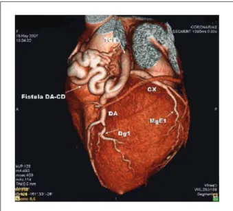
The transthoracic echocardiography (TTE) revealed normal global function, with the cardiac dimensions and systolic pressure of the pulmonary artery within normal standards. The TTE also revealed dilatation of the right coronary artery (RCA) and the left coronary artery (LCA). The LCA had a tortuous path with turbulent flow, leading into the pulmonary artery (PA). The coronary angiotomography showed that the anomalous artery branch with dilatation and significant tortuosity was located before the pulmonary artery, with its origin in the aorta, close to the emergence of the RCA, communicating with the anterior descending artery (ADA). This artery communicated with the PA in its mid third, emerging as a tortuous vessel (Figure 1). The percutaneous coronary angiography confirmed the previous findings. The myocardial perfusion imaging was normal.
Discussion
Coronary fistulae are characterized by anomalous communications between one or more branches of the coronary arteries, cardiac chambers’ coronary sinus, superior vena cava, and the pulmonary artery.
The coronary anomalies are occasional and usually an incidental finding during the coronary angiography, with an estimated incidence of 0.6 to 1.5% in patients undergoing examination1-3. Most of the coronary fistulae are congenital,
with causes that were probably acquired4. They represent a
broad spectrum of sizes and anatomical variations, and each of them has different clinical implications. Most frequently, they originate from the RCA (55%)5.
It seems that half of the cases are symptomatic, with symptoms being unspecific2,4. In patients with symptomatic
and bulky fistulae, the possible long-term complications include pulmonary hypertension and heart failure in cases of significant shunts; infectious endocarditis, thrombosis, or rupture of fistula; or myocardial ischemia secondary to the theft of myocardial flow1,4. However, studies of asymptomatic
patients free of interventions are scarce.
A consensus regarding the ideal treatment of fistulae does not exist, especially when treating moderate and asymptomatic fistulae4. This lack of consensus is due to the scarcity of clinical
follow-up of these patients. The few studies of conservative treatment with regular monitoring of the patients show good progress in the long-term5.
Fig. 1 - Coronary Computed Tomography Angiogram showing a large coronary
artery istula.
Key Words
Coronary Disease; Coronary Fistula; Coronary Abnormalities.
Potential Conflict of Interest
No potential conflict of interest relevant to this article was reported.
Sources of Funding
There were no external funding sources for this study.
Study Association
This study is not associated with any post-graduation program.
1. Libby P, Bonow RO, Mann DL, Zipes DP. Braunwald’s heart disease: a textbook of cardiovascular medicine.8th ed. Philadelphia: Elsevier Saunders; 2007.
2. Angelini P. Coronary fistulae: which ones deserve treatment, and what kind of treatment do they need? [Commentary]. Tex Heart Inst J. 2007; 34 (2): 202-3.
3. Cebi N, Schulze-Waltrup N, Frömke J, Scheffold T, Heuer H. Congenital coronary artery fistulas in adults: concomitant pathologies and treatment. Int J Cardiovasc Imaging. 2008; 24 (4): 349-55.
4. Luo L, Kebede S, Wu S, Stouffer GA. Coronary artery fistulae. Am J Med Sci. 2006; 332: 79-84.
5. Sherwood MC, Rockenmacher S, Colan SD, Geva T. Prognostic significance of clinical silent coronary artery fistulas. Am J Cardiol. 1999; 83: 407-11.
6. Mehta D, Redwood D, Ward DE. Multiple bilateral coronary arterial to
Most authors6-9 recommend surgery, fearing a possible risk
of adverse events. Others4,5,10, however, describe successful
conservative treatment in selected patients. They recommend regular clinical follow-up, with surgery if symptoms or an increase in the shunt occurs.
We suggest, therefore, clinical follow-up for asymptomatic patients and/or those without complications - ventricular dysfunction, ischemia, pulmonary hypertension - as in the case reported. In symptomatic patients or those with complications, however, percutaneous or surgical interventions are indicated, and these interventions are also better reported in the literature in these situations4.
References
pulmonary artery fistulae in an asymptomatic patient. Int J Cardiol. 1987; 16: 96-8.
7. Lowe JE, Oldham HN Jr, Sabiston DC Jr. Surgical management of congenital coronary artery fistulas. Ann Surg. 1981; 194: 373-80.
8. Macri R, Capulzini A, Fazzini L, Cornali M, Verunelli F, Reginato E. Congenital coronary artery fistula: report of five patients, diagnostic problems and principles of management. Thorac Cardiovasc Surg. 1982; 30: 167-71.
9. Armsby LR, Keane JF, Sherwood MC, Forbess JM, Perry SB, Lock JE. Management of coronary artery fistulae: patient selection and results of transcatheter closure. J Am Coll Cardiol. 2002; 39: 1026-32.
10. Cheung DLC, Au WK, Cheung HHC, Clement SW, Chin WTL. Coronary artery fistulas: long-term results of surgical correction. Ann Thorac Surg. 2001; 71: 190-5.
Arq Bras Cardiol 2009; 93(3) : e36-e37
Dourado et al Large Bilateral Coronary Artery Fistula