Jebmh.com
Original Article
J Evid Based Med Healthc, pISSN- 2349-2562, eISSN- 2349-2570/ Vol. 3/Issue 01/Jan. 04, 2016 Page 16
CORONARY ARTERY DOMINANCE PATTERN IN EAST-GODAVARI DISTRICT: A CADAVERIC
STUDY
Arindom Banerjee1, Anirban Das Gupta2
1Professor & HOD, Department of Anatomy, Konaseema Institute of Medical Sciences and Research Foundation,
Amalapuram.
2Professor, Department of Anatomy, Konaseema Institute of Medical Sciences and Research Foundation, Amalapuram.
ABSTRACT
With the advent of coronary angiography, coronary artery diseases can be well combated; but with time sedentary life style and stress as our constant partner have kept coronary artery disease as one of the major causes of death. Revascularization procedures demand a sound knowledge of the course of coronary arteries and their branches, both normal and their quite common variations. In this regard, posterior inter-ventricular artery (PIVA) deserves a special importance; PIVA determines the coronary dominance depending on its parent artery. Dominance can be right, left or of balanced type. Balanced type means that PIVA is derived from both right & left coronary arteries. Circulation can occur when both the coronary arteries emit a branch in that area. These and other variations form a very important repertoire of information based on which coronary bypass surgery and angioplasty can be safely and effectively performed. The aim of this study therefore is to document the coronary dominance pattern in this East Godavari district of Andhra-Pradesh.
60 adult human hearts were collected from museum of Anatomy department during the tenure of 5 years (2009 to 2014) and were preserved in 10% formalin. The hearts were dissected carefully to observe the posterior inter-ventricular artery in the posterior inter-ventricular sulcus of each heart and dominance pattern was recorded.
In our present study right dominance type was the commonest (46 out of 60) followed by left dominance (10 out of 60). Only 4 out of 60 were of the balanced type.
Present study, though not of the only member of its kind will definitely add up to the already existing vast knowledge, based on which various diagnostic and therapeutic intervention of coronary artery diseases can be done effectively and safely.
KEYWORDS
Coronary artery, Coronary dominance, Bypass surgery, Angioplasty.
HOW TO CITE THIS ARTICLE: Banerjee A, Gupta AD. Coronary artery dominance pattern in East-Godavari district: a cadaveric study. J Evid Based Med Healthc 2016; 3(1), 16-19. DOI: 10.18410/jebmh/2016/4
INTRODUCTION: The heart is supplied by two coronary arteries, right and left. The coronary arteries are the branches of ascending aorta. The two arteries form an oblique inverted crown, consisting of an anastomotic circle in the atrio-ventricular sulcus connected by marginal and inter-ventricular loops intersecting at the cardiac apex.[1] In
‘right dominance', (70%) the posterior inter-ventricular
artery is derived from the right coronary artery. In ‘left
dominance' (10%) the posterior inter-ventricular artery is derived from the left coronary artery. These people are likely to be affected by coronary diseases, because the entire left ventricle and left ventricular septum are under the nutritional control of the left coronary artery and obstruction of the left coronary artery may produce output failure of systemic
circulation. In the ‘balanced' pattern, (20%) posterior inter -ventricular branches are derived from both coronary
arteries. Individuals with ‘balanced' type of coronary
distribution are least affected by coronary diseases.[2]
There are many types of classification of coronary circulation. The first type was introduced by Banchi in 1904. Hettler defined the following types: left coronary artery dominance, right coronary artery dominance and co-dominance.[3]
Left dominance seems to be associated with higher mortality due to acute infarction and a higher incidence of arteriosclerosis. A recent post-mortem analysis showed a decreasing prevalence of left dominance with the increase of age, suggesting a worse prognosis for subjects with this dominance pattern.[4] The database of a registry of patients
undergoing cardiac catheterisation for acute coronary syndromes has demonstrated higher all-cause mortality in patients with left dominance.[5] In addition, a non-invasive
study with computed tomography coronary angiography screening of the coronary arteries in a heterogeneous group of patients with chest pain (with or without coronary artery disease) showed left dominance to be an independent predictor of non-fatal myocardial infarction (MI) and all-cause mortality.[6]
According to World Health Organization (WHO), coronary heart diseases constitute the main cause of death worldwide. Variation in the morphological pattern of coronary arteries and their major branches is an important factor in the assessment and treatment of coronary heart disease. The number of branches, their location and the myocardial mass irrigated are factors that determine the
Submission 14-12-2015, Peer Review 17-12-2015, Acceptance 29-12-2015, Published 02-01-2016. Corresponding Author:
Dr. Arindom Banerjee,
Professor & HOD, Department of Anatomy, College Block, KIMS & RF, NH-214, East Godavari District,
Jebmh.com
Original Article
J Evid Based Med Healthc, pISSN- 2349-2562, eISSN- 2349-2570/ Vol. 3/Issue 01/Jan. 04, 2016 Page 17
choice of therapy in all coronary artery bypass grafting (CABG) surgeries; thus surgery is preceded by a detailed analysis using angiographies.[7]
AIMS & OBJECTIVES: The aim of this study is to document the prevalence of coronary dominance pattern retrospectively from cadaveric hearts in this region of East-Godavari district of Andhra-Pradesh.
METHODS AND METHODS: The present cross sectional
study was carried out from data collected through cadaveric dissection during routine teaching for first year MBBS undergraduate students at the Department of Anatomy, Konaseema Institute of Medical Sciences and Research Foundation, Amalapuram over a period of three years.
A total of 60 adult human hearts irrespective of sex without having any obvious pathology were collected, numbered, and preserved in 10% formaldehyde solution. The thoracic cavity was opened and the pericardium was reflected. The great vessels were cut and the heart was removed out from the thoracic cavity. Then the visceral pericardium was removed carefully. The sub-pericardial fat on the hearts were dissected meticulously to see the coronary arteries and its branch in the posterior inter-ventricular sulcus.
RESULTS: In the 60 adult human hearts which were
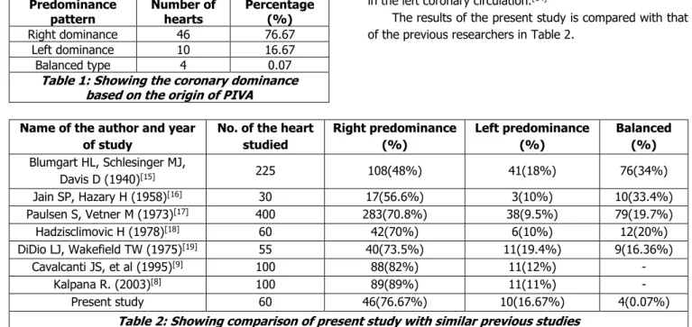
dissected, 46 (76.67%) of the hearts showed right predominance of heart, i.e. Posterior Inter-ventricular artery (PIVA) is given by RCA (Figure 1), while 10 (16.67%) of them showed the left predominance (Figure 2) and 4 (0.07%) hearts were of balanced type (Figure 3). Among the left dominant heart in all 10 heart specimens, the PIVA were the branch of left circumflex artery [Table 1].
Predominance pattern
Number of hearts
Percentage (%)
Right dominance 46 76.67
Left dominance 10 16.67
Balanced type 4 0.07
Table 1: Showing the coronary dominance based on the origin of PIVA
DISCUSSION: Normally in human heart, if the PIVA is a branch of RCA, it is termed as right predominance which occurred in 76.67% of hearts in our present study while left predominance was seen in 16.67% of the total hearts. In 0.07% of the hearts we found balanced type. A similar study done by Kalpana in 2003 observed right predominance is 89% and left predominance in 11% of the hearts but there was no balanced type. [8] According to Cavalcanti left
dominance was observed in 11.82% of specimens and right dominance in 88.18%. [9] In a study by Abuchaim DC et al [7]
in 25 human hearts the most common form of coronary circulation was found to be the right dominant. In a study in swine by Vieira TH et al [10] the arterial pattern was found to
be right dominance based on crux cordis and the subsinuosal interventricular branch. In a study on pigs by Sahni D et al
[11] the RCA was found to be dominance in all hearts. In a
genetic study with respect to the dominance pattern of coronary blood supply, two of three monozygotic twin pairs differed, while all dizygotic twin pairs were found to be concordant. [12]
Though right dominant circulation was more prevalent in our patients, Coronary Artery Disease was more frequent among those with left dominant circulation. This finding is in conformity with what is reported in the published literature. In another study it was found that the extent of coronary atherosclerosis does not depend on the type of dominant coronary artery [13] but in patients with Acute Coronary
Syndrome, left dominance is a significant and independent predictor of increased long-term mortality. [5] Similarly the
origin of the SA node artery is not related to coronary arterial dominance, but the origin of AV node artery is dependent on coronary arterial dominance. The presence of myocardial bridging is more related to coronary dominance, especially in the left coronary circulation.[14]
The results of the present study is compared with that of the previous researchers in Table 2.
Name of the author and year of study
No. of the heart studied
Right predominance (%)
Left predominance (%)
Balanced (%)
Blumgart HL, Schlesinger MJ,
Davis D (1940)[15] 225 108(48%) 41(18%) 76(34%)
Jain SP, Hazary H (1958)[16] 30 17(56.6%) 3(10%) 10(33.4%)
Paulsen S, Vetner M (1973)[17] 400 283(70.8%) 38(9.5%) 79(19.7%)
Hadzisclimovic H (1978)[18] 60 42(70%) 6(10%) 12(20%)
DiDio LJ, Wakefield TW (1975)[19] 55 40(73.5%) 11(19.4%) 9(16.36%)
Cavalcanti JS, et al (1995)[9] 100 88(82%) 11(12%) -
Kalpana R. (2003)[8] 100 89(89%) 11(11%) -
Present study 60 46(76.67%) 10(16.67%) 4(0.07%)
Table 2: Showing comparison of present study with similar previous studies
Although the left coronary artery always supplies a greater mass of myocardium than does the right, it is not
usually the ‘Dominance’ that can be a significant determinant
of prognosis in acquired coronary artery disease. In most
Jebmh.com
Original Article
J Evid Based Med Healthc, pISSN- 2349-2562, eISSN- 2349-2570/ Vol. 3/Issue 01/Jan. 04, 2016 Page 18
development or re-opening of collateral vessels is likely to be diminished.
Assigning anatomical definition to any coronary artery or its branches by any interventionist demands a sound anatomical knowledge of both normal and variant courses of coronary arteries and their branches. As our medical science takes a rapid pace towards development, constant addition of knowledge, however small, from various parts of world is mandatory for helping this development. Present study provides data from East Godavari region regarding prevalence of coronary circulation dominance, which may prove of some help to anatomists, radiologist and interventionists.
Fig. 1: Shows the posterior inter-ventricular artery (PIVA) arising from the right coronary artery and passing down along the posterior inter-ventricular
sulcus. This is an example of right dominance
Fig. 2: Showing the posterior inter-ventricular artery (PIVA) arising from the left coronary artery and passing down along the posterior inter-ventricular sulcus. This is
an example of left dominance
Fig. 3: Showing two posterior inter-ventricular arteries (PIVA). PIVA -1 arises from the left coronary artery and PIVA -2 from the right coronary artery. Both are passing down along the posterior inter-ventricular sulcus. This is an example of balanced type of coronary circulation. The middle cardiac vein is present in between the two PIVA
REFERENCES:
1. Michael A Gatzoulis. Heart and great vessels. In:
Susan Standring, editor in chief. Gray’s anatomy,
Churchill Livingstone Elsevier; 2008;40th ed:978-81. 2. Datta AK. Essentials 2 of human anatomy. (Thorax
and Abdomen). Lenin SARANEE, Kolkata: the Indian press pvt. Ltd. 2008;8th ed:81.
3. Gawlikowska-Sroka A, Miklaszewska D, Czerwi Å„ski F, et al. Analysis of the influence of heart size and gender on coronary circulation type. Feb 2010;69(1):35-41.
4. Knaapen M, Koch AH, Koch C, et al. Prevalence of left and balanced coronary arterial dominance decreases with increasing age of patients at autopsy. A postmortem coronary angiograms study. Cardiovasc Pathol. 2013;22:49-53.
5. Goldberg A, Southern DA, Galbraith PD, et al. Coronary dominance and prognosis of patients with acute coronary syndrome. Alberta Provincial Project for Outcome Assessment in Coronary Heart Disease (APPROACH) Investigators. Dec 2007;154(6):1116-22.
6. Veltman CE, de Graaf FR, Schuijf JD, et al. Prognostic value of coronary vessel dominance in relation to significant coronary artery disease determined with non-invasive computed tomography coronary angiography. Eur Heart J. 2012;33:1367-77.
7. Abuchaim DC, Spera CA, Faraco DL, et al. Coronary dominance patterns in the human heart investigated by corrosion casting. Rev Bras Cir Cardiovasc 2009;24(4):514-8.
8. Kalpana R. A study on principal branches of coronary arteries in humans. J Anat Soc India 2003;52:137-40. 9. Cavalcanti JS. Anatomic variations of the coronary
Jebmh.com
Original Article
J Evid Based Med Healthc, pISSN- 2349-2562, eISSN- 2349-2570/ Vol. 3/Issue 01/Jan. 04, 2016 Page 19
10. Vieira TH, Moura PC Jr, Vieira SR, et al. Anatomical indicators of dominance between the coronary arteries in swine. Morphologie. Mar 2008;92(296):3-6.
11. Sahni D, Kaur GD, Jit H, et al. Anatomy and distribution of coronary arteries in pig in comparison with man. Indian J Med Res. Jun 2008;127(6):564-70.
12. Fringsa AM, Mayera B, Böckera W, et al. Comparative coronary anatomy in six twin pairs with coronary artery disease. Heart 2000;83:47-50.
13. Balci B, Yilmaz O. Atherosclerotic involvement in patients with left or right dominant coronary circulation. Jun 2004;60(6):564-6.
14. Loukas M, Curry B, Bowers M, et al. The relationship of myocardial bridges to coronary artery dominance in the adult human heart. J Anat. Jul 2006;209(1):43-50.
15. Blumgart HL, Schlesinger MJ, Davis D. Studies on the relation of clinical manifestation of angina pectoris, coronary thrombosis and myocardial infarction to the pathological findings. Am Heart J 1940;19:1-18. 16. Jain SP, Hazary H. Coronary arterial pattern in man
and in some other mammals. J Anat Soc India 1958;7:5-8.
17. Paulsen S, Vetner M. Anatomical variations of the coronary arteries and origin of blood supply to sinoauricular and atrioventricular nodes determined on the basis of post-mortem coronary angiography. Acta Pathol Microbiol Scand A 1973;81:784-90. 18. Hadzisclimovic H. Coronary arteries and their
anastomosis in the new born. Folia anatomica 1978;8:89-100.