Safety of Ablation for Atrial Fibrillation with Therapeutic INR:
Comparison with Transition to Low-Molecular-Weight Heparin
Eduardo B. Saad
1, Ieda P. Costa
2, Rodrigo E. da Costa
1, Luiz Antonio O. Inácio Jr.
1, Charles Slater
1, Angelina
Camiletti
1, Dario G. de Moura Neto
1, Paulo Maldonado
1, Luiz Eduardo Camanho
1, Carisi A. Polanczyk
3Hospital Pró-Cardíaco, Setor de Arritmia Invasiva e Centro de Fibrilação Atrial1, Rio de Janeiro, RJ; Hospital de Messejana2, Fortaleza, CE; Hospital de Clinicas de Porto Alegre, Instituto para Avaliação de Tecnologias em Saúde (IATS) - Universidade Federal do Rio Grande do Sul3, Porto Alegre, RS, Brazil
Resumo
Background: The ideal management of oral anticoagulation (OAC) before and after catheter ablation for atrial fibrillation (AF) is still controversial.
Objetive: To compare two anticoagulation strategies for catheter ablation for AF: warfarin withholding and use of low-molecular-weight heparin (LMWH); and maintenance of warfarin and therapeutic INR (between 2.0 and 3.0).
Methods: 140 patients (pt) with persistent/permanent AF undergoing catheter ablation for AF were divided into two groups: Group I (70 pt), in which warfarin was withheld five days prior to the procedure and transition to LMWH was used (enoxaparin: 1 mg/kg 2x/day before ablation, and 0.5 mg/kg 2x/day after ablation); Group II (70 pt), in which warfarin was not withheld and the procedure was performed with therapeutic INR. Both groups received intravenous heparin (ACT > 350 seconds) during ablation.
Results: In Group I, one pt (1.4%) had a major hemorrhagic complication and four pts (5.7%) had minor hemorrhagic complications. In Group II, two pts (2.8%) had minor hemorrhagic complications and one pt had a major bleeding, which occurred after using LMWH due to INR < 2.0. None of the groups had thromboembolic complications or cardiovascular death over a period of 16 ± 8 months.
Conclusion: Catheter ablation for AF without withholding OAC and with therapeutic INR is a strategy that has similar safety and efficacy when compared with the traditional transition to LMWH, avoiding the potentially inadequate anticoagulation of the initial post-ablation period. (Arq Bras Cardiol 2011;97(4):289-296)
Keywords: Atrial fibrillation; catheter ablation; treatment outcome; withholding treatment; warfarin; heparin, low-molecular-weight
Mailing address: Eduardo B. Saad •
Av. Borges de Medeiros, 3607/303 – Lagoa – 22470-001 – Rio de Janeiro, RJ, Brazil
E-mail: [email protected]
Manuscript received February 21, 2011; revised manuscript received April 28, 2011; accepted May 16, 2011.
Introduction
Atrial fibrillation (AF) is the most common sustained arrhythmia in clinical practice1-3. Because of the low efficacy of antiarrhythmic drugs4-6, ablation was largely used in the past decade and has been consolidated as an effective and safe treatment alternative7-13. However, ablation involves extensive manipulation of the left atrium, and, thus, aggressive anticoagulation with heparin during the intervention has proved to be fundamental14-16.
Although stroke is one of the most feared complications associated with ablation, hemorrhagic complications are the most prevalent17, because of the need for anticoagulation in the context of multiple venous punctures.
The classical approach consists in withholding warfarin therapy three to five days prior to the procedure to normalize
INR, and in using transition therapy to low-molecular-weight heparin (LMWH) until the day prior to the procedure, warfarin being reinitiated immediately after the intervention11. That is a critical moment due to AF reversion and occurrence of extensive tissue damage in the left atrium during the procedure. However, that is also the time when the use of a full dose of LMWH (1 mg/kg of enoxaparin, twice a day) substantially increases the likelihood of hemorrhagic complications. That is why half of the usual dose of LMWH (0.5 mg/kg of enoxaparin, twice a day) is recommended, aiming at reducing the incidence of hemorrhagic complications. However, the patient is temporarily submitted to a potentially inadequate anticoagulation regimen.
To prevent possible complications related to that regimen, the strategy of performing the procedure without withholding warfarin (with therapeutic INR and no LMWH) has been proposed in centers of excellence18,19. This strategy represents
association of anticoagulant drugs and the occurrence of mechanical complications during the intervention.
This study aimed at assessing the safety and efficacy of catheter ablation for AF in patients undergoing OAC and with therapeutic INR.
Methods
Between January 2008 and August 2010, 140 patients with persistent or permanent AF refractory to at least one antiarrhythmic drug and undergoing catheter ablation at the Hospital Pró-Cardíaco (Rio de Janeiro state) were divided into two groups, according to the preference of the operator as follows: Group I (LMWH – 70 patients), suspension of warfarin and transition to LMWH; and Group II (70 patients), warfarin was maintained and ablation was performed with therapeutic INR (between 2.0 and 3.0).
In Group I, warfarin was withheld five days prior to the procedure, when enoxaparin (1 mg/kg, twice a day) was initiated. The last dose of LMWH was administered 24 hours prior to the procedure. The INR was measured on the day before the procedure, which was performed when the INR was lower than 2.0. Oral anticoagulation was restarted after ablation, and enoxaparin, at a reduced dose (0.5 mg/kg, twice a day), was initiated in the following morning and maintained until the INR > 2.0 was obtained again.
In Group II, OAC was not withheld. The INR was periodically assessed, mandatory measurements being performed as follows: five days prior to the procedure; on the day before the procedure; and on the day of the procedure. Blood typing was performed on hospital admission, and fresh plasma and recombinant coagulation factors were reserved for immediate reversion in case of hemorrhagic and/or mechanical complications. If the INR was not within the desired range, the procedure would be postponed, and the warfarin dose readjusted until the therapeutic range was achieved, when the procedure would be performed.
Ablation technique
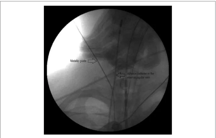
Under general anesthesia, all patients underwent transesophageal echocardiography during the procedure. The ablation technique consisted in the electrical isolation of the pulmonary vein antrum and of the superior vena cava guided by intracardiac echocardiography (ICE)15,20-22, in association with extensive damage to the left atrium in regions where complex fractionated atrial electrograms (CFAE) were obtained. The following venous accesses were obtained: two punctures of the right femoral vein; one puncture of the left femoral vein; and one puncture of the right internal jugular vein. The latter could be performed under fluoroscopic guidance by use of a catheter positioned in the vein through femoral access (Figure 1). The ICE probe (AcuNav, Mountain View, California) was positioned in the right atrium and a duodecapolar catheter was used to map the coronary sinus and the crista terminalis; when this was not possible, a decapolar catheter could be used to record the electrical signs in the coronary sinus. Immediately after obtaining the venous accesses, systemic heparinization was
initiated to achieve an activated coagulation time (ACT) > 350 seconds (aim: between 350 and 400 seconds); only then the ICE-guided transseptal access was obtained (Figure 2). After double transseptal puncture, a circular decapolar catheter (20 mm) for mapping (Lasso, Biosense Webster, Baldwin Park, California) and an ablation catheter with external irrigation and 3.5-mm tip (Biosense Webster, Diamond Bar, California) were positioned. Maximum power and temperature were 35W and 43ºC, respectively, with a flow of 30 mL/min. Esophageal temperature was monitored by inserting a multipolar thermometer (Figure 3), and when it exceeded 39ºC, the application was interrupted and reinitiated at lower power.
After all radiofrequency damage to the left atrium, electrical cardioversion was performed if sinus rhythm was not resumed during the applications. After sinus rhythm resumption, a high dose of intravenous isoprenaline (20 µg/ min) was infused during ten minutes, aiming at assessing the reconnection of the veins and at triggering ectopic foci located outside the pulmonary veins, which, when present, were then mapped and ablated. Finally, the cavotricuspid isthmus was ablated to prevent atrial flutter.
Right after withdrawing the sheaths from the left atrium, systemic heparinization was reverted with protamine to achieve an ACT < 200 seconds. Vascular compression was performed for 20 minutes, and a compressive dressing was maintained for six hours.
Follow-up after the procedure
Patients were discharged from the hospital on the morning following the procedure, and were followed up at the outpatient clinic with visits 30, 90 and 180 days after that, when they underwent ECG and 24-hour Holter. Pulmonary vein computed tomography or magnetic resonance angiography was performed prior to the procedure and three months after that for detecting occasional stenosis.
Patients were instructed to check their pulse on a daily basis and to immediately report any symptom or irregularity observed, having, then, immediate access to the medical team.
Antiarrhythmic drugs were maintained for four weeks and OAC for, at least, three months, being withheld in the absence of symptoms or of sustained arrhythmias, as long as the thromboembolic risk was not greater than moderate (CHADS2 < 2). In such cases, OAC was maintained or individually discussed with each patient.
All patients provided written informed consent, and the study was approved by the Committee on Ethics and Research of the Hospital Pró-Cardíaco.
Variables analyzed
Figure 1 – Puncture of the right internal jugular vein under luoroscopic vision. The ablation catheter is introduced from the right femoral vein to the right jugular vein, serving as a luoroscopic marker, reducing the likelihood of an accidental puncture of the carotid artery. After puncturing the vein, the metallic guide is introduced to provide access to the 8F sheath (traditional Seldinger technique).
Statistical analysis
Descriptive statistical analysis of the numerical variables was performed through mean and standard deviation, and inferential analysis was performed by use of Student t test for independent samples. The categorical variables were analyzed by using Fisher exact and Pearson chi-square tests. The significance value of p< 0.05 was adopted.
Results
The study sample comprised 140 patients (mean age, 72 ± 8.2 years; 83%, males) undergoing catheter ablation.
Transesophageal echocardiography revealed blood flow delay in the left atrial cavity in 56 Group I patients (80%) and in 64 Group II patients (91%) (p = ns). However, previous history of thrombus in the left atrial auricle was observed in two Group I patients (2.8%) and 12 Group II patients (17%; p < 0.05).
In Group I, the mean size of the left atrium was 44 ± 7mm, and the mean ejection fraction was 60% ± 15%. In 35% of the patients, structural cardiopathy was observed, and the most common etiology was ischemia (67%). Thirteen patients (18.6%) were classified as having a CHADS2 score of 1, while 27 (38.6%), 26 (37.1%), and four patients (5.7%) were classified as having a CHADS2 score of 2, 3, and 4, respectively. On the day of the procedure, all patients had an
INR lower than 2.0. The mean dose of the bolus of venous heparin used to achieve the target ACT was 16,000 ± 4,000 IU. The major clinical characteristics are shown in Table 1.
Group I showed more major hemorrhagic complications: one patient (1.4%) developed a large inguinal hematoma on the fourth day after the procedure, requiring blood transfusion. Four patients (5.7%) had minor hemorrhagic complications (inguinal hematoma), requiring suspension of LMWH.
In Group II, the mean size of the left atrium was 45 ± 6.2mm, and the mean ejection fraction was 58% ± 11%. In 33% of the cases, structural cardiopathy was observed, and the most common etiology was ischemia (72%). Eleven patients (15.7%) were classified as having a CHADS2 score of 1, while 29 (41.5%), 25 (35.7%), and five patients (7.1%) were classified as having a CHADS2 score of 2, 3, and 4, respectively (p = ns). The mean INR on the day of admission was 2.3 ± 0.2. The mean bolus dose of venous heparin used to achieve the target ACT was significantly lower (7,000 ± 2,000 IU; p < 0.01).
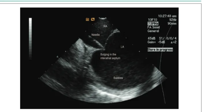
Figure 2 – Intracardiac echocardiography-guided transseptal puncture. The transseptal puncture needle is placed exactly in the thin portion of the interatrial septum (fossa ovalis) through the right atrium (RA); when pressing, tenting of the septum is clearly observed. After its perforation, the simple injection of saline solution generates microbubbles in the left atrium (LA), demonstrating the correct position of the needle in the LA without requiring iodine contrast medium.
Figure 3 – Fluoroscopic positioning of the catheters during ablation of the right inferior pulmonary vein (RIPV). The circular mapping catheter is placed in the vein
Table 1 – Major clinical characteristics observed in both groups
Variable Group IN = 70 Group IIN = 70 p
Age (years) 76 ± 7,4 73 ± 5,6 0,68
Male sex (%) 85% 81% 0,5
LA size (mm) 44 ± 7 45 ± 6,2 0,43
LVEF (%) 55 ± 15 61 ± 11 0,18
Structural cardiopathy (%) 35 33 0,72
CHADS 2 ≥ 2 81,4% 84,3% 0,5
LVEF – left ventricular ejection fraction; LA – left atrium.
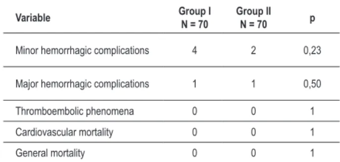
Table 2 – Clinical outcomes in both groups in the long-term follow-up
Variable Group I
N = 70
Group II
N = 70 p
Minor hemorrhagic complications 4 2 0,23
Major hemorrhagic complications 1 1 0,50
Thromboembolic phenomena 0 0 1
Cardiovascular mortality 0 0 1
General mortality 0 0 1
nor OAC suspension. One patient (1.4%) needed LMWH because of INR < 2.0 on the day following the procedure, and showed a large hematoma in the right thigh, requiring blood transfusion because of the development of unstable angina due to significant anemia.
N e i t h e r t h r o m b o e m b o l i c c o m p l i c a t i o n s n o r cardiovascular death were observed in the groups over 16 ± 8 months of follow-up. The variables analyzed are shown in Table 2.
Discussion
This study is the first national experience about ablation for AF without withholding OAC (therapeutic INR) in a population at moderate thromboembolic risk. This represents a paradigm change, because invasive procedures in patients undergoing OAC have always been considered of high risk for hemorrhagic complications. However, the data presented do not corroborate that hypothesis, since patients undergoing OAC showed no increase in complications.
Previously published studies
In 2007, Wazni et al18 reported a study on the safety and efficacy of that strategy in 310 patients with persistent AF divided into three groups: Group I, in which warfarin was withheld before the procedure, and transition to full-dose LMWH (1 mg/kg of enoxaparin, twice a day) was used in the pre- and post-ablation period; Group II, with the same protocol, but a reduced dose of LMWH (0.5 mg/kg of enoxaparin, twice a day); and Group III, in which warfarin was not withheld and the procedure was performed with INR between 2.0 and 3.0. That clinical assay showed the safety and strategy of ablation with the use of OAC, with a significantly lower rate of mild bleedings (hematomas that do not require drainage – 22% versus 19% versus 5%, respectively, p < 0.001) and of major bleedings (hematoma requiring drainage, bleeding requiring transfusion, or pericardial tamponade – 8.5% versus 0% versus 0%, respectively, p < 0.001) in Group III, and no increase in the risk of major complications (ischemic stroke or pericardial effusion). Such data support the recommendation of lower doses of LMWH in the post-ablation period, in face of the
significant increase in major complications when full doses are used, despite the concern regarding thromboembolic complications when subtherapeutic levels are achieved.
Later, Hussein et al19 have published a study with a significant number of patients with paroxysmal and persistent AF, in which the strategy of maintaining warfarin during ablation was also adopted. The study comprised 3,052 patients referred for ablation for AF with INR > 1.8 on the day of the intervention (mean INR, 2.53 ± 0.62). Once more, a low incidence of hemorrhagic complications was observed with that strategy (1.1% – most events of little clinical significance). The results of the present study have also shown a low incidence of thromboembolic and hemorrhagic neurological events, whose rates were 0.09% (ischemic stroke) and 0.03% (hemorrhagic stroke), respectively. The incidence of pericardial effusion was only 0.29%, most of which mild, requiring no drainage. In addition to protecting against embolic events, that strategy did not show a greater incidence of hemorrhagic complications, corroborating the
findings previously reported by Wasni et al18.
Cappato et al17 have published a world record in which 16,309 patients of 182 centers (24 countries) underwent interventions between 2003 and 2006. In that study, warfarin was withheld before the procedure, and anticoagulation performed with intravenous or subcutaneous heparin or LMWH, after ablation. The greatest rate of complications was 4.5% (741 patients), and the most frequent were cardiac tamponade (1.31%), femoral pseudoaneurysm (0.93%), and transient ischemic attack (0.71%). When compared with those results, the strategy of continuing warfarin showed a lower incidence of severe hemorrhages and thromboembolic complications.
Correlation with the indings of the present study
The findings in this study are in accordance with those of the literature, with a low index of complications in the group of therapeutic INR. No embolic complication was observed.
It is worth noting that all studies published have been performed in high-volume and experienced centers, and always using ICE. That method is of fundamental importance when that strategy is adopted, because it provides real-time transseptal access under direct visualization without the use of iodine contrast medium, monitoring of the catheter position and contact with the atrial wall, and early detection of mechanical complications (such as cardiac tamponade), which make the procedure safer22,24. The following should be considered impacting factors regarding safety of the procedure: operator’s experience and ability; caution during venous accesses; and manipulation of the catheters inside the left atrium.
Recently, Page et al25 have published a study with a similar design, comparing the ablation procedure in 109 patients using LMWH with 89 patients whose warfarin was maintained (mean INR, 2.3 ± 0.5). In that study, however, ICE was not used. Two patients (2.2%) in the group of therapeutic INR required pericardial drainage due to cardiac tamponade.
Intracardiac echocardiography is the only method capable of detecting intracardiac thrombi. Although its use has not been proven to significantly reduce the incidence of clinical embolic events, recent evidence suggests that they can be present in up to 10% of the cases during ablation for AF26-28. That is why our protocol involves the aggressive use of venous heparin to achieve an ACT over 350 seconds, initiated prior to transseptal access. That strategy has been adopted after one case in which a thrombus was visualized in the circular catheter immediately after access to the left atrium, even before beginning radiofrequency applications29. At that time, heparin was only initiated after
obtaining both transseptal accesses. By using ICE, both transseptal accesses are safely obtained even under intense anticoagulation (oral and venous).
Ablation for AF is associated with a low risk for symptomatic cerebral thromboembolism. However, the possibility of silent cerebral ischemia, similarly to that which occurs in open surgical procedures, is feared. Gaita F. et al30 have recently published their experience using magnetic resonance imaging (MRI) as a diagnostic tool and have demonstrated that the intensity of anticoagulation during the procedure, expressed as ACT, was one of the factors associated with the occurrence of lesions without clinical expression. Of the patients with an ACT < 250 seconds, 17% had a positive MRI; when ACT > 250 seconds, MRI was positive in only 9% (p = 0.01).
Implications
To safely use that strategy, in addition to the measures previously mentioned, it is worth emphasizing that the heparin dose necessary to achieve the ideal ACT (between 350 and 400 seconds) was significantly lower in Group
II (without withholding OAC) than in Group I, and that interaction between warfarin and heparin seemed to occur, making patients more sensitive to lower heparin doses. It is clear that, when choosing that strategy, monitoring ACT before initiating the manipulation is fundamental to prevent unacceptably high levels of anticoagulation and their possible complications.
It is also worth emphasizing that the maintenance of OAC as a therapeutic strategy reduces the cost associated with the use of LMWH. This cost, often covered by the patient, represents a hindrance to the ablation procedure; however, recent data have shown no reduction in total costs of the
treatment per patient (including hospitalization)25.
Thus, we believe that it is fundamental to look for adequate anticoagulation in the pre- and post-ablation period, without increasing the risks related to the procedure. The strategy of not withholding warfarin and consequently performing ablation with therapeutic INR is an interesting and safe therapeutic proposal. In addition, it is worth emphasizing the use of ICE in such cases, as we believe that the advantages associated with that method significantly contribute to the safety and efficacy of that strategy, despite its additional cost, certainly greater than that of LMWH.
Limitations
The non-randomized design of this report does not allow the elimination of the preferential selection of patients at a higher risk for maintaining OAC as the strategy for ablation. The major limitation was that the groups were not randomized for one or the other strategy, but, when comparing the clinical characteristics of both groups, they were very similar. In addition, the relative small size of the sample might have contributed to the lack of thromboembolic phenomena in the group using LMWH, despite its reduced dosage.
Conclusion
Catheter ablation for AF without withholding OAC and with therapeutic INR has similar safety and efficacy when compared with the traditional transition to LMWH, avoiding the potentially inadequate anticoagulation of the initial post-ablation period.
Potential Conflict of Interest
No potential conflict of interest relevant to this article was reported.
Sources of Funding
There were no external funding sources for this study.
Study Association
References
1. Lip GY, Kakar P, Watson T. Atrial fibrillation--the growing epidemic. Heart. 2007;93(5):542-3.
2. Camm AJ, Kirchhof P, Lip GY, Schotten U, Savelieva I, Ernst S, et al. Guidelines for the management of atrial fibrillation: the Task Force for the Management of Atrial Fibrillation of the European Society of Cardiology (ESC). Eur Heart J. 2010;31(19):2369-429.
3. Wann LS, Curtis AB, January CT, Ellenbogen KA, Lowe JE, Estes NA 3rd, et
al. 2011 ACCF/AHA/HRS focused update on the management of patients with atrial fibrillation (updating the 2006 guideline): a report of the American College of Cardiology Foundation/American Heart Association Task Force on Practice Guidelines. Circulation. 2011;123(1):104-23.
4. Roy D, Talajic M, Dorian P, Connolly S, Eisenberg MJ, Green M, et al. Amiodarone to prevent recurrence of atrial fibrillation. Canadian Trial of Atrial Fibrillation Investigators. N Engl J Med. 2000;342(13):913-20. 5. Calkins H, Reynolds MR, Spector P, Sondhi M, Xu Y, Martin A, et al. Treatment
of atrial fibrillation with antiarrhythmic drugs or radiofrequency ablation: two systematic literature reviews and meta-analyses. Circ Arrhythm Electrophysiol. 2009;2(4):349-61.
6. Singh BN, Singh SN, Reda DJ, Tang XC, Lopez B, Harris CL, et al. Amiodarone versus sotalol for atrial fibrillation. N Engl J Med. 2005;352(18):1861-72. 7. Jais P, Cauchemez B, Macle L, Daoud E, Khairy P, Subbiah R, et al. Catheter
ablation versus antiarrhythmic drugs for atrial fibrillation: the A4 study. Circulation. 2008;118(24):2498-505.
8. Pappone C, Rosanio S, Augello G, Gallus G, Vicedomini G, Mazzone P, et al. Mortality, morbidity, and quality of life after circumferential pulmonary vein ablation for atrial fibrillation: outcomes from a controlled nonrandomized long-term study. J Am Coll Cardiol. 2003;42(2):185-97.
9. Pappone C, Augello G, Sala S, Gugliotta F, Vicedomini G, Gulletta S, et al. A randomized trial of circumferential pulmonary vein ablation versus antiarrhythmic drug therapy in paroxysmal atrial fibrillation: the APAF Study. J Am Coll Cardiol. 2006;48(11):2340-7.
10. Crijns HJ, Van Gelder IC, Van Gilst WH, Hillege H, Gosselink AM, Lie KI. Serial antiarrhythmic drug treatment to maintain sinus rhythm after electrical cardioversion for chronic atrial fibrillation or atrial flutter. Am J Cardiol. 1991;68(4):335-41.
11. Calkins H, Brugada J, Packer DL, Cappato R, Chen SA, Crijns HJ, et al. HRS/ EHRA/ECAS expert Consensus Statement on catheter and surgical ablation of atrial fibrillation: recommendations for personnel, policy, procedures and follow-up. A report of the Heart Rhythm Society (HRS) Task Force on catheter and surgical ablation of atrial fibrillation. Heart Rhythm. 2007;4(6):816-61. 12. Haissaguerre M, Jais P, Shah DC, Takahashi A, Hocini M, Quiniou G, et al.
Spontaneous initiation of atrial fibrillation by ectopic beats originating in the pulmonary veins. N Engl J Med. 1998;339(10):659-66.
13. Wilber DJ, Pappone C, Neuzil P, De Paola A, Marchlinski F, Natale A, et al. Comparison of antiarrhythmic drug therapy and radiofrequency catheter ablation in patients with paroxysmal atrial fibrillation: a randomized controlled trial. JAMA. 2010;303(4):333-40.
14. Wazni OM, Rossillo A, Marrouche NF, Saad EB, Martin DO, Bhargava M, et al. Embolic events and char formation during pulmonary vein isolation in patients with atrial fibrillation: impact of different anticoagulation regimens and importance of intracardiac echo imaging. J Cardiovasc Electrophysiol. 2005;16(6):576-81.
15. Kanj M, Wazni O, Natale A. Pulmonary vein antrum isolation. Heart Rhythm. 2007;4(3 Suppl):S73-9.
16. Ren JF, Marchlinski FE, Callans DJ, Gerstenfeld EP, Dixit S, Lin D, et al. Increased intensity of anticoagulation may reduce risk of thrombus during atrial fibrillation ablation procedures in patients with spontaneous echo contrast. J Cardiovasc Electrophysiol. 2005;16(5):474-7.
17. Cappato R, Calkins H, Chen SA, Davies W, Iesaka Y, Kalman J, et al. Updated worldwide survey on the methods, efficacy, and safety of catheter ablation for human atrial fibrillation. Circ Arrhythm Electrophysiol. 2010;3(1):32-8.
18. Wazni OM, Beheiry S, Fahmy T, Barrett C, Hao S, Patel D, et al. Atrial fibrillation ablation in patients with therapeutic international normalized ratio: comparison of strategies of anticoagulation management in the periprocedural period. Circulation. 2007;116(22):2531-4.
19. Hussein AA, Martin DO, Saliba W, Patel D, Karim S, Batal O, et al. Radiofrequency ablation of atrial fibrillation under therapeutic international normalized ratio: a safe and efficacious periprocedural anticoagulation strategy. Heart Rhythm. 2009;6(10):1425-9. 20. Callans DJ, Wood MA. How to use intracardiac echocardiography for
atrial fibrillation ablation procedures. Heart Rhythm. 2007;4(2):242-5. 21. Kanj MH, Wazni OM, Natale A. How to do circular mapping catheter-guided pulmonary vein antrum isolation: the Cleveland Clinic approach. Heart Rhythm. 2006;3(7):866-9.
22. Saad EB, Costa IP, Camanho LE. [Use of intracardiac echocardiography in the electrophysiology laboratory.]. Arq Bras Cardiol. 2011;96(1):e11-7. 23. Di Biase L, Burkhardt JD, Mohanty P, Sanchez J, Horton R, Gallinghouse GJ, et al. Periprocedural stroke and management of major bleeding complications in patients undergoing catheter ablation of atrial fibrillation: the impact of periprocedural therapeutic international normalized ratio. Circulation. 2010;121(23):2550-6.
24. Marrouche NF, Martin DO, Wazni O, Gillinov AM, Klein A, Bhargava M, et al. Phased-array intracardiac echocardiography monitoring during pulmonary vein isolation in patients with atrial fibrillation: impact on outcome and complications. Circulation. 2003;107(21):2710-6.
25. Page SP, Siddiqui MS, Finlay M, Hunter RJ, Abrams DJ, Dhinoja M, et al. Catheter ablation for atrial fibrillation on uninterrupted warfarin: can it be done without echo guidance? J Cardiovasc Electrophysiol. 2011;22(3):265-70.
26. Zhou L, Keane D, Reed G, Ruskin J. Thromboembolic complications of cardiac radiofrequency catheter ablation: a review of the reported incidence, pathogenesis and current research directions. J Cardiovasc Electrophysiol. 1999;10(4):611-20.
27. Ren JF, Marchlinski FE, Callans DJ. Left atrial thrombus associated with ablation for atrial fibrillation: identification with intracardiac echocardiography. J Am Coll Cardiol. 2004;43(10):1861-7.
28. Saad EB, Marrouche NF, Natale A. Ablation of atrial fibrillation. Curr Cardiol Rep. 2002;4(5):379-87.
29. Martelo S, D’Avila A, Ferreira F, Saad EB. Implantation of bilateral carotid artery filters to allow safe removal of left atrial thrombus during ablation of atrial fibrillation. J Cardiovasc Electrophysiol. 2006;17(10):1140-1. 30. Gaita F, Caponi D, Pianelli M, Scaglione M, Toso E, Cesarani F, et al.