INTRODUCTION
Herpes simplex keratitis (HSK) is a major cause of vision
morbi-dity worldwide(1,2). Despite improvements in diagnosis and medical
treatment, recurrence of HSK is common and leads to severe corneal scarring and sometimes descemetocele formation or perforation. In some cases, corneal transplantation is needed to recover corneal cla-rity and rehabilitation of visual acuity. Penetrating keratoplasty (PK) is a conventional surgical procedure for treatment of HSK-induced
corneal scarring(3-6); however, the success of corneal transplantation
due to herpetic corneal scarring is lower as compared to other common indications, such as keratoconus, corneal dystrophy, and bullous
keratopathy(7). The reduced survival rate of corneal grafts in patients
undergoing PK for herpetic eye disease is mainly attributed to the
recurrence of herpetic infection and immune reaction. The postoperative course in these cases can be complicated by high rates of re -currence, graft rejection, and graft failure(8,9).
The purpose of this study was to evaluate the outcome of PK for corneal scarring caused by HSK and to determine whether the type of corneal scar affects the treatment outcome.
METHODS
The protocol of this retrospective study was approved by the Ethical Committee of Ankara Training and Research Hospital (Ankara, Turkey) and was conducted in compliance with the tenets of the De-claration of Helsinki. This study enrolled 55 patients with HSK-related
The outcome of penetrating keratoplasty for corneal scarring due
to herpes simplex keratitis
O resultado da ceratoplastia penetrante para cicatriz de córnea consequente à ceratite por herpes simplex
Yesim AltAY1, semA tAmer1, AbdullAh sencer KAYA1, Ozgur bAltA1, AYse burcu1, Firdevs OrneK1
Submitted for publication: January 11, 2016 Accepted for publication: October 23, 2016
1 Department of Ophthalmology, Ankara Training and Research Hospital, Ankara, Turkey.
Funding: No specific financial support was available for this study.
Disclosure of potential conflicts of interest: None of the authors have any potential conflict of interest to disclose.
Corresponding author: Yesim Altay. 48. Cadde Yonca Apt. 33/31 - Cukurambar - Ankara - 06520 Turkey - E-mail: [email protected]
Approved by the following research ethics committee: Ankara Training and Research Hospital (#5024 on 27.05.2015-595).
ABSTRACT
Purpose: To determine the outcomes of penetrating keratoplasty (PK) for treatment of corneal scarring caused by Herpes simplex virus (HSV) keratitis, and whether the corneal scar type affects treatment outcome.
Methods: A retrospective analysis of patients who underwent PK for HSV-related corneal scarring between January 2008 and July 2011 was performed. The patients were categorized into two groups. Group 1 consisted of patients with a quiescent herpetic corneal scar and group 2 consisted of patients who developed a corneal descemetocele or perforation secondary to persistent epithelial defects with no active stromal inflammation. The mean follow-up was 21.30 ± 14.59 months. The main parameters evaluated were recurrence of herpetic keratitis, graft rejection, graft failure, visual acuity, and graft survival rate.
Results: There were 42 patients in group 1 and 13 in group 2. Preoperative BCVA varied from hand movements to 0.7 logMAR. Postoperatively, 34 patients (61.8%) achieved visual acuity of 0.6 logMAR or more. Recurrence of HSV keratitis was noted in 12 (28.57%) eyes in group 1 and 4 (30.76%) eyes in group 2 (p=0.40). Graft rejection occurred in 4 eyes (9.52%) in group 1 and in 3 (23.07%) eyes in group 2 (p=0.58). The 1-, 2-, and 3-year graft survival rates were 91.9%, 76.0%, and 65.1% in group 1, and 89.5%, 76.0%, and 63.6% in group 2 (p=0.91), respectively. Conclusıons: Although there were different recurrence and graft rejection rates for two groups, the graft survival rates at 3 years were similar. According to our results, without inflammation, corneal herpetic scarring with a descemetocele or perforation achieved similar graft survival rates with quiescent herpetic corneal scars.
Keywords: Acyclovir/therapeutic use; Keratitis, herpetic; Keratoplasty, penetrating; Cornea; Graft survival
RESUMO
Objetivo: Determinar os resultados da ceratoplastia penetrante (PK) para o trata-mento da cicatriz da córnea consequente à ceratite por Herpes simplex vírus (HSV), e se o tipo de cicatriz na córnea afeta o resultado cirúrgico.
Métodos: Foi realizada análise retrospectiva dos pacientes, submetidos à PK para a cicatriz da córnea relacionados com o HSV entre janeiro de 2008 e julho de 2011. Os pacientes foram divididos em dois grupos. Grupo 1 consistiu de pacientes que tiveram cicatriz corneana herpética quiescente e grupo 2 consistiu de pacientes que desenvolveram descemetocele ou perfuração córnea secundária a defeitos epiteliais persistentes sem inflamação estromal ativa. O seguimento médio foi de 21,30 ± 14,59 meses. Os principais parâmetros avaliados foram recorrência de ceratite herpética, rejeição de enxerto, falência do enxerto, acuidade visual e taxa de sobrevida do enxerto.
Resultados: Foram avaliados 42 pacientes do grupo 1 e 13 doentes do grupo 2. Acui-dade visual pré-operatória variou de movimentos das mãos (HM) para 0,7 logMAR. No pós-operatório, 34 pacientes (61,8%) atingiram acuidade visual de 0,6 logMAR ou melhor. Doze olhos (28,57%) tiveram recorrência de HSV ceratite no grupo 1, e quatro olhos (30,76%) tiveram recorrência no grupo 2 (p=0,40). A rejeição do enxerto ocorreu em 4 olhos (9,52%) no grupo 1, e em 3 olhos do grupo 2 (23,07%; p=0,58), taxa de sobrevivência do enxerto foi de 91,9% a 1 ano, 76,0% aos 2 anos e 65,1% aos 3 anos no grupo 1, e 89,5% a 1 ano, 76,0% aos 2 anos e 63,6% aos 3 anos no grupo 2 (p=0,91).
Conclusões: Embora diferentes taxas de recorrência e de rejeição do enxerto foram encontradas nos dois grupos, a taxa de sobrevida do enxerto em 3 anos foi semelhan-tes nos dois grupos. De acordo com nossos resultados, em casos sem inflamação, a cicatriz herpética da córnea com descemetocele ou perfuração demonstra as taxas de sobrevivência do enxerto semelhantes às da cicatriz corneana herpética quiescente.
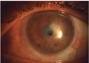
corneal scarring who underwent PK (PK) between January 2008 and July 2011, and who had a minimum postoperative follow-up of 18 months. The preoperative diagnosis of herpetic keratitis was based on the history of typical recurrences and clinical manifestations, such as episodes of epithelial dendritic or stromal ulceration with or without inflammatory edema, which responded well to the antiviral therapy. The patients were categorized into two groups. Group 1 consisted of patients who had a quiescent herpetic corneal scar (Fi-gure 1) and group 2 consisted of patients who developed a corneal descemetocele or perforation secondary to persistent epithelial de-fects with no active stromal inflammation (which was defined as the presence of corneal perforation or descemetocele without typical stromal inflammatory edema of the surrounding cornea in otherwise quiet eyes) (Figure 2).
Preoperatively, the following parameters were recorded for all patients: sex, age, interval without herpetic activation before PK, and best-corrected visual acuity (BCVA). The BCVA of patients was divided into three classifications based on logMAR values: logMAR ≤0.6, logMAR >0.6 to <1.2, and logMAR ≥1.2.
The standard PK procedure was used in all cases. The recipient bed was created by trephination using a trephine of 7.0-7.5 mm in
diameter and an oversized fresh button (0.25-0.50 mm) was used for grafting. The recipient-graft junction was fixed with 16 continuous or separate sutures of 10-0 nylon.
Postoperatively, the patients were treated with topical 0.3% oflo-xacin and 0.1% dexamethasone sodium phosphate eye drops 6 times daily. Topical steroids were used for 12 months in tapering doses. If intraocular pressure increased, topical antiglaucoma medications were added. All patients received oral acyclovir at 400 mg 5 times daily for the first 3 months, which was tapered to 400 mg twice daily for the following 12 months. Rejection episodes were treated with 0.1% dexamethasone sodium phosphate eye drops every 3 h, which was tapered off over several weeks depending on the clinical response, and the dose of oral acyclovir was adjusted from 200 to 400 mg 5 ti-mes daily and then tapered off over 2-3 months. Recurrence of herpes simplex stromal keratitis was treated with oral acyclovir at 400 mg 5 times with 0.1% dexamethasone sodium phosphate eye drops twice daily. In severe cases, daily oral steroid therapy at 40 mg was initiated.
All patients were followed-up at 2 weeks and 1, 3, 6, 12, and 18 months after surgery, and every year thereafter. At each follow-up visit, visual acuity, graft status, and any signs of graft rejection or re currence of herpetic infection were recorded. Corneal graft rejec-tion was defined as the development of an epithelial or endothelial (Khodadoust) rejection line, spread of corneal edema and keratic pre-cipitates limited to a previously clear graft, and an anterior chamber reaction with decreased vision. Herpetic epithelial recurrence was diagnosed on clinical findings; that is, dendritic or geographic ulcera-tion or a nonhealing epithelial defect. Recurrence of herpetic stromal keratitis was also diagnosed on occurrence of stromal swelling and infiltration, and the presence of keratic precipitates not limited to the graft. Graft failure was defined as irreversible edema and cloudiness of the graft.
Statistical analysis was performed using SPSS software (ver. 15; IBM-SPSS, Inc., Chicago, IL, USA). The effect of the type of herpetic cor neal scar on graft survival, graft rejection, and recurrence was investigated using the log-rank test (Mantel-Cox). Kaplan-Meier survi-val estimates were calculated. The BCVA survi-values at the end of follow-up were compared between the groups using the chi-square test. A probability (p) value of <0.05 was considered statistically significant.
RESULTS
The study enrolled 55 patients, which included 42 patients (9 women/33 men) with a quiescent herpetic corneal scar (group 1) and 13 (2 women/11 men) with a descemetocele or perforated herpetic corneal scar (group 2). The mean patient age was 44.51 ± 14.91 years in group 1 and 41.76 ± 14.33 years in group 2. The mean quiescent pe-riod between the last HSV infection and corneal transplantation was 21.77 ± 22.31 months in group 1 and 16.30 ± 20.57 months in group 2. Before surgery, there was no statistically significant difference between the two groups regarding age, sex, and the mean quiescent period between the last HSV infection and corneal transplantation (p=0.56, 0.63, and 0.49, respectively) (Table 1). Preoperative BCVA varied from hand movements to 0.7 logMAR for the entire cohort. Most (93%) of the patients in the herpetic scar group and all (100%) of those in the descemetocele group had a BCVA of ≥1.2 logMAR.
The surgical technique and postoperative management were similar in all patients. The mean donor diameter was 7.50 mm (range, 7.25-8.0 mm). The recipient bed was undersized by 0.25 or 0.50 mm. The mean postoperative follow-up period was 19.80 ± 12.91 months in group 1 and 26.15 ± 18.85 months in group 2 (p=0.17).
Recurrence of HSV keratitis was found in 12 (28.57%) eyes, which was generally epithelial or epithelial together with stromal keratitis in the quiescent herpetic scar group and the recurrence-free survival rate in this group was 89.3% at 1 year, 65.6% at 2 years, and 42% at 3 years (Figure 3). When limited to 13 patients with a herpetic corneal scar with a descemetocele or perforation, recurrence was found in 4 (30.76%) eyes and recurrence-free survival in this group was 100% at 1 year, 83.3% at 2 years, and 63.6% at 3 years. The difference between
groups was not statistically significant (p=0.40) (Figure 4) (Table 1).
Figure 1. Slit-lamp biomicroscopy image showing a quiescent herpetic corneal scar.
Graft rejection occurred in 4 eyes of 42 patients (9.52%) in group 1 (including the 2 eyes with recurrent HSV keratitis) and the re-jection-free survival was 97% at 1 year, 87.5% at 2 years, and 65.7% at 3 years. In group 2, graft rejection occurred in 3 eyes of 13 (23.07%) patients and the rejection-free survival was 83.3% at 1 year, 76.4% at 2 years, and 63.6% at 3 years. The difference between groups was not statis tically significant (p=0.58). All rejections in both groups were the endothelial rejection type (Figure 5).
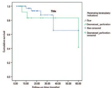
Graft failure occurred in 8 (19.04%) eyes at a mean duration of 24.5 months (range 8-60 months) in group 1, among which the causes for graft failure included rejection in 2 patients and recurrence of HSV keratitis in 6 patients. In group 1, the graft survival rate was 91.9% at 1 year, 76.0% at 2 years, and 65.1% at 3 years. In group 2, graft failure developed in 4 of 13 eyes (30.76%) and the graft survival rate was 89.5% at 1 year, 76.0% at 2 years, and 63.6% at 3 years. The difference
between groups was not statistically significant (p=0.91) (Figure 6).
Of the entire cohort, 34 (61.8%) patients achieved visual acuity of 0.6 logMAR or better, whereas 11 eyes (20%) had a postoperative BCVA worse than 1.2 logMAR because of graft rejection or failure. At the end of the follow-up, 28 (66.7%) patients in the herpetic corneal
Figure 3. Kaplan-Meier graphic of recurrence-free survival.
Figure 4. Slit-lamp biomicroscopy image showing recurrence of epithelial HSV-keratitis.
Table 1. Preoperative characteristics of patients and overall survival rates of groups
Herpetic corneal scar
Herpetic corneal scar with perforation
Log rank (Mantel-Cox)
n=42 n=13 P
Gender
Male 33 (78.6%) 11 (84.6%) 0.63 (NS)
Female 09 (21.4%) 02 (15.4%)
Age 44.51 ± 14.91 41.76 ± 14.33 0.56 (NS)
Mean follow-up 19.80 ± 12.91 26.15 ± 18.85 0.17 (NS)
Mean inactive duration 21.77 ± 22.31 16.30 ± 20.57 0.49 (NS)
Recurrence 12/42 4/13 0.40 (NS)
Recurrence-free survival 71.4% 69.2%
Rejection 4/42 3/13 0.58 (NS)
Rejection-free survival 90.5% 76.9%
Graft failure 8/42 4/13 0.91 (NS)
Graft failure-free survival 81% 69.2%
NS= not significant.
Figure 5. Kaplan-Meier graphic of rejection-free survival.
scar group and 6 (46.2%) in the descemetocele group had a BCVA of <0.6 logMAR, 7 (16.7%) patients in the scar group and 3 (23.1%) in the descemetocele group had a BCVA between 0.6 and 1.2 logMAR, and 7 (16.7%) patients in the scar group and 4 (30.8%) in the desceme-tocele group had a BCVA of >1.2 logMAR. There were no statistically
significant differences between groups (p=0.38, chi-square test).
DISCUSSION
In this study, the long-term outcomes of PK for treatment of HSK-induced corneal scarring are reported. In general, corneal surgeons hesitate to perform corneal transplantation for the management of herpetic corneal disease because the long-term success is relatively poor due to immunologic graft failure. HSK can also recur in corneal allografts and is sometimes difficult to distinguish from graft rejec-tion. Recurrent HSV infection usually follows reactivation of latent virus in the trigeminal ganglion, although the use of corticosteroids
after PK may be a triggering factor(10,11). Due to inflammation,
recurrence of HSK after PK is associated with an increased risk of graft re -jection. In addition, in the case of combined HSK recurrence and graft rejection, drug application is more difficult(4). The use of oral an tivirals for prophylaxis can significantly reduce the recurrence rate of herpes. Nonetheless, these grafts remain at a higher risk for failure from im-munologic rejection. Acyclovir, an acyclic purine nucleoside analog,
is the most commonly used antiviral medication(12-15).
In this study, recurrence of epithelial keratitis or epithelial toge-ther with stromal keratitis was the major type of recurrence of HSV
keratitis, similar to that reported in previous studies(16,17). The
recur-rence rate was 28.57% in group 1 and 30.76% in group 2, but this
difference was not statistically significant (p=0.40). Several studies
have reported different recurrence rates of HSV keratitis after PK(12,16,18). For example, Wu et al. reported a recurrence rate of 20.6% after PK for herpetic corneal scarring and 1-, 2-, and 3-year recurrence-free
survival rates of 88.9%, 85.1%, and 77.9%, respectively(16); however,
this study did not enroll patients with HSK-induced corneal leucoma with a history of corneal perforation and the patients received oral acyclovir at 200 mg 5 times daily for the first 3 months, which was
then tapered to 400 mg twice daily for a further 12-18 months(17). Awan
et al. reported recurrence of herpetic corneal scarring in 33% of 18 patients following deep anterior lamellar keratoplasty and oral acyclovir at 200 mg twice daily for 12 months. In that study, the mean
preoperative disease-free duration was 2 years(18). The difference in
recurrence rates and recurrence-free survival between those studies can be mainly attributed to preoperative disease-free duration and use of prophylactic antiviral therapy.
In the present study, the rejection rate was 9.52% in the quiescent herpetic scar group and 23.07% in the descemetocele or perforated herpetic scar group. Rejection-free survival rates at 1, 2, and 3 years were 97.0%, 87.5% and 65.7% in group 1, and 83.3%, 76.4% and 63.6% in group 2, respectively. Although the rejection rates seemed higher in group 1 than in group 2, rejection-free survival was similar
at 3 years between groups (p=0.58). Garcia et al. reported 1-, 2-, and
3-year rejection-free survival rates of 83.6%, 77.2%, and 74.5%, res-pectively, in those patients who received oral 400 mg acyclovir twice
daily(15). They measured host vascularity by the number of involved
quadrants and found no association with allograft rejection or failure. The rejection-free survival rates at 1 and 2 years in group 1 of this study were higher than those reported by Garcia et al., which may have resulted from the dose and duration of oral acyclovir treatment; however, the rejection rate in group 2 in this study was higher than in group 1, which can be attributed to perforation-induced corneal inflammation and/or to the small number of subjects in group 2. This study has some shortcomings: 1) it is a retrospective study, and 2) host vascularity was not recorded preoperatively.
A total of 8 grafts (19.04%) failed in group 1, one half due to graft rejection and the other half due to recurrence. When limited to only
13 patients in group 2, 4 (30.76%) grafts failed. The failure rate of
group 1 was markedly lower than that in the study by Moyes et al.(17),
where the failure rate was 30%. A possible explanation for this diffe-rence is the routine use of prophylactic oral acyclovir in our patients, as several other studies reported lower graft failure rates with the
routine use of prophylactic oral acyclovir(4,19). Another possibility is
that the mean preoperative disease-free duration in the present study was 21.77 ± 22.31 months in group 1 and 16.30 ± 20.57 months in group 2. Unfortunately, these data were not available for comparison for the cohort of Moyes et al.; however, Garcia et al. reported a failure rate of 11.4% with a 56-month disease-free duration before surgery,
which could have led to better postoperative graft survival(15).
In this study, although the recurrence and graft rejection rates differed between the two groups, the 3-year graft survival rates were similar (65.7% and 63.6%, respectively). In a study by Killingsworth et al., 15 patients who required therapeutic PK for HSK were divided into two groups. The first group included patients with severe stromal keratitis who did not respond to medical treatment and patients who progres-sed to perforation. The second group consisted of patients who de-veloped corneal perforation without active inflammation. Only 4 of the 11 patients in the first group achieved clear grafts, whereas all 4
patients in the second group achieved clear grafts(20). Likewise,
accor-ding to our results, without inflammation, corneal herpetic scarring with a descemetocele or perforation, achieved close graft survival and quiescence of the herpetic corneal scar.
CONCLUSION
It has already been reported that corneal grafts had a significantly high risk of rejection and failure in cases with underlying herpetic di-sease. Epstein et al. and Gittins-Nunez et al. found that the incidence of graft failure after rejection episodes was significantly greater in
grafts for herpes, as compared to grafts for keratoconus(21,22). In this
study, without inflammation, cases with corneal herpetic scarring with a descemetocele or perforation achieved similar graft survival rates with quiescent herpetic corneal scarring. The preoperative disease-free interval and dose and duration of prophylactic systemic acyclovir therapy are major factors affecting the graft survival.
REFERENCES
1. Knickelnein JE, Hendricks RL, Charukamnoetkanok P. Management of Herpes Simplex Virus stromal keratitis: an evidence-based review. Surv Ophthalmol. 2009;54(2):226-43. 2. Liesegang TJ. Herpes simplex virus epidemiology and ocular importance. Cornea. 2001;
20(1):1-13.
3. Ing JJ, Ing HH, Nelson LR, Hodge DO, Bourne WM. Ten-tear postoperative results of penetrating keratoplasty. Ophthalmology. 1998;105(10):1855-65.
4. Tambasco FP, Cohen EJ, Nguyen LH, Rapuano CJ, Laibson PR. Oral acyclovir after penetrating keratoplasty for herpes simplex keratitis. Arch Ophthalmol. 1999;117(4): 445-9.
5. Halberstadt M, Machens M, Gahlenvek KA, Bohnke M, Gerweg JG. The outcome of corneal grafting in patients with stromal keratitis of herpetic and non-herpetic origin. Br J Ophthalmol. 2002;86(6):646-52.
6. Shtein RM, Garcia DD, Musch DC, Elner VM. Herpes simplex virus keratitis: histopa-thologic inflammation and corneal allograft rejection. Ophthalmology. 2009;116(7): 1301-5.
7. Thompson RW Jr, Price MO, Bowers PJ, Price FW Jr. Long-term graft survival after pe-netrating keratoplasty. Ophthalmology. 2003;110(7):1396-402.
8. Lomholt JA, Baggesen K, Ehlers N. Recurrence and rejection rates following corneal transplantation for herpes simplex keratitis. Acta Ophthalmol Scand. 1995;73(1):29-32. 9. Ficker LA, Kirkness CM, Rice NS, Steele ADM. Long-term prognosis for corneal grafting
in herpes simplex keratitis. Eye (Lond.). 1988;2(Pt 4):400-8.
10. Langston DP. Oral acyclovir suppresses recurrent epithelial and stromal herpes sim-plex. Arch Ophthalmol. 1999;117(3);391-2.
11. Beyer CF, Hill JM, Byrd TJ, Kaufman HE. Herpes simplex dendritic keratitis after kerato-plasty. Am J Ophthalmol. 1991;112(3):355-6.
12. Ghosh S, Jhanjı V, Lamoureux E,Taylor HR, Vajpayee RB. Acyclovir therapy in preven-tion of recurrent herpetic keratitis following penetrating keratoplasty. Am J Ophthal mol. 2008;145(2):198-202.
14. Jansen AF, Rijneveld WJ, Remeijer L, Völker-Dieben HJ, Eggink CA, Geerards AJ, et al. Five-year follow-up on the effect of oral acyclovir after penetrating keratoplasty for herpetic keratitis. Cornea. 2009;28(8):843-5.
15. Garcia DD, Farjo Q, Musch DC, Sugar A. Effect of prophylactic oral acyclovir after pe-netrating keratoplasty for herpes simplex keratitis. Cornea. 2007;26(8):930-4. 16. Wu SQ, Zhou P, Zhang B, Qıu WY, Yao YF. Long-term comparison of full-bed deep
la-mellar keratoplasty with penetrating keratoplasty in treating corneal leucoma caused by herpes simplex keratitis. Am J Ophthalmol. 2012;153(2):291-9.
17. Moyes AL, Sugar A, Musch DC, Barnes RD. Antiviral therapy after penetrating kerato-plasty for herpes simplex keratitis. Arch Ophthalmol. 1994;112(5):601-7.
18. Awan MA, Roberts F, Hegarty B, Ramaesh K. The outcome of deep anterior lamellar
keratoplasty in herpes simplex virus-related corneal scarring, complications and graft survival. Br J Ophthalmol. 2010;94(10):1300-3.
19. Barney N, Foster CS. A prospective randomized trial of oral acyclovir after penetrating keratoplasty for herpes simplex keratitis. Cornea. 1994;13(3):232-6.
20. Killingsworth DW, Stern GA, Driebe WT, Knapp A, Dragon DM. Results of therapeutic penetrating keratoplasty. Ophthalmology. 1993;100(4):534-41.
21. Epstein RJ, Seedor JA, Dreizen NG, Stulting RD, Waring GO 3rd, Wilson LA, et al. Pene-trating keratoplasty for herpes simplex keratitis and keratoconus: Allograft rejection and survival. Ophthalmology. 1987;94(8):935-44.
22. Gittins-Nunez LO, Diaz Del Castillo-Martin E, Huerta-Albanil I, Rios-Prado R, Soto-Da-vila MA. [Risk factors and rejection frequency in patients undergoing penetrating keratoplasty]. Rev Med Inst Mex Seguro Soc. 2016;54(2):190-5. Spanish.