Subclinical Coronary Artery Disease in Patients with Type 1
Diabetes
Mellitus
Undergoing Hemodialysis
Dinaldo Cavalcanti de Oliveira
1,2, Fabio Sandoli de Brito Junior
1,2, Rosley Weber Alvarenga Fernandes
1,2, João Roberto
de Sa
1,2, Valter Correia de Lima
1,2Hospital do Rim e Hipertensão - Fundação Oswaldo Ramos1; Hospital São Paulo - Universidade Federal de São Paulo2, São Paulo, SP - Brazil
Summary
Background: In patients with type diabetes mellitus, atherosclerosis occurs earlier in life and coronary artery disease (CAD) constitutes the major cause of death.
Objective: Evaluate the prevalence and anatomic characteristics of coronary artery disease (CAD) in type diabetic patients with chronic renal failure undergoing hemodialysis.
Methods: This is a descriptive study of 20 patients with type diabetes mellitus undergoing hemodialysis without known CAD. CAD was assessed by quantitative coronary angiography (QCA) and intravascular ultrasound (IVUS). QCA was performed in all lesions ≥30%, visually. All proximal 18-mm segments of the coronary arteries were analyzed by IVUS. All other coronary segments with stenosis ≥30% were also analyzed.
Results: Angiography detected 29 lesions ≥30% in 15 patients (75%). Eleven (55%) of the lesions were ≥50% and 10 (50%) ≥70%. Thirteen patients had all 3 major arteries interrogated by IVUS. Atherosclerosis was present in all patients and in all 5 proximal 8-mm segments analyzed. The mean vessel diameter of these segments was significantly larger at the IVUS than at the QCA, for all vessels. IVUS images of 25 (86.2%) of the 29 lesions ≥30% were obtained. Fibrotic plaques were common (48%) and 60% had intermediate vessel remodeling.
Conclusion: CAD was present in all vessels of all type diabetic patients undergoing hemodialysis. These findings are in agreement with other autopsy, angiography and IVUS studies. Additionally, they indicate the need for additional epidemiological and imaging studies to better understand and treat such a complex and serious clinical condition affecting young people. (Arq Bras Cardiol 2009;93():4-9)
Key words: Coronary artery disease; diabetes mellitus, type ; ultrasonography, interventional; renal insufficiency, chronic.
Mailing address: Dinaldo Cavalcanti de Oliveira •
Rua Abílio Soares, 625 / 64 A, Paraíso, 04005-002, São Paulo, SP - Brazil E-mail: [email protected], [email protected]
Manuscript received on January 26, 2008; revised manuscript received on June 19, 2008; accepted on June 19, 2008.
Introduction
In patients with type 1 diabetes mellitus, atherosclerosis occurs earlier in life and coronary artery disease (CAD) constitutes the major cause of death1. By the age of 55 years,
the cumulative mortality rate due to CAD has been reported to be 30 to 40%2, which is much higher than the overall mortality
rate of 4% in non-diabetic subjects. In fact, in type 1 diabetic patients, the mortality risk by CAD is increased 4 to 9 fold in men and 4 to 29 fold in women3. This risk is even higher
when proteinuria or renal failure is present4,5. Tuomilehto et
al6 followed 5,148 type 1 diabetic patients for more than 10
years and found that once chronic renal failure occurs, CAD develops earlier and much more often6.
Despite growing epidemiological and clinical interest, very few studies have been currently undertaken to characterize CAD in these individuals7-11, in opposition to type 2 diabetic
patients. The present study was designed to evaluate the
prevalence, the qualitative and quantitative characteristics of CAD in type 1 diabetic patients with chronic renal failure undergoing hemodialysis.
Methods
Patients with type 1 diabetes mellitus undergoing hemodialysis without known CAD were prospectively included in this descriptive and observational study. All patients underwent biochemical and standard hematological work-up. Cardiovascular assessment included electrocardiogram, Doppler-echocardiogram and an ischemic test.
The study protocol was approved by the Ethics Committee of the Federal University of Sao Paulo and written informed consent was obtained from all participants.
Quantitative coronary angiography and intravascular ultrasound analysis
Nitroglycerin (300 µg) was given immediately before angiograms and intravascular ultrasound (IVUS) interrogations.
normal arteries, lesions < 30% or lesions ≥ 30%. Quantitative coronary angiography (QCA) was performed in all lesions ≥ 30% using an automated edge detection algorithm (QCA CMS System, MEDIS Medical Imaging Systems, Leiden, Netherlands). Lesion length, minimal lumen diameter, reference diameter and percent diameter stenosis were measured at these sites.
IVUS imaging was performed using mechanical systems (Clear View or Galaxy, Boston Scientific, Sunnyvale, CA, USA) consisting of 30-MHz ultrasound catheters with a 0.5 mm/s motorized pull-back system. IVUS recording was initiated at the most distal segment of each vessel with diameter greater than 2 mm. Accordingly, at least half of the total vessel length was studied. The S-VHS recorded tape was used for offline measurements using dedicated IVUS analysis software (QIVA, Pie Medical, Netherlands). For this analysis, the most proximal 18-mm segment of the major coronary arteries was arbitrarily selected. Additionally, all other segments in the coronary tree presenting stenosis ≥ 30% at the angiography were analyzed.
The following measurements were performed at the proximal 18-mm coronary segments: vessel, lumen and plaque cross-sectional areas (CSA) and maximum plaque thickness, at every millimeter12. Based on these measurements,
vessel, lumen and plaque volumes were calculated. Minimal
lumen CSA was also obtained. Atherosclerotic plaques were considered present when intimal thickness exceeded 0.3 mm13. The axial distribution of the plaque along each of these
coronary segments was assessed by calculating its coefficient of variation, by dividing the mean plaque CSA by the standard deviation of the plaque CSA.
At the segments selected for analysis for the presence of stenosis ≥ 30%, the lumen, vessel and plaque CSA were measured at the site with the minimum lumen CSA. Plaque burden was calculated as: (plaque CSA, / vessel CSA at the lesion site) x 100. Proximal and distal reference sites were defined as those cross-sections with the least amount of disease and largest lumen within 10 mm of the lesion boundary. At these sites, vessel and lumen CSA were measured. The averaged lumen CSA of the proximal and distal references was considered the reference lumen CSA. The lumen area stenosis was calculated as: [(reference lumen CSA minus lumen CSA at the lesion site / reference lumen CSA) x 100]. Vessel remodeling was determined based on the following definition: adaptive remodeling was considered to be present when vessel CSA at the lesion site was larger than vessel CSA at the proximal reference; constrictive remodeling was considered to be present when vessel CSA at the lesion site was smaller than vessel CSA at the distal reference and intermediate remodeling was present when vessel CSA at the lesion site was intermediate between vessel CSA at the proximal and distal references14. A remodeling
index was calculated as vessel CSA at the lesion site divided by vessel CSA at the proximal reference15.
At the lesion site, plaque composition was classified as soft, fibrotic or fibrocalcific, according to its characteristics12.
QCA and IVUS measurements were performed at the Core Laboratory of Hospital Israelita Albert Einstein (São Paulo, Brazil).
Statistical analysis
Quantitative data are presented as the mean value ± SD and qualitative data are presented as frequencies.
To study the association of the presence of stenosis ≥ 30% with age and duration of type 1 diabetes, the Student’s t test was applied. The same test was used to compare mean vessel diameter of the proximal 18-mm at the IVUS and angiography.
The search for an association between plaque maximum thickness and plaque volume in the most proximal 18-mm segments of the coronary arteries and age, serum cholesterol, body mass index, hemodialysis duration, and diabetes duration was carried out by calculating Pearson’s linear correlation coefficient.
All significance tests were two-tailed, and p values < 0.05
were considered significant.
Results
Study population
From February to November 2003, 20 patients were recruited for the study. Demographic and clinical data are depicted in Table 1. Only two patients (10%) presented myocardial ischemia at the functional tests.
Angiographic analysis
Visual assessment of the coronary angiograms detected 29 lesions ≥ 30% in 15 (75%) patients [1.93 ± 1.03 lesions per patient (Table 2)]. Eleven (55%) of them had lesions ≥ 50% and 10 (50%) had lesions ≥ 70%. One patient presented a proximal left anterior descending (LAD) artery occlusion. One of the patients with a positive ischemic test had stenosis in all three vessels [LAD: 50%, left circumflex (LCX): 80% and right coronary artery (RCA): 40%]. The other one presented stenosis in two vessels (LAD: 70% and RCA: 50%).
Intravascular ultrasound analysis
IVUS imaging of the most proximal 18-mm segments of at least one of the major coronary arteries was obtained from all patients. Thirteen patients had all 3 major arteries interrogated, 5 patients had 2 and 2 patients had only one. Atherosclerosis was present in all patients and in all 51 most proximal 18-mm segments analyzed (18 LAD, 18 LCX and 15 RCA). These segments presented
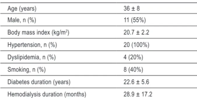
Table 1 – Patients’ demographic and clinical characteristics
Age (years) 36 ± 8
Male, n (%) 11 (55%)
Body mass index (kg/m2) 20.7 ± 2.2
Hypertension, n (%) 20 (100%)
Dyslipidemia, n (%) 4 (20%)
Smoking, n (%) 8 (40%)
Diabetes duration (years) 22.6 ± 5.6
Table 2 - Angiographic data
CAD Prevalence (n: 20 patients)
Lesions < 30%* 5 (25%)
30-49%*, n (%) 4 (20%)
50-69%*, n (%) 1 (5%)
≥ 70%*, n (%) 10 (50%)
Lesion Severity and QCA (n: 29 lesions)
30-49%*, n 11 (38%)
50-69%*, n 6 (20%)
≥ 70%*, n 12 (42%)
Reference Diameter, mm 2.88 ± 0.78
Minimal Lumen Diameter, mm 1.64 ± 0.51
Diameter stenosis, % 45 ± 13
Lesion length, mm 9.6 ± 13.3
Lesion location (n=29)
Proximal LAD: 4 (14%) Proximal LCX: 2 (7%)
Proximal RCA: 3
(10%)
Mid LAD: 11 (38%) Mid LCX: 4 (14%) Mid RCA: 4 (14%)
Distal LAD: 0 (0%) Distal LCX: 0 (0%) Distal RCA: 1 (3%)
* Visual analysis; QCA - quantitative coronary angiography; LAD - left anterior descending artery; LCX - left circumlex artery; RCA - right coronary artery.
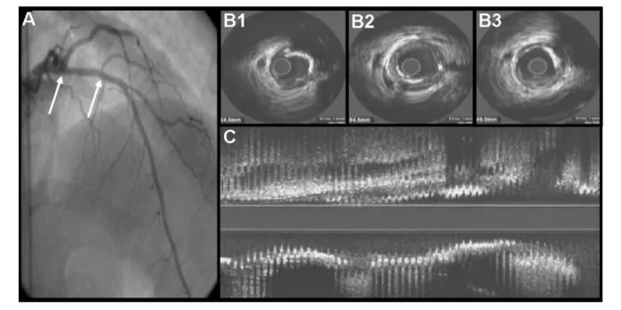
Figure 1 -A - Left coronary artery angiography displaying the left anterior descending artery with mild atherosclerosis. The arrows indicate the 18-mm most proximal
segment; B - Three cross-sectional IVUS frames of this segment showing large plaque mass and great arc of calcium; C - IVUS long view of the same segment showing atherosclerosis disease uniformly distributed (Axial coeficient of variation of plaque area: 41).
abundant and diffusely distributed atherosclerosis (Fig. 1). In 9 (17.6%) segments, minimal lumen CSA was ≤ 4 mm2. Plaque
volume, maximum plaque thickness, minimal lumen CSA, and axial coefficient of variation of the plaque area for these segments are shown in Table 3.
The mean vessel diameter of the proximal segments was significantly larger at the IVUS than at the QCA, in all vessels: [18 LAD segments: 4.24 ± 0.45 vs. 3.30 ± 0.59 (p<0.001)]; [18 LCX segments: 3.64 ± 0.60 vs. 3.20 ± 0.56 (p=0.02)]; [15 RCA segments: 4.35 ± 0.56 vs. 3.82 ± 0.56 (p=0.01)].
IVUS images of 25 (86.2%) of the 29 lesions ≥ 30% were obtained. Fibrotic plaques were the most common (48%) and the majority presented intermediate vessel remodeling (60%). Plaque characteristics, plaque burden, lumen area stenosis and minimal lumen CSA of these lesions are shown in Table 3.
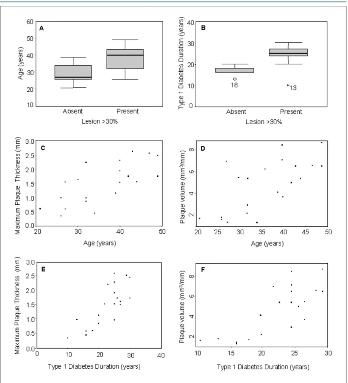
Age and diabetes duration were related to greater plaque thickness, plaque volume and the presence of lesions ≥ 30% (Fig. 2).
Discussion
The most remarkable finding of this study was the diffuse vessel involvement and the large amount of atherosclerosis identified at the IVUS in this selected population of type 1 diabetic patients with end-stage renal disease (ESRD). We verified a relatively uniform plaque distribution along all the most proximal 18-mm segments, as shown by the low plaque area axial coefficient of variation (33.6 ± 10%). Additionally, plaque quantification in these segments demonstrated the existence of large plaque masses by both the axially corrected plaque volume (4.6 ± 2.4 mm3/mm) and maximum intimal
thickness (1.49 ± 0.71 mm).
Figure 2 -A - Age according to the presence or absence of lesion ³ 30% (p=0.003); B - Duration of type 1 diabetes according to the presence or absence of lesion ³ 30%
(p=0.03); C - Correlation between age and plaque maximum thickness. Pearson’s linear coeficient = 0.71 (CI = 0.39–0.87); D - Correlation between age and plaque volume. Pearson’s linear coeficient = 0.68 (CI = 0.34 – 0.86); E - Correlation between duration of type 1 diabetes and plaque maximum thickness. Pearson’s linear coeficient = 0.79 (CI = 0.53 – 0.91); F - Correlation between duration of type 1 diabetes and plaque volume. Pearson’s linear coeficient = 0.77 (CI = 0.51 – 0.90).
of fibrinolysis and coagulation. Diabetic nephropathy is associated with an atherogenic lipoprotein profile, including elevated low-density lipoprotein, very low-density lipoprotein, and lipoprotein (a) and decreased high-density lipoprotein. Furthermore, a hypercoagulable state, characterized by increased plasminogen activator inhibitor-I, factor VII and
plasma fibrinogen levels has been described. Reduced renal function can lead to the accumulation of advanced glycosylation end-products in the circulation and tissues, which may accelerate atherosclerosis16.
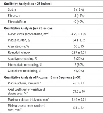
Table 3 - Intravascular ultrasound analysis
Qualitative Analysis (n = 25 lesions)
Soft, n 3 (12%)
Fibrotic, n 12 (48%)
Fibrocalciic, n 10 (40%)
Quantitative Analysis (n = 25 lesions)
Lumen cross sectional area, mm2 4.29 ± 1.95
Plaque burden, % 64 ± 13.2
Area stenosis, % 58 ± 15
Remodeling index 0.87 ± 0.21
Adaptive remodeling, % 5 (20%)
Intermediate remodeling, % 15 (60%)
Constrictive remodeling, % 5 (20%)
Quantitative Analysis of Proximal 18 mm Segments (n=51)
Plaque volume, mm3/mm * 4.6 ± 2.4
Axial coeficient of variation of
plaque area, %* 33.6 ± 10
Maximum plaque thickness, mm* 1.49 ± 0.71 Minimal lumen cross sectional
area, mm2 * 5.1 ± 2.1
* Analysis per patient.
reported in type 1 diabetic patients who underwent angiography for CAD screening before simultaneous pancreas-kidney transplantation8.
The amount and extent of atherosclerosis found at the IVUS in our investigation are in agreement with the results reported in a necropsy study of 9 individuals with average age of 29 years at the time of death17. In this study, 90.5 cm (47%) of the total 191
cm of coronary artery segments assessed had more than 50% of cross-sectional area stenosis, compared with only 2 cm of the 155 cm in the control group of similar age and sex. Another coronary IVUS study in type 1 diabetes also reported that all patients had CAD11. Interestingly, these results were obtained in a less sick
population when compared to ours, because individuals with creatinine > 150 µmol/l and overt nephropathy (urinary albumin excretion > 300 mg/24 h) were excluded.
Larsen et al18 showed that the degree of subclinical coronary
atherosclerosis was significantly more severe in patients with type 1 diabetes than in controls. The mean plaque area (by IVUS) was ≥ 40% in 71% of diabetic arteries when compared to 33% of control arteries (p < 0.001).
Importantly, our study showed the predominance of atherosclerotic lesions with characteristics of stability, represented by their fibrotic composition with negative or intermediate remodeling. However, even considering the lower percentage of potentially vulnerable plaques (lipidic composition with positive remodeling), the enormous amount of disease makes it a frequent finding in the coronary tree of type 1 diabetic patients with ESRD, which may explain the high rates of adverse events in this population.
When correlating clinical variables with coronary atherosclerosis, we observed that age and diabetes duration were related to greater plaque thickness, plaque volume and the presence of lesions ≥ 30%. We did not find any correlation between coronary atherosclerosis and serum cholesterol, smoking, body mass index, and the hemodialysis procedure. For a few of these variables, the lack of correlation might be due to beta error. In another study19, a 1% increase in mean HbA
1c over
18 years implied in a 6.4% increase in plaque burden, 15 mg increase in total cholesterol implied in 10% increase in plaque burden and a 10-year increase in age implied in a 16.2% increase in plaque burden.
It is known that the angiography underestimates vessel diameter and atherosclerosis extent when compared with IVUS, and there are three predictors for this discrepancy: vessel diameter by angiography < 3 mm, proximal segments and diabetes, presumably type 2. Our study is the first to address this issue in type 1 diabetic patients20.
Magnetic resonance imaging and computed tomography (through the evaluation of coronary artery calcium content) can identify subclinical CAD16, 21. The calcium score has been used to
evaluate the prevalence and prognosis of CAD in type 1 diabetic patients22,23.However, in patients with ESRD, the association
between coronary calcium and coronary atherosclerosis is less well established24.
Currently, a better CAD characterization in type 1 diabetic patients with ESRD undergoing hemodialysis has become more important, in view of the increasingly availability of simultaneous pancreas-kidney transplantation. This modern treatment may improve cardiovascular outcome in these patients. In fact, it has been reported that CAD progression is reduced based on the results of angiography performed before transplantation and four years later25.
This study has two main limitations: the small sample size, and the lack of information regarding long-term glycemic control.
Conclusion
In summary, subclinical CAD is present in all vessels of all type 1 diabetic patients undergoing hemodialysis. It indicates the need for additional epidemiological and imaging studies to better understand and treat such a complex and serious clinical condition affecting young people.
Potential Conflict of Interest
No potential conflict of interest relevant to this article was reported.
Sources of Funding
There were no external funding sources for this study.
Study Association
References
1. Libby P, Nathan D, Abraham K, Brunzell JD, Fradkin JE, Haffner SM, et al. Report of the National Heart, Lung, and Blood Institute-National Institute of Diabetes and Digestive and Kidney Diseases Working Group on Cardiovascular Complications of Type 1 Diabetes Mellitus. Circulation. 2005; 111: 3489-93.
2. Krolewski AS, Warram JH, Rand LI, Kahn RC. Epidemiologic approach to the etiology of type 1 diabetes mellitus and its complications. N Engl J Med. 1987; 317: 1390-8.
3. Laing SP, Swerlow AJ, Slater SD, Burden AC, Morris A, Waugh NR, et al. Mortality from heart disease in a cohort of 23,000 patients with insulin-treated diabetes. Diabetologia. 2003; 46: 760-5.
4. Jensen T, Borch-Johnsen K, Kofoed-Enevoldsen A, Deckert T. Coronary heart disease in young type 1 (insulin-dependent) diabetic patients with and without diabetic nephropathy: incidence and risk factors. Diabetologia. 1987; 30: 144-8.
5. Foley RN, Culleton BF, Parfrey PS, Harnett JD, Kent GM, Barre PE. Cardiac disease in diabetic end-stage renal disease. Diabetologia. 1997; 40: 1307-12.
6. Tuomilehto J, Borch-Johnsen K, Molatirus A, Molarius A, Forsen T, Rastenyte D, et al. Incidence of cardiovascular disease in type 1 (insulin dependent) diabetic subjects with and without diabetic nephropathy in Finland. Diabetologia. 1998; 41: 784-90.
7. Manske CL, Wilson RF, Wang Y, Thomas W. Prevalence of, and risk factors for, angiographic determined coronary artery disease in type I diabetic patients with nephropathy. Arch Intern Med. 1992; 152: 2450-5.
8. Oliveira DC, Gusmão G, Nakamoto A, Souza FL, Sá JR, Pestana JO, et al. Prevalence of coronary artery disease in type 1 diabetic patients candidates for double transplantation (kidney and pancreas). Arq Bras Cardiol. 2005; 84: 108-10.
9. Ramanathan V, Goral S, Tanriover B, Feurer JD, Kazancioglu R, Shaffer D, et al. Screening asymptomatic diabetic patients for coronary artery disease prior to renal transplantation. Transplantation. 2005; 79: 1453-8.
10. Senior PA, Welsh RC, McDonald CG, Paty BW, Shapiro AMJ, Ryan EA. Coronary artery disease is common in non uremic, asymptomatic type 1 diabetic islet transplantation candidates. Diabetes Care. 2005; 28: 866-72.
11. Larsen JR, Brekke M, Sandvik L, Arnesen H, Hanssen KF, Jorgensen KD. Silent coronary atheromatosis in type 1 diabetic patients and its relation to long term glycemic control. Diabetes. 2002; 51: 2637-41.
12. Mintz GS, Nissen SE, Anderson WD, Bailey SR, Erbel R, Fitzgerald PJ, et al. American College of Cardiology Clinical Expert Consensus Document on Standards for Acquisition, Measurement and Reporting of Intravascular Ultrasound Studies (IVUS). A report of the American College of Cardiology Task Force on Clinical Expert Consensus Documents. J Am Coll Cardiol. 2001; 37: 1478-92.
13. Nissen SE, Gurley JC, Erines CL, Grines CL, Booth DC, McClure R, Berk M, et
al. Intravascular ultrasound assessment of lumen size and wall morphology in normal subjects and patients with coronary artery disease. Circulation. 1991; 84: 1087-99.
14. Tauth J, Pinnow E, Sellebarger T, Sullebarger JT, Basta L, Gursoy S, et al. Predictors of coronary arterial remodeling patterns in patients with myocardial ischemic. Am J Cardiol. 1997; 80: 1352-5.
15. Sabaté M, Kay P, Feyter PJ, van Domburg RT, Deshpande NV, Ligthart JM, et al. Remodeling of atherosclerotic coronary arteries varies in relation to location and composition of plaque. Am J Cardiol. 1999; 84: 135-40.
16. Kim WY, Astrup AS, Stuber M, Tarnow L, Falk E, Botnar RM, et al. Subclinical coronary and atherosclerosis detected by magnetic resonance imaging in type 1 diabetes with and without diabetic nephropathy. Circulation. 2007; 115: 228-35.
17. Crall FV, Roberts WC. The extramural and intramural coronary arteries in juvenile diabetes mellitus: analysis of nine necropsy patients aged 19 to 38 years with onset of diabetes before age 15 years. Am J Med. 1978; 64: 221-30.
18. Larsen JR, Tsunoda T, Tuzcu EM, Schoenhagen P, Brekke M, Arnesen H, et al. Intracoronary ultrasound examinations reveal significantly more advanced coronary atherosclerosis in people with type 1 diabetes than in age- and sex-matched non-diabetic controls. Diab Vasc Dis Res. 2007; 4: 62-5.
19. Tuzcu EM, Kapadia SR, Tutar E, Ziada KM, Hobbs RE, McCarthy PM, et al. High prevalence of coronary atherosclerosis in asymptomatic teenagers and young adults: evidence from intravascular ultrasound. Circulation. 2001; 103: 2705-10.
20. Moussa I, Kobayashi Y, Adamian M, Hirose M, Di Mario C, Moses J, et al. Characteristics of patients with a large discrepancy in coronary artery diameter between quantitative angiography and intravascular ultrasound. Am J Cardiol. 2001; 88: 294-6.
21. Gottlieb I, Lima JAC. Should all high-risk patients be screened with computed tomography angiography ? Circulation. 2008; 117: 1318-32.
22. Costacou T, Edmundowicz D, Prince C, Conway B, Orchard TJ. Progression of coronary artery calcium in type 1 diabetes mellitus. Am J Cardiol. 2007; 100: 1543-7.
23. Thilo C, Standal E, Knez A, Reiser M, Steinbeck G, Haberl R, et al. Coronary calcification in long-term type 1 diabetic patients: a study with multi slice spiral computed tomography. Exp Clin Endocrinol Diabetes. 2004; 112: 561-5.
24. Achenbach S, Daniel WG. Computed tomography of the heart. In: Libby P. Braunwald’s heart disease: a textbook of cardiovascular medicine. 8th ed. Philadelphia: Saunders Elsevier Company; 2008. p. 415-35.