SYSTEMATICS, MORPHOLOGY AND PHYSIOLOGY
Comparative Study of the Stridulatorium Sulcus, Buccula and Rostrum of
Nymphs of Triatoma klugi Carcavallo et al, Triatoma vandae Carcavallo et
al and Triatoma williami Galvão et al (Hemiptera: Reduviidae)
Maria B a Silva, JoSé JurBerg, CleBer galvão, Helene S BarBoSa
Lab Nacional e Internacional de Referência em Taxonomia de Triatomíneos, Lab de Biologia Estrutural, Instituto Oswaldo Cruz, Fiocruz, Av Brasil 4365, 21040-900, Rio de Janeiro, RJ, Brasil
Edited by Eunice Galati – FSP/USP Neotropical Entomology 39(1):035-045 (2010)
ABSTRACT - Ultrastructural analysis of the ventral region of the head - rostrum, buccula and stridulatorium sulcus - of 1st, 3rd and 5th instars of Triatoma klugi Carcavallo et al, Triatoma vandae Carcavallo et al, and Triatoma williami Galvão et al, are described in here. Morphological differences in the analyzed structures for all three Triatoma species studied were detected under scanning electron
microscopy, allowing their grouping by their morphological similarities. Species-specific differences
at each nymphal development stage were analyzed as well.
KEY WORDS: Triatominae, morphology, nymphal development, T. oliveirai complex
Chagas disease is considered a predominantly rural endemic parasitic disease transmitted by triatomine bugs which constitutes a serious problem in Latin America due to its impact in society as well as in economy. This disease is caused by infection with the protozoan Trypanosoma cruzi (Chagas 1909), mainly transmitted through triatomine bugs’ faeces. These bugs have as main biological feature their obligate haematophagy at all nymphal and adult stages (Carcavallo et al 2000). They belong to Triatominae subfamily, and have a wide geographic distribution in the Americas, from Salt Lake region (USA), to the South areas of Patagonia, although some species may also occur in the Old World (Galvão et al 2003).
Triatominae is currently composed of 140 species (Schofield & Galvão 2009) within several specific complexes belonging
to the genus Triatoma Laporte, which are very homogenous with regard to their morphologic traits and generally associated with geographical areas. Triatoma klugi Carcavallo et al, T. vandae Carcavallo et al, and T. williami Galvão et al are in the T. oliveirai complex together with T. matogrossensis Leite & Barbosa, T. guazu Lent & Wygodzinsky, T. jurbergi Carcavallo et al,T. baratai Carcavallo & Jurberg, T. deaneorum Galvão et al and T. oliveirai Neiva et al, which is the nominotypical species (Noireau et al 2002).
The importance of the rostrum in the Triatominae genus characterization has been underlined since the 20s and the 30s by Pinto (1927, 1931). Carcavallo et al (1994) studied 11 species of Panstrongylus using SEM and considered P. herreri, P. humeralis, and P. lignarius as related species. Subsequently, Carcavallo et al (1995, 1997) compared various anatomic areas of Psammolestes Bergroth and reported important differences among species. The same
tool was used by this author to study 97 of 110 Triatominae species (Carcavallo et al 1997). Other structural studies reported structural differences between the buccula and the gula of 5th instars of T. williami and Triatomagersteaeckeri (Stål) (Ferro et al 1997).
In the present study, the structure of the stridulatory sulcus, buccula, and rostrum during the T. klugi,T. vandae and T. williami nymphal development was evaluated using a scanning electron microscope (SEM).
Material and Methods
Insects. All five instars of T. klugi, T. vandae and T.
williami were obtained from laboratory colonies which were established with insects from Nova Petrópolis (RS), Itiquira (MT) and Goiania (GO). Vouchers were deposited at the Rodolfo U Carcavallo Collection, which is part of the Entomological Collection of the Instituto Oswaldo Cruz.
Scanning electron microscopy (SEM). Three nymphs of
the 1st, 3rd and 5th instar of these three species were selected after eclosion and/or ecdysis. First instars were collected soon after hatching, killed by freezing, dehydrated at 37°C for 24h,
and fixed on double-faced band on metallic stubs. Due to the
Results
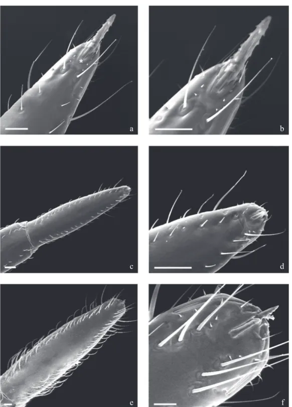
Triatoma williami Galvão et al
The ultrastructural analysis of the rostral 3rd segment in the terminal region of the 1st and 3rd instars shows long and short sensilla located at the apical plaque end that is a
glabrous area. Two long sensilla are located at the end, and 2+2 spiculae which are found beside the two long sensilla. There are 1+1 circular sensilla located close to the 1+1 lateral rift (Figs 1a, b, f), both located just below the two long sensillae previously mentioned. In 3rd and 5th instars the 2+2 spiculae remain but this segment carries its own sensilla wose distribution is irregular, arranged in parallel or in a line along
Fig 1 Triatoma williami - ventral view: rostrum and apical plate. a, b) 1st instar; c, d) 3 rd instar; e, f) 5th instar. Note the increased number and types of sensillae during the nymphal development. Bars: 20 µm
a
c
e
b
d
the lateral boundaries (Figs 1c, e). The authors noted that the insertions of sensilla are larger on the extreme apex, but short and small sensilla may also be found close to the apical plaque with 1+1 sensilla and especially along the lateral rifts 3+3 there are short sensilla which gradually diminish from the anterior to the posterior region (Figs 1a-f).
The buccula located on the posterior region of the rostrum shows a “U” form with thick edges on the anterior region, becoming gradually thinner towards to the posterior edge. In the central internal area, the buccula presents pleats with papillae as well as striae in its anterior edge. The internal area
at the central region of the buccula of first instars does not
have granulations or rifts (Fig 2a). There is, however, a rift in the central internal area of 3rd and 5th instars (Figs 2a-c).
The ultrastructure of the posterior region of the rostrum area of 1st instars shows a central area close to the acetabulum cavity of the 1st pair of legs having a wrinkled aspect, showing some elementary parallel lines and unnumbered papillae, which form a stridulatory sulcus (Figs 3a, b). On the anterior edge, close to the proesternum, long sensillae-like bristles may be seen in all stages. The stridulatory sulcus becomes rectangular on the 3rd instars and its sharp posterior edge reaches the 1st pair of feet. Acetabulum cavity showing delicate striae on its surface. The lateral edges show a profusion of papillae and setiferous tubercles of different sizes (Fig 3). On the 5th instar, this structure is larger and there is an increase in the number of papillae and setiferous tubercles (Figs 3d, e).
Triatoma klugi Carcavallo et al
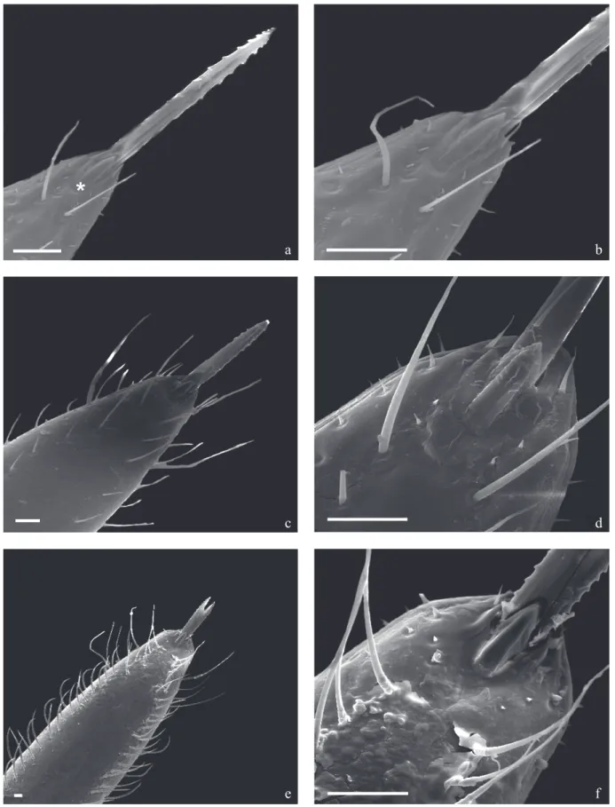
The 3rd segment of the T. klugi rostrum also shows a larger number and size of sensillae as nymphal develpoment progress. The apical plaque is delimited by 1+1 long sensilla detached on their anterior portion and 2+2 small spiculae right below the lance-like structure, delimited by 1+1 long and thin sensilla inserted in the circular and short basis tubercles (Figs 4a, b). Also observed next to the rostrum apex 1+1 were lateral rifts and 1+1 circular structures in the internal posterior portion, which are probably sensillae (Figs 4a-f).
The T. klugi buccula has a “U” shape with thick boundaries which have a granulated and spiral internal boundary. The internal central area shows a pleat on its anterior boundary with some papillae and striae on its posterior portion but no rifts on the 1st instar (Fig 5a). In the 3rd instar this structure shows only one rift and papillae distributed on the posterior and in the internal central areas (Fig 5b). In the 5th instar, the buccula shows external lateral boundaries with a sharp “sulcus” and its internal boundaries spiralled and richly granulated ending up in a prominent area at the basis of the rostrum insertion (Fig 5c). The buccula has a sole rift in the internal central granulated area, presenting three striae in the anterior region delimited by a long and short rift. It also shows some bristles inserted into round tubercles on the side of this structure and a richly granulated area between its posterior boundary and the beginning of the gullet. The basis of the
first segment of the rostrum displays a conspicuous area
similar in shape to the buccula, which are both superposed when resting for all stages observed (Figs 5a-c).
Fig 2 Triatoma williami - ventral view: buccula (the small arrows show sulcus and the arrow heads the rifts). a) 1st instar; b) 3rd instar; c) 5th instar. Bars: 20 µm
b a
Fig 3 Triatoma williami - ventral view: proesternumum and stridulatory sulcus. a,b) 1st instar (the small arrow shows rudimentary horizontal striae; the large arrows show two sensillae); c) 3rd instar; d, e) 5th instar. Bars: 20 µm
a
c
d
b
Fig 4 Triatoma klugi - ventral view: Rostrum and apical plate. a, b) 1st instar; c, d) 3 rd instar; e, f) 5th instar. Bars: 20 µm
a
c
e
b
d
The stridulatory sulcus shows a “V” shape up to the 1st instar with a small striated area delimited by numerous papillae (Figs 6a, b). These papillae will give space to the setiferous tubercles on the following instars. In the following stages the stridulatory sulcus shows straight anterior and thin posterior boundaries. Still on the anterior boundary, close to the prosternum 1+1 long bristles similar to sensilla can be seen in all stages (Figs 6a-f). On the 3rd and 5th instars, the deep long “sulcus” has a very thin posterior edge presenting innumerable setiferous tubercles and some papillae (Figs 6c-f).
Triatoma vandae Carcavallo et al
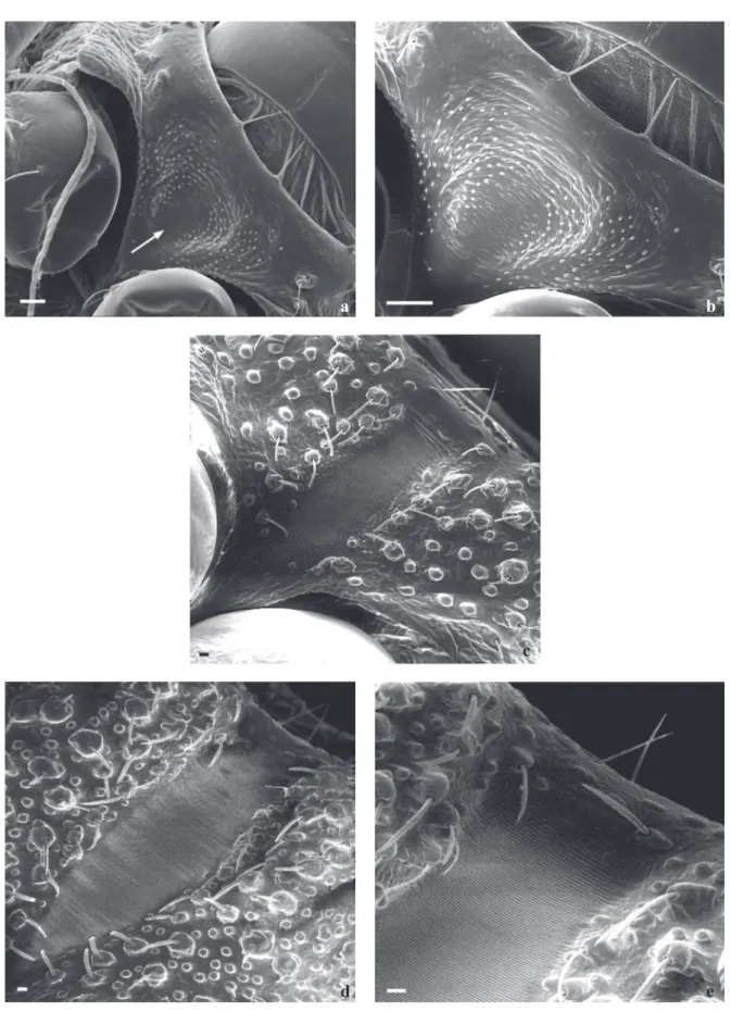
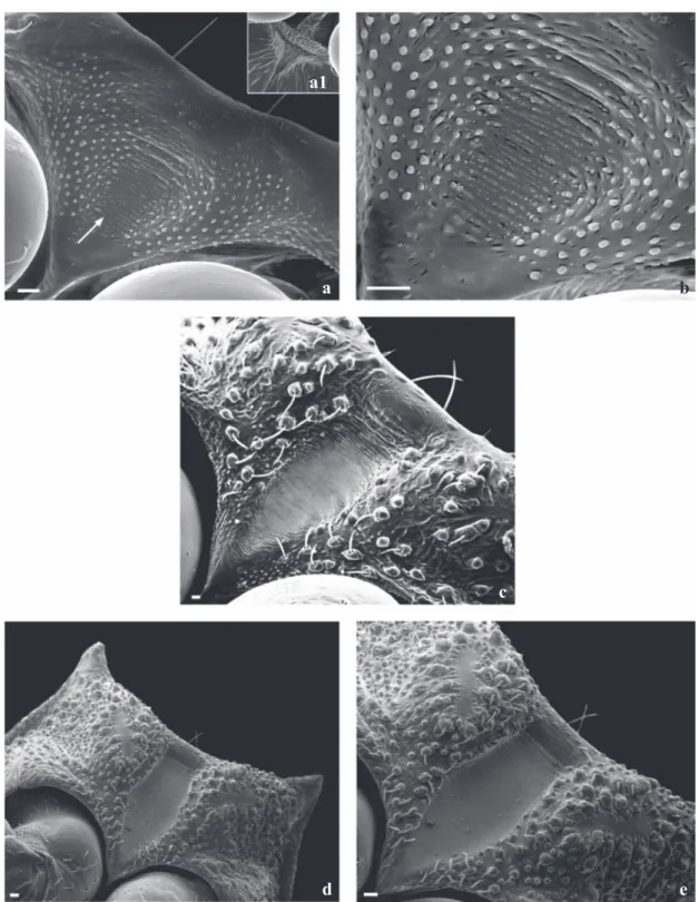
The analysis of the 3rd segment of the rostrum of T. vandae during the first two instars showed the presence of differentiated sensilla which increase in quantity and length as the insect grows. The apical plaque is delimitated by 2+2 long detached sensillae that progressively grow from the basis to the apex. Close below were also observed 1+1 spiculae and 1+1 long and thin sensilla inserted in circular, short tubercle basis (Figs 7a, b). Moreover, there a re 1+1 lateral rifts and 1+1 circular structures in its internal posterior portion, close to the tip of the rostrum which may be sensillae (Figs 7b, d, f). Additionally, there are 3+3 sensillae in line which decrease in size from the anterior to the posterior area of the rostrum (Fig 7b). Following the same pattern of the other two species studied in this paper, the ultrastructural images of the buccula show no granule or rifts in the internal central area for the 1st, 3rd and 5th instars. However, striae and granules in the anterior area of the insertion part of the 1st insertion of the rostrum were observed (Figs 8a-c).
The sketch of the future stridulatorium sulcus shows in
the first instar a highly organized area where the papillae
are laterally disposed delimiting a area with striae abruptly ending together with the papillae between the prothorax
thighs (Figs 9a, b). Right below the acetabulum, the final
portion of the future stridulatorium sulcus, ends up in a triangular area thickly reticulated (Fig 9a1). A more detailed analysis demonstrates the systematic organization of the papillae together with the striae (Fig 9b). On the 3rd instar the “V” shape structure is delimitated by few bristles which are more concentrated in the posterior area (Fig 9c). In 5th instars, the sulcus becomes triangular in the apex and square-shaped in its posterior half (Figs 9d, e).
Diagnosis
The following differences were observed among the nymphs of the species studied:
1st instars. The amount of papillae and striae found in the
proesternum area (future stridulatory sulcus):
1) In a comparative view T. klugi shows more density of papillae and striae than T. vandae, and both have more than T. williami; 2) T. vandae and T. klugi show the future stridulatory sulcus more expanded in the proesternum area, while T. williami presents this structure in a more central portion of the proesternum.
Fig 5 Triatoma klugi - ventral view: buccula (the small arrows show sulcus and the arrow heads the rifts). a) 1st instar; b) 3rd instar; c) 5th instar. Bars: 20 µm
a
b
Apical plaque.Triatoma williami and T. klugi present only two long sensillae at the end of the apical plaque and four spiculae at the basis of this structure, which are delimitated by two long and thin sensillae inserted in tubercles of
circular and short basis. On the other hand, T. vandae shows a structure delimitated by 1+1 long sensillae detached at
its final portion and 2+2 small spiculae right below on the
basis delimitated by 1+1 long and thin sensillae inserted in Fig 6 Triatoma klugi - ventral view: proesternumum and stridulatory sulcus. a, b) 1st instar (the small arrow shows rudimentary horizontal striae; the large arrows show two sensillae); c, d) 3rd instar; e, f) 5th instar. Bars: 20 µm
a
c
e
b
d
tubercles of circular short basis.
Buccula. Triatoma klugi shows papillae in the internal central
area, while T. williami and T. vandae have none.
5th instar. Thebuccula shows one rift in the T. klugi and in
the T. williami, but this was not observed in the T. vandae.
Discussion
Galvão et al (2005) published a table listing specific Fig 7 Triatoma vandae - ventral view: rostrum and apical plate. a, b) 1st instar; c, d) 3 rd instar; e, f) 5th instar. Bars: 20 µm
a
c
e
b
d
names, used approaches and references of papers dedicated to the morphology of triatomine eggs and nymphs, showing how little is known on the morphology of triatomine bugs. The analysis by scanning electron microscopy is an additional tool for systematic studies in entomology, which allows to distinguish similar species,
cryptic species and specific complexes.
Several researchers are trying to use new tools, as ontogenetic morphometrics (Galvão et al 2005, Rocha et al 2005) besides the SEM analysis (Carcavallo et al
1998), which may be combined with those identified by
Lent & Wygodzinsky (1979), leading to the establishment
of specific identification keys. These authors also called
the attention for the importance the stridulatory sulcus
may have on species identification, as it may vary in
size, shape, number of rifts and in the distance between the rifts among species. The importance of this structure was underlined by Barth in 1953 and its functionality by Galíndez-Girón et al (1998).
Gonçalves et al (1985) made a comparative study of the stridulatory sulcus of T. maculata (Erichson) and T. pseudomaculata Corrêa & Espínola concluding this structure was appropriate to distinguish between both species. The stridulatory sulcus was also informative to distinguish T. guazu from T. jurbergi at both 1st and 5th instars (Silva 2000, Silva et al 2002). In the present study, T. williami and T. klugi nymphs of 3rd instar differ on ornamentation and quantity of rifts in the internal central area. In the extreme apex of the rostrum 3rd segment, the labial apical plaque (Quadri 1951, Parsons 1966, Cobben 1978) has proved to be an important parameter of differentiation between species. In the present study this structure was useful to distinguish T. williami and T. klugi,from T. vandae.
Silva (1999, 2000) and Silva et al (2003) analysed the buccula of T. guazu and T. jubergi at all nymphal stages and concluded that this structure differed between both species, being suggested as an additional taxonomic trait. Catalá (1996) described ten sensillae on the rostrum of eight species of Triatomine and concluded that the chaetigerous sensillae is more abundant in the rostrum in six lines along the main axe of the 2nd and 3rd segments. She also observed that these sensillae increased substantially in quantity in the 3rd segment and also verified that its quantity and distribution were not different in nymphs or adults.
In conclusion, the ultrastructural study of the rostrum, stridulatorium sulcus and buccula presented reliable differences that allow the differentiation among nymphs of species of species of the T. oliverai complex, contributing to an earlier diagnosis of these species.
Acknowledgments
The authors thank Bruno Ávila for the image processing. This work was supported by grants from Conselho Nacional
de Desenvolvimento Científico e Tecnológico (CNPq) and
SVS-Ministério da Saúde from Brazil and CDIA - Chagas Disease Intervention Activities-European Community. Fig 8 Triatoma vandae - ventral view: buccula (the small
arrows show sulcus and the arrow heads the rifts). a) 1st instar; b) 3rd instar; c) 5th instar. Bars: 20 µm
a
b
References
Brener Z, Andrade Z, Barral Netto P (1999) Trypanosoma cruzi
e doença de Chagas. Rio de Janeiro, Ed. Guanabara Koogan S.A., 431p.
Carcavallo R U, Galíndez-Girón I, Catalá S, Jurberg J, Lent H, Galvão
Fig 9 Triatoma vandae - ventral view: proesternumum and stridulatory sulcus. a, b) 1st instar (the small arrow shows rudimentary horizontal striae; the large arrows show two sensillae); a1) End region of stridulatory sulcus; c) 3rd instar; d, e) 5th instar. Bars: 20 µm
C, Barata J M S, Valderrama A (1998). Some anatomic structures studied with scanning electron microscopy (SEM). Algumas estruturas anatômicas estudadas com microscopia eletrônica de varredura (MEV). Vol. I. p.299-393. InCarcavallo R U, Galíndez-Girón I, Jurberg J, Lent H, Atlas of Chagas’ disease vectors in the Americas. Atlas dos vetores da doença de Chagas nas Américas. Rio de Janeiro, Editora FIOCRUZ, 1217p.
a a1
c
d
b
relationships within the oliveirai complex (Hemiptera: Reduviidae: Triatominae). Infec Gen Evol. 2: 11-17.
Parsons M C (1966) Labial skeleton and musculature of the Hydrocorisae (Heteroptera). Can J Zool 44:1051-1084.
Pinto C (1931) Valor do rostro e das antenas na caracterização dos triatomíneos.Bol Biol 19: 45-137.
Rocha D S, Patterson J S, Sandoval C M, Jurberg J, Angulo V M, Esteban L, Galvão C (2005) Description and ontogenetic morphometrics of nymphs of Belminus herreri Lent & Wygodzinsky (Hemiptera: Reduviidae, Triatominae). Neotrop Entomol 34: 491-497.
Schofield C J, Galvão C (2009) Classification, evolution, and species
groups within the Triatominae. Acta Tropica 110: 88-100.
Silva M B A (2000) Morfologia e morfometria de Triatoma guazu
Lent & Wygodzinsky, 1979 e Triatoma jurbergi Carcavallo, Galvão e Lent, 1998, através de microscopia óptica e eletrônica de varredura (Hemiptera, Reduviidae). Dissertação de mestrado, Fundação Oswaldo Cruz, Rio de Janeiro, xiv + 93p.
Silva M B A, Barbosa H S, Carcavallo R U, Galvão C, Jurberg J (1999) Placas apicais do lábio das ninfas de 1º estádio de
Triatoma guazu Lent & Wygodzinsky, 1979 e Triatoma jurbergi
Carcavallo, Galvão & Lent, 1998 (Hemiptera, Reduviidae), vetores da doença de chagas. Entomol Vect6: 663-668.
Silva M B A, Barbosa H S, Galvão C, Jurberg J, Carcavallo R U (2003) Comparative study of stridulatorium sulcus, buccula and rostrum of Triatoma guazu Lent & Wygodzinsky, 1979 and
Triatoma jurbergi Carcavallo, Galvão e Lent, 1998 nymphs by
scanning electron microscopy (Hemiptera, Reduviidae).Mem Inst Oswaldo Cruz 98: 335-344.
Silva M B A, Barbosa H S, Jurberg J, Galvão C, Carcavallo R U (2002) Comparative ultrastructural analysis of the antennae of Triatoma guazu Lent & Wygodzinsky, 1979 and Triatoma jurbergi Carcavallo, Galvão e Lent, 1998 during the nymphal stage development (Hemiptera, Reduviidae). J Med Entomol 39:705-715.
Silva M B A, Jurberg J, Barbosa H S, Rocha D S, Carcavallo R U, Galvão C (2005) Morfologia comparada dos ovos e ninfas de
Triatoma vandae Carcavallo, Jurberg, Rocha, Galvão, Noireau
& Lent, 2002 e Triatoma williami Galvão, Souza & Lima, 1965 (Hemiptera, Reduviidae, Triatominae). Mem Inst Oswaldo Cruz 100: 549-561.
Silva M B A, Jurberg J, Galvão C, Barbosa H S, Carcavallo R
U (1998) Same insuficiently know cuticular structures of the
Triatoma guazu Lent & Wygodzinsky, 1979. Mem Inst Oswaldo
Cruz 93(suppl. II): 345.
Silva MBA, Jurberg J, Galvão C, Carcavallo R U (2000) Descrição dos ovos e ninfas de Triatoma guazu Lent & Wygodzinsky, 1979 (Hemiptera, Reduviidae, Triatominae) vistos através de microscopia óptica e eletrônica de varredura. Entomol Vect 7: 311- 334.
Zacharuk R Y (1980) Ultrastructure and function of insect chemosensilla.Ann Rev Entomol 25: 27-47.
Received 28/III/08. Accepted 24/VII/09.
Carcavallo R U, Jurberg J, Lent H, Noireau F, Galvão C (2000) Phylogeny of the Triatominae (Hemiptera: Reduviidae). Proposals for taxonomic arrangements. Entomol Vect7(Supl 1): 1-99.
Carcavallo R U, Jurberg J, Rocha D S, Galvão C, Noireau F, Lent H (2002) Triatoma vandae sp. do complexo oliveirai encontrada no estado do Mato Grosso, Brasil (Hemiptera: Reduviidae: Triatominae). Mem Inst Oswaldo Cruz 97: 649-654.
Catalá S (1996) Sensila associated with the rostrum of eight species of Triatominae. J Morphol 228:195-201.
Cobben R H (1978) Evolucionary trends in Heteroptera Part II. Mouth part-structures and feeding strategies. Meded Landbouwhoge School Wageningen78: 5-407.
Ferro Z P A, Barbosa H S, Jurberg J, Carcavallo R U (1997) The buccula and gula of Triatominae nymphs by scanning electron microscopy (Hemiptera: Reduviidae).Acta Microsc 6:572-573.
Galvão C, Carcavallo R U, Rocha D S, Jurberg J (2003) A checklist of the current valid species of subfamily Triatominae Jeannel, 1919 (Hemiptera, Reduviidae) and their geographical distribution, with nomenclatural and taxonomic notes. Zootaxa 202: 1-36.
Galvão C, McAloon M, Rocha D S, Schaefer C W, Patterson J, Jurberg J (2005). Description of eggs and nymphs of
Linshcosteus karupus (Hemiptera: Reduviidae: Triatominae).
Ann Entomol Soc Am 98: 861-872.
Garcia M H H M, Souza L, Souza R C M, Paula A S, Borges E C,
Barbosa S E, Schofield C J, Diotaiuti L (2005) Occurrence and
variability of Panstrongylus lutzi in the State of Ceará, Brazil. Rev Soc Bras Med Trop38:410-415.
Gonçalves T C M, Jurberg J, Costa M C, Souza W (1985) Estudo morfológico comparativo de ovos e ninfas de Triatoma maculata
(Erichson,1848) e Triatoma pseudomaculata Correa & Espínola, 1964 (Hemiptera, Reduviidae, Triatominae). Mem Inst Oswaldo Cruz 80:276-276.
Galíndez-Girón I, Rocha D S, Lent H, Carcavallo R U, Jurberg J, Galvão C, Barbosa H S, Martínez A, Barata J M S, Rosa J A da (1998) Nynphal stages. Estádios ninfais. Vol. II. p.449-513. InCarcavallo R U, Galíndez-Girón I, Jurberg J, Lent H, Atlas of Chagas’ disease vectors in the Americas. Atlas dos vetores da doença de Chagas nas Américas. Rio de Janeiro, Editora FIOCRUZ, 1217p.
Jurberg J, Silva M B A, Galvão C, Rocha D S, Barbosa H S, Carcavallo R U (2002) Descrição dos ovos e ninfas de Triatoma jurbegi Carcavallo, Galvão & Lent, 1998 vistos através de microscopia óptica e eletrônica de varredura (Hemiptera, Reduviidae). Mem Inst Oswaldo Cruz 97:209-216.
Lent H, Wygodzisnky P (1979) Revision of the Triatominae
(Hemiptera - Reduviidae) and their significance as vectors of
Chagas’ disease. Bull Amer Mus Natur Hist 163: 123-520.
Mc Iver S (1975) Structure of cuticular mechanoreceptors of arthropods. Ann Rev Entomol 20: 381-397.