Deolinda Isabel Fernandes da Silva
Mestrado em Bioquímica
Departamento de Química e Bioquímica 2014
Orientador
Susana Seixas, PhD, IPATIMUP
Alpha-1-Antitrypsin
deficiency, exploring
the role of SERPINA1
rare variants and
searching for genetic
modifiers of associated
diseases
(Granulomatosis with
Polyangiitis)
No me d o A u to r, l etra A ria l Bold tamanh o 10 , just if ica d o à es q u er d aTodas as correções determinadas pelo júri, e só essas, foram efetuadas. O Presidente do Júri,
Agradecimentos
“Somos a junção de vários pedaços e perder algum é como uma amputação” Pedro Chagas Freitas
O culminar desta etapa deve-se à junção de todos os pedacinhos que fui construindo ao longo desta minha caminhada iniciada bem lá atrás, quando iniciei o percurso escolar, não seria justo resumi-la a estes dois últimos anos apenas, pois seria a perda de muito do que sou. Por isso começo por agradecer à minha FAMÍLIA, Pais, Irmãos, Cunhados e Sobrinhos que sempre estiveram ao meu lado, me apoiaram em todas a minhas decisões, certas ou erradas mas que me trouxeram até aqui, me deram conselhos e me ajudaram em tudo o que precisei. Eles são o meu suporte e é nesta união que busco forças para nunca desmoronar ou desistir. Na realidade não há palavras que possam descrever aquilo que significam ou que fazem por mim, mas aqui fica o meu simples gesto de agradecimento a todos eles.
Agradeço à Susana Seixas, minha orientadora, pela oportunidade de desenvolver este projeto no IPATIMUP, por todo o apoio, compreensão e disponibilidade que sempre demostrou ao longo deste ano e por todos os ensinamentos que me transmitiu. Devo ainda agradecer a oportunidade e confiança para o desenvolvimento do projeto em colaboração com o laboratório de Munique.
À Sílvia, Patrícia e Andreia, minhas colegas de laboratório, pelas ajudas no trabalho, pelo companheirismo, amizade, simpatia e boa disposição demostrados ao longo deste ano de permanência no instituto.
To Dieter Jenne, my advisor in Munich, for his help and availability, for the opportunity to be there and learn so much and know other ways to work, other country and its culture and all the teachings that gave me.
To Heike, the lab technician, that followed all my work and taught me all that I needed, and for the friendship.
To Natascha, Therese and Lisa, my colleagues in Munich lab, for help in developing the work and for the friendship while I was there.
Por último, mas não menos importante, a todos os meus amigos, sem ser necessário enumera-los, que a par da família são muito importantes e que sei que posso sempre contar quando alguma dificuldade surgir. E também a todas as pessoas que conheci ao longo deste trajeto que de uma forma ou de outra me deixaram um ensinamento e me ajudaram a construir enquanto pessoa.
Resumo
O estudo do genoma humano em muito tem contribuído para o entendimento das bases moleculares que estão na origem das doenças genéticas. Neste sentido, a criação de bases de dados que reúnem todas as variações genéticas do genoma humano, identificadas até ao momento, tem-se revelado um ponto-chave para perceber como estas influenciam as características humanas atuais, o risco e progressão de doenças, assim como a resposta a diversos tratamentos médicos. A deficiência de alfa-1-antitripsina (DAAT) é uma doença monogénica causada por mutações no gene SERPINA1 que geralmente culminam numa baixa concentração da proteína SERPINA1 no soro. Esta doença afeta um número considerável de indivíduos, sendo mais comum em populações de origem Europeia, onde está associada com um elevado risco de desenvolver patologias respiratórias, como a doença pulmonar obstrutiva crónica (DPOC) ou o enfisema pulmonar. Desde a identificação da DAAT em 1963 foram identificados múltiplos alelos com diferentes implicações na concentração de proteína no soro, e com subsequentes diferenças ao nível das manifestações clínicas da doença. Os alelos mais comuns são os M (M1, M2, M3 e M4) que estão relacionados com níveis normais de proteína (0,9 a 2 g/L), e os alelos S (Glu264Val) e Z (Glu342Lys) cujos níveis de proteína no soro variam entre 50-60% e 10-15% do normal, respetivamente. Estes são também os dois variantes mais associados com a patologia clínica. Os casos mais severos da doença são geralmente verificados em indivíduos homozigóticos para o alelo Z, cuja acentuada deficiência da proteína no soro resulta da acumulação intracelular em polímeros nos hepatócitos. Por esta razão a DAAT é também associada com um risco de doença hepática em consequência dos efeitos tóxicos dos polímeros Z nas células. A polimerização do alelo Z foi também proposta como uma das causas possíveis para a origem de uma resposta auto-imune nos casos de vasculite. A granulomatose com poliangite (GPA) é um síndrome multissistémico prevalente entre pacientes com DAAT e caracterizada por inflamações granulomatosas nos pequenos vasos. Outros mecanismos que têm sido apontados na origem da GPA incluem o desequilíbrio proteolítico entre a proteinase 3 (PR3) e a sua principal inibidora no soro a SERPINA1, bem como uma possível hereditariedade de genes auto-imunes transmitidos conjuntamente com genótipos de DAAT devido à sua proximidade no cromossoma 14q32.1. O gene SERPINA2 localizado 12kb a jusante do gene SERPINA1 possui uma sequência de DNA muito similar a este e embora tenha sido durante muito tempo
considerado um pseudogene, o gene SERPINA2 possui uma forma ativa de função desconhecida que é expressa em diferentes tecidos incluindo nos leucócitos.
O principal objetivo deste trabalho foi identificar e caracterizar alelos raros de DAAT na população Portuguesa. No sentido de obter mais informação sobre as bases moleculares e a variabilidade intra-haplotípica de cada variante combinou-se a sequenciação de DNA de uma região de ~8kb do gene SERPINA1 com a genotipagem de dois microssatélites flanqueantes CAn (~7,5kb a jusante) e GTn (~207kb a montante). De entre os 51 casos de DAAT sequenciados foram identificadas 13 mutações patogénicas num total de 14 alelos raros: MMalton (Phe52del;
n=18), MPalermo (Phe52del; n=9), I (Arg39Cys; n=7), Q0Ourém (Leu353framStop376; n=4),
PLowell (Asp256Val; n=3), MHerleen (Pro369Leu; n=2), MWurzburg (Glu342Lys; n=1), Q0Lisbon
(Thr68Ile; n=1), T (Glu264Val; n=1), Q0Gaia (Leu263Pro; n=1), PGaia (Glu162Gly; n=1),
Q0Oliveira do Douro (Arg281framStop297; n=1), Q0Vila Real (Met374framStop392; n=1) e
Q0Faro (IVSIC+3Tins; n=1). Os últimos cinco alelos são novos e descritos pela primeira
vez no presente trabalho. Os alelos Q0Gaia e PGaia resultam ambos de substituições de
aminoácidos com repercussões na estrutura da proteína. Os variantes Q0Oliveira do Douro e
Q0Vila Real resultam de pequenas deleções nucleotídicas que estão na origem da
alteração da matriz de leitura e da inserção de codões de terminação prematura. O alelo Q0Faro afeta o normal processamento do mRNA por alteração de um local de
splice.
A análise da variação haplotípica permitiu avaliar os alelos anteriormente descritos em populações de ancestralidade Europeia e elucidar a origem dos alelos raros MMalton e MPalermo em bases moleculares distintas M2 e M1, respetivamente. A
avaliação do espetro mutacional aponta para o facto de uma percentagem das mutações do gene de SERPINA1 ocorrem em regiões hipermutáveis, enquanto os motivos repetitivos tendem a acumular mutações do tipo inserções e deleções (indels), os dinucleótidos CpG apresentam um número elevado de substituições nucleotídicas preferencialmente de CGTG e de CGCA. Por outro lado, a análise da distribuição das mutações patogénicas e não patogénicas na região codificante do gene de SERPINA1 mostra que as mutações patogénicas se tendem a concentrar em importantes domínios funcionais da molécula, por oposição às mutações não patogénicas que se encontram dispersas uniformemente por toda a sequência de SERPINA1.
Numa segunda parte do trabalho procuramos avaliar o gene SERPINA2 como um potencial candidato para a associação observada entre a SERPINA1 e a GPA. Apesar de preliminares, os nossos resultados apontam para uma maior
homogeneidade haplotípica nos controlos face aos casos de GPA, o que levanta a hipótese do haplótipo mais frequentemente associado com alelo Z (SERPINA2/V3) apresentar um fator protetor. No âmbito deste trabalho, foram ainda realizados alguns ensaios experimentais de expressão de SERPINA2 em células humanas, HEK293, e em células de Drosophila Schneider S2. Embora a expressão da SERPINA2 tenha sido obtida em ambos os sistemas celulares, nas células Schneider S2 foram conseguidos níveis de proteína mais elevados em grande parte devido à acumulação intracelular da SERPNA2 nas células HEK293.
Palavras-chave: Deficiência de alfa-1-antitripsina, SERPINA1, alelos raros, haplótipos, mutações patogénicas, Granulomatose com poliangite, SERPINA2.
Abstract
The study of the human genome has greatly contributed to the understanding of the molecular basis of genetic diseases. In this sense, the creation of databases that gather all the genetic variations in the human genome identified so far, has proved a key point to understand how these influence current human traits, disease risk and progression, as well as the response to various clinical treatments. The alpha-1-antitrypsin deficiency (AATD) is a monogenic disease caused by mutations in the
SERPINA1 gene, which generally culminate in low serum levels of the SERPINA1
protein. The disease affects a considerable number of individuals, being more frequent in populations of European descended, where it is associated with a high risk of developing respiratory diseases, such as chronic obstructive pulmonary disease (COPD) or lung emphysema. Since the description of AATD in 1963 multiple alleles were identified and these were found to correlate with different serum levels and clinical manifestations of the disease. The most common alleles are M (M1, M2, M3 and M4) which are associated with normal protein levels (0.9 to 2 g/L) and the S (Glu264Val) and Z (Glu342Lys) alleles whose protein levels range between 50-60% and 10-15% of the normal, respectively. The last two are also the most commonly associated variants with respiratory complaints. The most severe cases of the disease are usually observed in Z homozygous in which the serum deficiency is determined by the intracellular accumulation of Z polymers in the hepatocytes. For this reason AATD has also been associated with an increased risk of liver disease as a result of toxic effects of the Z polymers within the cells. The polymerization of the Z allele has been proposed to underlie an autoimmune response in the case of vasculitis. Granulomatosis with poliangite (GPA) is a multisystemic syndrome affecting patients with AATD, which is characterized by a small vessels granulomatous inflammation. Other mechanisms that have been suggested to play a role in GPA include the proteolytic imbalance between proteinase 3 (PR3) and its major inhibitor in serum, SERPINA1, and the co-inheritance of autoimmune genes and AATD genotypes mainly due to its close proximity in chromosome 14q32.1. SERPINA2 is located 12kb downstream of the SERPINA1 and share with this a high DNA sequence similarity. Although SERPINA2 has been regarded as a pseudogene for a long time it has an active isoform of unknown function that is expressed in different tissues including in leukocytes.
The main goal of this work was to characterize rare alleles of AATD in the Portuguese population. In order to obtain additional information about the molecular basis and the intrahaplotipic variability of each variant, we combined the DNA sequencing of 8 kb region of the SERPINA1 with the genotyping of two flanking microsatellite CAn (~7.5 kb downstream) and GTn (~207 kb upstream). Among the 51 AATD cases analysed we have identified 13 pathogenic mutations in a total of 14 rare alleles: MMalton (Phe52del n = 18) MPalermo (Phe52del n = 9), I (Arg39Cys, n = 7) Q0Ourém
(Leu353framStop376 n = 4) PLowell (Asp256Val, n = 3) MHerleen (Pro369Leu, n = 2)
MWurzburg (Glu342Lys, n = 1) Q0Lisbon (Thr68Ile n = 1), T (Glu264Val, n = 1), Q0Gaia
(Leu263Pro n = 1) PGaia (Glu162Gly, n = 1) Q0Oliveira doDouro (Arg281framStop297 n = 1)
Q0Vila Real (Met374framStop392, n = 1) and Q0Faro (IVSIC+3Tins, n = 1). The last five
alleles are novel and are described for the first time in this work. The Q0Gaia and PGaia
alleles both result from amino acid substitutions affecting the protein structure. The Q0Oliveira do Douro and Q0Vila Real variants result from small deletions causing alterations of
the reading frame and the insertion of premature termination codons. The Q0Faro allele
affects the normal mRNA processing by disrupting a splice site.
The haplotype variability analysis allowed the evaluation of the alleles previously described in other populations with European ancestry and to elucidate the independent origin of MMalton and MPalemo alleles in the M2 and M1 molecular basis,
respectively.
The assessment of the mutational spectrum indicates that a proportion of the
SERPINA1 mutations occur in hypermutable regions and while short repetitive motifs
tend to accumulate insertions and deletions (indels), the CpG dinucleotides display a large number mutations preferably from CGTG and CGCA. Furthermore, the analysis of the distribution of pathogenic and non-pathogenic mutations across
SERPINA1 coding region shows that pathogenic mutations tend to cluster in crucial
functional domains of the molecule, in opposition to the non-pathogenic mutations, which are more uniformly spread throughout the SERPINA1 sequence.
In the second part of the work, we evaluate SERPINA2 as a potential candidate gene for the reported association between SERPINA1 and GPA. For this purpose, we conducted a sequencing study in control samples (ZZ individuals without disease), and GPA cases. Our preliminary results point to a higher haplotype homogeneity in controls than in GPA cases. We hypothesized that the haplotype more often associated to the Z allele (SERPINA2 / V3) may provide a protective factor to GPA. In this work, we have also performed several experimental assays of SERPINA2 expression in human HEK293 cells and in Drosophila Schneider S2 cells. Despite SERPINA2 expression
was achieved in both cellular systems, in Schneider S2 cells higher levels of protein were collected due to intracellular accumulation of SERPINA2 in HEK293 cells.
Key-words: Alpa-1-antitrypsin deficiency, rare alleles, SERPINA1, haplotypes, pathogenic mutations, Granulomatosis with polyangiitis, SERPINA2.
Contents
Agradecimentos ... i Resumo ... ii Abstract ... v Contents ... viii List of Tables ... x List of Figures ... xi Abbreviations ... xii 1. Introduction ... 11.1 The SERPIN Superfamily ... 3
1.2 The Alpha-1-antitrypsin (SERPINA1) ... 4
1.3 SERPINA1 variation and human disease ... 9
1.4 SERPINA2 ... 14
2. Aims ... 16
3.Material and Methods ... 18
3.1 Severe AATD by rare SERPINA1 variants ... 19
3.1.1 Samples ... 19
3.1.2 DNA extraction ... 19
3.1.3 PCR and sequencing ... 19
3.1.4 PCR and Microsatellite analysis ... 20
3.1.5 Data analysis ... 21
3.1.6 Characterization of Q0Faro allele ... 21
3.1.7 SERPINA1 conservation ... 22
3.2 SERPINA2 ... 23
3.2.1 Samples ... 23
3.2.2 PCR and DNA sequencing ... 23
3.2.3 Cloning of SERPINA2 ... 24
3.2.4 Transfection and protein extraction ... 25
3.2.5 Western blot ... 25
4.Results and Discussion ... 27
4.1 SERPINA1 mutational spectrum ... 28
4.1.1 Rare alleles causing AATD ... 28
4.1.1.1 Novel mutations ... 29
4.1.1.1.2 Small Deletions... 33
4.1.1.1.3 Splice site mutation ... 35
4.1.1.2 Previously described mutations ... 39
3.1.2 Mutational spectrum of SERPINA1 ... 47
4.1.3 Conservation and functional implications ... 57
4.2 SERPINA2 ... 60
4.2.1 SERPINA2 genotyping in GPA cases and controls ... 60
4.2.2 Expression of SERPINA2 ... 62
5.Conclusions ... 64
6.References ... 67
List of Tables
Table 1: SERPINA1 serum levels in different genotypes and corresponding risk of
developing emphysema or liver disease. ... 9
Table 2: Accession numbers for SERPINA1 cDNA sequences. ... 23
Table 3: Rare alleles of SERPINA1associated with AATD identified in the current work ... 28
Table 4: Molecular base of Q0Faro, M1Ala213 and M2 alleles. ... 37
Table 5: Haplotipic characterization of common and rare alleles. ... 44
Table 6: SERPINA1 mutation spectrum ... 48
Table 7: SERPINA2 haplotypes identified in ZZ controls from Portugal and Birmingham. ... 60
Table 8: SERPINA2 haplotypes identified in GPA samples from Birmingham and Bochum. ... 61
Table A 1: Conditions for SERPINA1 amplification and sequencing………...74
List of Figures
Figure 1: SERPINA1 structure. ... 5
Figure 2: Schematic representation of SERPINA1. ... 6
Figure 3: Phylogeny of SERPINA1 common alleles.. ... 7
Figure 4 : Distribution of S and Z alleles in the European continent. ... 8
Figure 5: Pathophysiology of SERPINA1 and its deficiency (AATD). ... 11
Figure 6: The SERPINA1 locus shows differential associations with anti-PR3 positive patients. ... 14
Figure 7: Distribution of SERPINA2 non-functional alleles in 52 human populations. .. 15
Figure 8: Schematic representation of SERPINA1 amplification…. ... 20
Figure 9: Location of the two microsatellites used in the haplotipic characterization of SERPINA1 rare alleles. ... 21
Figure 10: Schematic representation of SERPINA2 amplification.. ... 24
Figure 11: Sequence alignment of SERPINA1 orthologs ... 30
Figure 12: Three dimensional structure of SERPINA1 ... 31
Figure 13: Electrophoretic patterns of PGaia and common SERPINA1 alleles. ... 32
Figure 14: Q0Oliveira do Douro allele ... 33
Figure 15: Q0Vila Real allele.. ... 34
Figure 16. Electropherogram of Q0Faro allele.. ... 35
Figure 17: Alternative SERPINA1 transcripts (14n) of mononuclear phagocytes. ... 36
Figure 18: Detection of Arg101His and Ala213Val variation in the cDNA from mononuclear phagocytes of the two Q0Faro subjects.. ... 38
Figure 19: Schematic representation of SERPINA1 alternative splicing as deduced from the exon composition of mRNA species produced by mononuclear-phagocytes in M alleles and Q0Faro and Q0Porto.. ... 39
Figure 20: Origin of T variant from S and M2 or M3 alleles. ... 43
Figure 21: Graphic distribution of SERPINA1 residue conservation based on alignments of 18 vertebrates species and mutational spectrum.. ... 59
Abbreviations
AAT Alpha-1-antitrypsin
AATD Alpha-1-antitrypsin deficiency ANCA Antineutrophil Cytoplasmic Antibody cDNA Complementar DNA
CHO Chinese Hamster Ovary
COPD Chronic Obstructive Pulmonary Disease DNA Deoxyribonucleic acid
EB Elution Buffer
EDTA Ethylenediaminetethaacetic acid ELANE2 Elastase
ER Endoplasmic Reticulum Fw Forward
GPA Granulomatosis with Polyangiitis HEK Human Embryonic Kidney IEF Isoelectric Focusing INDELS Insertions and Deletions Mh MHerleen
ML Maximum Likelihood Mm MMalton
MPA Microscopic Polyangiitis Mpa MPalermo
Mw MWurzburg
NCBI National Center for Biotechnology Information NEB Naïve Empirical Bayes
NMD Nonsense mRNA Decay
PBS-T Phosphate buffer solution with Tween 20 PCR Polymerase Chain Reaction
Pl PLowell
PR3 Proteinase 3 Q0F Q0Faro
Q0l Q0Llisbon
Q0OD Q0Oliveira do Douro
Q0VR Q0Vila Real
RCL Reactive Center Loop
RNase Ribonuclease
RT-PCR Reverse Transcription PCR Rv Reverse
SERPIN Serine Proteinase Inhibitors SERPINA1 Alpha-1-antitrypsin
The understanding of the molecular basis of human diseases has been a major challenge for the scientific community for several decades, as well as the finding of their causes, susceptibility factors and treatment to improve the life quality of the patients.
In this sense, the progress achieved in the field of human genetics and the efforts made in the last years to generate a reference human genome sequence (Human Genome Project) and more recently, a database with thousands of sequenced individuals from different geographic regions (1000 Genomes Project) are remarkable1,2. Altogether, these projects allowed to create a detailed catalogue of the human genetic variation, which to date represents a fundamental tool to address how genetics influence current human traits, disease risk and progression and the response to differentmedical treatments1.
The human genome comprises the following categories of sequence variants: single nucleotide polymorphisms (SNPs), insertions and deletions (INDELs) which may range from 1bp to 10kb in length and larger structural variants (also known as copy number variation), which extend from 10 kb to several megabases2. Another class of genetic variants includes minisatellites and microsatellites2,which are tandem repeated DNA sequences of 6 bp to 100bp units and 1 to 5 bp units, respectively3. The most common category of variants involves a mutation in a single base in the DNA (SNPs), whereas other categories include the loss (deletion) or the gain (duplication or insertion) of one or multiple nucleotide(s). Sequence variants may have different repercussions according to their localization in a gene. If a mutation occurs in the coding region of a gene they can (1) have no effect or (2) result in an altered protein product unable to perform its regular function or (3) cause the protein premature termination. Otherwise if a variant occurs in a regulatory region, it may compromise the regular expression of a gene or even inactivate the entire gene. Importantly, mutations might also be classified as a “loss of function” if they drastically affect the normal activity of a gene or as a “gain of function” mutation, if the mutated gene acquires a novel property or function4.
However, in most cases the understanding of the impact of the different categories of variants in human health and disease is far from being completed.
Genetic diseases affect thousands of individuals around the world and in most cases these are expected to result from a mutation, which occurred in a germ cell and was then transmitted to the following generations. If the mutation has a strong effect and occurs in a single gene the disease will be monogenic (or Mendelian disease) and the inheritance pattern, might be autosomal or linked dominant, autosomal or
X-linked recessive or Y-X-linked or mitochondrial5. Most of these diseases are detected at low frequencies in the random population (rare diseases less 1%). Once the underlying genetic alteration has been identified, it is easier to understand the molecular pathogenesis of the disease and to design a genetic test for the diagnosis of the disease. Classical examples of monogenic diseases include alpha-1-antitrypsin deficiency (AATD), cystic fibrosis, phenylketonuria and Huntington’s disease, which are all correlated with the occurrence of pathogenic mutations in single genes6,7,8,9.
On the other hand, if several mutations with smaller effects occur in multiple genes, the disease is defined as complex or multifactorial and in general, these diseases may affect a larger percentage of the population. In this case, interactions among genes and between genes and the environment are likely to have an important role in the disease phenotype and in the molecular mechanism of the disease, thus making the design of screening tools much more difficult10. Here chronic obstructive pulmonary disease (COPD), antineutrophil cytoplasmic antibodies (ANCA) associated vasculitis, diabetes and Crohn’s disease are some examples of complex diseases associated with genetic variants distributed over multiple loci11-14. Worth of note, in most cases of complex diseases a significant proportion of their heritability still remains unexplained5.
1.1 The SERPIN Superfamily
SERPINs (serine proteinase inhibitors) are a superfamily of functional diverse protease inhibitors, sharing a conserved tertiary structure, which is determined by about 350 to 400 amino acids and has a molecular weight of 40-50 KDa15,16. Hundreds of SERPINs were already found in viruses, prokaryotes, plants, and animals where they are involved in many diverse physiological processes17. For example, in vertebrates SERPINs have key roles as protease inhibitors in blood coagulation, fibrinolysis, inflammation, angiogenesis, apoptosis and in complement activation. However, some SERPINs have developed other non-inhibitory functions, and act as molecular chaperones, hormone carriers or as storage proteins15.
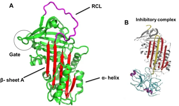
The archetypical SERPIN structure has three β-sheets (A,B,C), nine α-helices (A-I), and a flexible stretch of approximately seventeen residues between β sheet A and C named the reactive center loop (RCL) (Figure 1A), which is normally exposed to the solvent acting as a pseudo-substrate for proteases18. SERPINs ability to inhibit a specific protease is determined by the amino acid composition of the RCL, in particular those located at residues P1-P1’. Once a protease binds to the RCL, it establishes a
covalent ester linkage between the protease residue Ser-195 and the backbone carbonyl of the P1 residue leading to the cleavage of the P1-P1’ peptide bond. Such event initiates a major conformational rearrangement in SERPINs, and the molecule undergoes a complete transition from a “stressed” to a “relaxed” state (S to R transition). Briefly, immediately after the cleavage, the RCL is rapidly inserted into the β-sheet A (shutter region), caring the protease to the opposite site of the SERPIN molecule, distorting the catalytic domain of the protease and consequently the entire molecule (Figure 1B). This distortion avoids the breakdown of the acyl-enzyme intermediate, resulting in an irreversible SERPIN protease complex16, 19.
1.2 The Alpha-1-antitrypsin (SERPINA1)
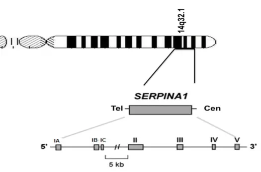
One of the most studied SERPINs is alpha-1-antitrypsin (SERPINA1 or AAT), a 52-kDa plasma glycoprotein with 394 amino acids synthesized at high levels by hepatocytes, and at lower concentrations by intestinal epithelial cells, neutrophils, lung epithelial cells and macrophages20. The protein is encoded by the SERPINA1 gene located at chromosome 14q32.1, which covers approximately 12.2 kb, and has four coding exons, three untranslated exons and six introns (Figure 2). The untranslated region of SERPINA1 comprises exons IA to IC and controls SERPINA1 expression through three alternative transcription initiation sites (Figure 2). While transcription may start in exons IA or IB in macrophages (middle and beginning of the exon, respectively), the transcription in hepatocytes is initiated only at exon IC (middle of the exon)20.
SERPINA1 is an important acute phase protein and the major serine protease inhibitor of human plasma, where it shows strong affinity towards neutrophil elastase (ELANE2) and proteinase 3 (PR3). However, recent studies have shown that SERPINA1 is also an irreversible inhibitor of kallikreins 7 and 14 and it has the ability to inhibit intracellular and cell-surface proteases such as matriptase and caspase-321. This inhibitory activity is mainly conferred by methionine 358 and serine 359 residues which correspond to RCL P1-P1’, respectively. The principal site of SERPINA1 activity is in lung, where the protein protects the fragile connective tissue of the lower respiratory tract from the uncontrolled proteolysis triggered by neutrophils during inflammation6. Importantly, in recent years SERPINA1 has emerged as a complex and multifunctional protein combining inhibitory properties with immunomodulatory and
anti-inflammatory activities against neutrophils, lymphocytes, macrophages, monocytes, mast cells and epithelial cells22.
Mutations in the SERPINA1 gene are the main cause for alpha-1-antitrypsin deficiency (AATD), an autosomal-codominant disorder, characterized by reduced protein serum levels and affecting 1 in 2000 to 1 in 7000 individuals of European descent24. The disease was first described by Laurell and Eriksson in 1963, whenthey noticed the absence of the SERPINA1 band by plasma protein gel electrophoresis in patients with COPD and lung emphysema25. Indeed, AATD patients have a significant higher risk of developing pulmonary disease, like early-onset emphysema and chronic obstructive pulmonary disease (COPD), which is correlated with the uncontrolled activity of neutrophil elastase in the lungs. Another major clinical manifestation of AATD is liver cirrhosis as a result of the cytotoxic effect of protein accumulation in the hepatocytes26. Presently, there are more than 125 variants of SERPINA1 identified and a considerable large number of those variants may be associated with abnormal protein plasma levels. Accordingly,SERPINA1 alleles are classified as: 1) “deficient” if
they are associated with a significant reduction in plasma levels, either because the synthesized protein is misfolded and retained within hepatocytes or because it has poor stability, leading always to reduced secretion; 2) “null” alleles (Q0), if there are no Figure 1: SERPINA1 structure. SERPINA1 comprising three β-sheets and nine α-helices. A –The shutter region (β-sheet A) is highlighted in red and the RCL is shown in magenta. B – Stable protease inhibitor complex. (Adapted from Khan et al. 16 Whisstock et al. 23)
protein traces in the plasma27. While, deficiency variants can be easily identified by isoelectric focusing (IEF) techniques, where different letters are assigned to different gene products according to the migration velocity in the protein electrophoresis gel (M to medium; S to slow; F to fast and Z very slow); null variants are characterized by the absence of a visible band in the IEF gels28.
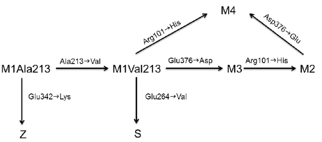
In European populations only the alleles M1, M2, M3, M4, S and Z reach polymorphic frequencies (>1%). The M alleles are considered the normal ones with 100% levels of the plasma protein (0.9 to 2 g/L) and the S and Z alleles, the common deficiency variants, are associated with 50-60% and 10-15% of normal plasma concentrations, respectively. The analysis of the molecular basis of the M, S and Z alleles allowed the reconstruction of the phylogenetic relationships between SERPINA1 common variants (Figure 3). The M1 can be subdivided in two subtypes, the M1Ala213 (ancestral allele) and the M1Val213. The M3 differs from the M1Val213 by an amino acid replacement at codon 376 (Glu376Asp) and the M2 has another substitution at codon 101 (Arg101His). The M4 shares with M2 the Arg101His substitution but lacks the Glu376Asp found in M3 and M2 variants (Figure 3)29. The S allele results from a Glu264Val mutation, in exon III, in a M1Val213 background. The Glu264Val causes the disruption of a salt bridge (Glu264-Lys387), highly conserved among SERPINs and linking the C terminus of the G α-helix to a β-strand in the hydrophobic core of the
Figure 2: Schematic representation of SERPINA1. The gene is located in SERPIN 14q32.1 cluster and it is organized in 3 untranslated exons (IA-IC), 4 coding exons (II-V) and 6 introns.
molecule18. This mutation is known to alter the stability of the molecule and to increase the susceptibility of the protein to polymerize, however it is only associated with disease when it is heterozygous with the Z allele30. Conversely, the Z allele arose by a Glu342Lys substitution in exon V, within a M1Ala213 allele. The Glu342Lys causes the disruption of another crucial salt bridge (Glu342-Lys290), which in turn affects the stability of A β-sheet. Importantly the Z mutation has more serious repercussions in protein folding than the S mutation because it leads to the spontaneous polymerization and accumulation of polymerised fibrils in the endoplasmic reticulum of hepatocytes, with subsequent cell damage18, 31.
The polymorphism of SERPINA1 has been widely studied in several populations due to its importance in human health32. The M alleles (M1, M2 and M3) are present in ethnical diverse populations, such as Europeans, Africans and Amerindians, with some differences in their frequencies. The M4 allele has also been reported in multiple samples from diverse geographic regions, but this variant is less studied than the other M subtypes because it is difficult to discriminate using only isoelectric focusing techniques. In contrary to M alleles, S and Z variants are only present in populations of European descent, or in cases of miscegenation with Europeans. The S allele is broadly distributed among the European continent, but its values tend to increase from northeast to southwest reaching the higher frequencies in the Iberian Peninsula, with an increase of more than 70 cases per 1000 individuals. This distribution suggests that the S mutation may have arisen in the north of the Iberian Peninsula in prehistoric times and then spread eastwards by population movements (Figure 4A). This hypothesis is supported by the 8500-16500 years estimate of the S allele obtained for
the Portuguese population, which are close to the end of the last glaciations (18000 years ago) and the European population expansion from the glacial refuge in the Iberian Peninsula32, 33.
The Z mutation has a different distribution among European populations, the frequency is higher in the north-east, more precisely in Scandinavia and the Baltic region where it can reach average values of about 2-4 %34 while in south populations it varies between 0.19 and 0.30% (Figure 4B). In summary, it has been suggested that the Z mutation occurred in the southern Scandinavia and Baltic regions about 2000-5000 years ago and spread later throughout the continent during Neolithic times 21, 33, 35,
36,37.
Figure 4 : Distribution of S and Z alleles in the European continent. A – Frequency of S alleles per 1000 inhabitants. B- Frequency of Z alleles per 1000 inhabitants (Adapted from Blanco et al.37)
1.3 SERPINA1 variation and human disease
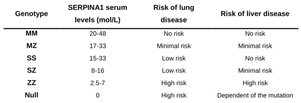
Lung emphysema is the principal clinical manifestation of AATD, which is associated with SERPINA1 concentrations below a protective threshold of 11 µM (0.50g/L)6,38. Although different SERPINA1 genotypes may lead to such reduced serum levels (Table 1) the most common genotype is the ZZ, which represents approximately 95% of the cases of severe AATD39. The SZ genotype is also associated with a lower risk of lung disease since protein levels are close to the 11 µM6.
Table 1: SERPINA1 serum levels in different genotypes and corresponding risk of developing emphysema or liver disease. (adapted from Bals et al. 6)
Genotype SERPINA1 serum
levels (mol/L)
Risk of lung
disease Risk of liver disease
MM 20-48 No risk No risk
MZ 17-33 Minimal risk Minimal risk
SS 15-33 Low risk No risk
SZ 8-16 Low risk Minimal risk
ZZ 2.5-7 High risk High risk
Null 0 High risk Dependent of the mutation
Among ZZ individuals emphysema is the most common cause of death (58-72%) and tends to appear in early ages, around 40 to 50 years in smokers or around 60 to 70 years in non-smokers40. Pathogenesis of the lung disease in ZZ individuals has been mostly associated with the unopposed activity of neutrophil elastase and the elastolytic damage of the lung extracellular matrix39. However, recent findings suggest that the polymerization of Z variant may occur in other tissues beyond hepatocytes, including in peripheral tissues such, as the lung. There, SERPINA1 polymers may delay or arrest the neutrophils within the lung interstitium promoting their cellular adhesion and, degranulation with the subsequent release of proteolytic enzymes. These events cause additional damage in the extracellular matrix and contribute to the spread of the focus of inflammation throughout the lung lobules. Such physiological response activates the production of several inflammatory mediators, which amplifies the recruitment of neutrophils to the damaged tissue and further increases the proteolysis of the extracellular matrix. Furthermore, the lack of a functional SERPINA1 also contributes to an uncontrolled activation of apoptotic cascades hence causing
alveolar cell death and the characteristic panlobular distribution of emphysema (Figure 5)41.
Besides its role in lung emphysema the Z allele is also an important risk factor for chronic obstructive pulmonary disease (COPD), which is defined as the presence of airflow obstruction that is not fully reversible (American Thoracic Society,2003)42. In ZZ individuals the lung emphysema may be preceded by a COPD phase, however in COPD there is growing evidence for a MZ genotype representing a significant risk factor, too. In general MZ individuals show more pronounced breathlessness and wheezing when compared with MM genotype, and the MZ subjects have a 2.2% higher probability of being hospitalized when the disease manifests than MM individuals. Like in emphysema, the smoking history is also an important environmental risk factor for COPD and the disease frequently appears at early ages in MZ smokers than non-smokers42.
Liver disease is another common clinical manifestation of AATD observed among ZZ individuals. Here, the key factor for disease development is the formation of SERPINA1 polymers and the cytotoxic effect of SERPINA1 granules in the endoplasmic reticulum (ER) of hepatocytes. In spite of being correctly transcribed and assembled on the ribosome, the Z protein once translocated to the ER lumen folds slowly and inefficiently leading to an abnormal conformation in which multiple molecules aggregate to form polymers. Under normal circumstances cellular mechanisms are activated to direct misfolded proteins to a series of proteolytic intracellular pathways of degradation. However, in cell lines derived from ZZ individuals affected by severe liver disease, a lag in ER degradation of SERPINA1 is observed43. Several environmental and genetic factors are currently thought to influence the balance between the accumulation of cytotoxic SERPINA1 aggregates and the activity of different intracellular mechanisms of protein degradation. The increased synthesis during systemic inflammation, the increment of body temperature32, and viral infections like hepatitis B and C are known to promote the increase of Z load and its polymerization, and to cause ER stress in hepatocytes6. In addition, alcohol and large fat consumption are other factors causing liver injury also contributing to disease progression6. On the other hand, recent studies have also identified several polymorphisms at different genes enrolled in the pathways of Z allele degradation predisposing to liver disease associated with AATD (Figure 5)44.
The liver disease in ZZ homozygous can arise in childhood and/or adulthood. In children, the most common pathology is neonatal hepatitis, characterized by the occurrence of conjugated hyperbilirubinemia, hepatomegaly and elevated
Figure 5: Pathophysiology of SERPINA1 and its deficiency (AATD). SERPINA1 synthesis, secretion and circulation into the lungs of healthy individuals (top). How the liver and lungs are adversely affected in the classical form of disease (below) (Figure from Ghouse et al.44)
transaminases and it can be simply explained by the immaturity of the hepatic metabolism. In adults, the disease manifests as chronic hepatitis, cirrhosis, portal hypertension or hepatocellular carcinoma and it is partially correlated with a decline of liver function with aging32.
Only 17% of ZZ subjects have hepatic dysfunction earlier in life and among these only a third (7% of all ZZ individuals) have severe disease, culminating in fatal cirrhosis at young ages32. Similarly, in adults only a small fraction of ZZ individuals develops liver disease (15% to 20%), and in most cases these are men over 50 years with cirrhosis. In rare instances SZ heterozygotes may also show signs of liver disease associated with AATD. Nevertheless, some rare alleles have been shown to cause intrahepatic SERPINA1 accumulation and in the particular case of MMalton allele, liver
disease was inclusively reported in cases of heterozygosity with M non-deficiency alleles6.
Besides pulmonary and liver diseases other disorders have been consistently associated with AATD to a lesser extent, this is the case of panniculitis and vasculitis such as Wegener’s granulomatosis.
Panniculitis is a skin disorder characterized by inflammation and necrotizing lesions in subcutaneous tissues, which preferably manifests in trunk and proximal extremities. Many conditions may cause panniculitis including AATD which currently represents the most important genetic risk factor for the disease. In general, patients with severe AATD caused by ZZ homozygosity seem to be the most affected group but cases of panniculitis complicated by AATD have been observed in SZ, SS, MZ and even in MS genotypes45. In the AATD panniculitis associated syndrome skin lesions due to neutrophilic inflammation in subcutaneous nodules and tissue inflammation can persist for months progressing from an acute to chronic stage. Several mechanisms were proposed to drive AATD-associated panniculitis including the accumulation of neutrophils and Z polymers in affected tissues and the subsequent implications in the inflammatory processes. Here, Z polymers are thought to contribute directly to neutrophil recruitment and chronic inflammation46.
The antineutrophil cytoplasmic antibody (ANCA)-associated vasculitis is a complex systemic disorder of small vessels, which is subdivided in three major clinical syndromes, namely granulomatosis with polyangiitis (GPA), formerly known as Wegener’s granulomatosis, microscopic polyangiitis (MPA), and Churg-Staruss syndrome47. GPA is the most prevalent ANCA syndrome among AATD patients and it is defined as a multisystemic disorder characterised by necrotising granulomatous inflammation and pauci-immune small-vessel vasculitis. In the initial phase of disease affected patients have upper airways symptoms, such as nasal discharge, sinusitis or epistaxis caused by the granulomatous inflammation. Other organs may also be implicated in the initial stages of the disease such as the trachea, bronchi and lung parenchyma. Later on, the disease progresses to a vasculitis phenotype stage with
arthralgias, cutaneous vasculitis, mononeuritis or polyneuritis. In addition, more than 70% of the patients also have renal complications as a result of necrotising glomerulonephritis48. The most common molecular signature of GPA patients is the presence of auto-antibodies against proteinase 3 (PR3), an elastase-like serine protease normally synthesized by neutrophils and located in granules and on their surface47,49. Although the relation between GPA and AATD still remains poorly understood, three mechanisms for the pathogenesis of the disease have been proposed. The first suggests the proteolytic imbalance between PR3 and SERPINA1, as an adverse process once the vasculitis syndrome is initiated, since SERPINA1 is the major inhibitor of PR3 in the extracellular fluids. Here, the low levels of SERPINA1 in the plasma could play a role in the development of autoimmunity through the circulating levels of unopposed PR3, acting as antigen. The second mechanism proposes a coinheritance of autoimmune genetic variants along with the SERPINA1 deficiency genotypes due to its close proximity on chromosome 14q32.1. The last hypothesis assumes that the Z polymerization in the liver may trigger autoimmune vasculitis responses42. The Z polymerization together with low plasma levels may result in an ineffective inhibition of normally controlled proteolytic enzymes or lead to the formation of circulating immune complexes that cause diffuse systemic vasculitis50, 51.
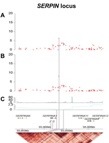
In a recent genome-wide association study for ANCA associated vasculitis performed in cases and controls from Northern Europe, SERPINA1 was confirmed as one of the most prominent genes associated with GPA and PR3 ANCA vasculitis. Indeed, the SNP with the strongest P-value (rs7151526) was located upstream of the
SERPINA1 gene and in linkage disequilibrium with the Z allele. The absence of a
significant association with the disease independently of Z allele indicates that the causal variant is either the Z allele itself or another variant strongly associated with the Z allele (Figure 6)52.
Figure 6: The SERPINA1 locus shows differential associations with anti-PR3 positive patients. A – Association of SNPs at the SERPIN locus with all ANCA associated vasculitis, B – with anti-PR3 positive cases only. C – The genomic architecture of the SERPIN locus indicates cumulative genetic distance from the most associated SNP and haplotype block structure. The grey lines indicate the genomic location of the most associated SNP together with the defining S and Z alleles. (Adapted from Lyons et al.52)
1.4 SERPINA2
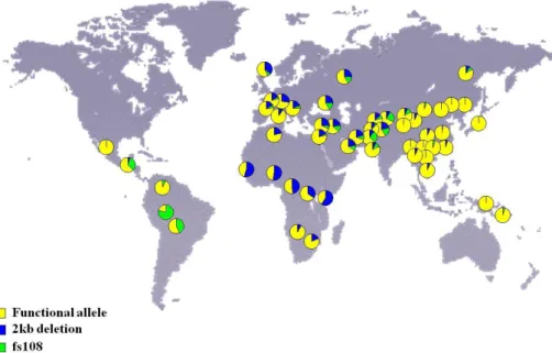
The nearest SERPINA1 neighbours are positioned 52 kb upstream (SERPINA11) and 12 kb downstream (SERPINA2). The latter has a high DNA sequence similarity to SERPINA1 but it has been regarded as a pseudogene due to a 2kb deletion encompassing exon IV and part of exon V53.However, an active isoform of SERPINA2 segregates within human populations and it has been shown to be expressed in vitro and in vivo in leukocytes54. In addition, the active SERPINA2 is conserved in primates, where it originated by gene duplication from SERPINA1 and diverged into a SERPIN with a distinct inhibitory activity. In SERPINA2, the P1 residue of the RCL (P1-P1’) is a tryptophan instead of a methionine54.
The frequency of the active SERPINA2 largely differs across human populations and it is predicted to vary between Europeans and Africans. While in
Europeans the active isoform is more frequent in African and Amerindians the disrupt
SERPINA2 form is the most prevalent (Figure 7)53. Furthermore, several genetic
variants were identified in the full SERPINA2 including premature stop codons (Leu108framStop and Leu277framStop) and four amino acid replacement variants (Ile280Thr, Leu308Pro, Glu320Lys and Pro387Leu). Previously the Leu308Pro and the Pro387Leu were predicted to alter protein structure based on computational tools53. However, no clear differences were detected between three SERPINA2 variants (V1: Pro308-Lys320; V2: Leu308-Glu320; and V3: Pro308-Glu320) used for the transfection of mammalian cell lines54.
Figure 7: Distribution of SERPINA2 non-functional alleles in 52 human populations. (Human Genome Diversity Panel samples)
The current work will focus on the characterization of SERPINA1 rare variants underlying cases of AATD in Portugal and on the analysis of the functional repercussions, haplotype background and geographical distribution of different
SERPINA1 variants. For this purpose we took advantage from our sample collection
comprising a few dozens of AATD cases caused by rare SERPINA1 alleles and from publically available databases of genome variation. We combined the DNA sequencing of SERPINA1 with the genotyping of two flanking microsatellites to assess the molecular basis of each rare allele as well as their intrahaplotipic variability. In addition, we compile the published information from SERPINA1 variation and from 1000Genomes and NHLBI GO Exome Sequencing Project to evaluate the distribution of rare alleles in human populations and correlate the level of each mutation with pathogenicity and SERPINA1 patterns of residue conservation.
In a second part of the work, we will explore the hypothesis of SERPINA2 as a potential candidate gene for the observed genetic association with GPA. Theoretically, a non-functional SERPINA2 variant could contribute to a higher susceptibility to GPA by increasing the chance of bacterial infections (unopposed bacterial proteases) or by an uncontrolled activity of endogenous proteases. To achieve our goal, we started by increasing the density of sequence variants within SERPINA2 in both GPA cases and controls (ZZ individuals without the disease) on one hand, and on the other hand by further investigating the inhibitory properties of SERPINA2. To this end, we expressed SERPINA2 in novel host cell models (HEK293 and Schneider S2 cells).
3.1 Severe AATD by rare SERPINA1 variants
3.1.1 Samples
Our sample included 51 unrelated individuals and a few relatives (father, mother and/or siblings) requested to perform SERPINA1 genotyping after a first screening of AATD by quantitative analysis of serum levels (radial immunodiffusion or nephelometry). The SERPINA1 genotyping was done previously as a part of the AATD diagnostic service at IPATIMUP, which combines the serum protein analysis by isoelectric focusing55 and the analysis of common mutations (Arg101His, Ala213Val, Glu264Val and Glu342Lys) by multiplex PCR56. All cases were found to carry rare
SERPINA1 variants. Blood samples were collected using EDTA as anticoagulant and
then frozen separately as serum and cellular fraction (leukocytes and erythrocytes).
3.1.2 DNA extraction
DNA was isolated from the frozen blood cellular fraction. DNA was extracted using the Generation Capture Column Kit (Qiagen) according manufacturer’s protocol.
3.1.3 PCR and sequencing
The DNA amplification was done in three different PCR reactions as illustrated in Figure 8. Briefly, SERPINA1 gene was subdivided into three different fragments of about 2.6 kb (F1) and 2.7 kb (F2 and F3). The first fragment comprised the promoter region spanning from exons IA to IC, the second fragment included exons II and III, and finally the third fragment included exons IV and V. Fragment 2 and 3 had a small overlap in intron III.
Amplification of SERPINA1 fragments from genomic DNA was done by long PCR using the cycling conditions described in Table A1 (Appendix) and the following reagents: 0.5 µM of forward and reverse primers, 200 µM of dATP, 200 µM of dTTP, 200 µM of dGTP, 200 µM of dCTP, 4% of DMSO, 1.75mM of MgCl2, 1U of Long PCR
Enzyme Mix (Thermo Scientific) and 10x Long PCR Buffer and approximately 120ng of DNA. The sequencing of the three gene fragments was done using ABI BigDye Terminator version 3.1 cycle sequencing chemistry (Life Technologies), and electrophoresis analysis was done on an ABI 3130 automated sequencer. All sequences were assembled and analysed using the Phred-Phrap-Consed package57. All putative polymorphisms and software-derived genotype calls were visually inspected and were individually confirmed using Consed. Details about sequencing primers are presented in Table A1 (Appendix).
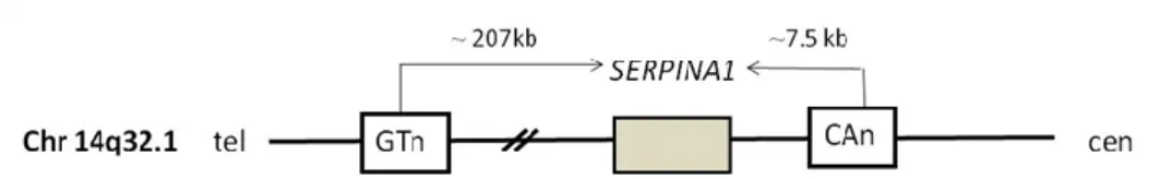
3.1.4 PCR and Microsatellite analysis
Haplotype characterization of the SERPINA1 rare alleles included the analysis of two different microsatellites. A CAn repeat located 7.5 kb downstream of SERPINA1 and a GTn repeat located 207 kb upstream of SERPINA1, as showed in Figure 9. The amplification of the two microsatellites was done using fluorescently labelled primers as previously described33. Microsatellite amplicons were separated by electrophoresis in a 3130 ABI Sequencer and the analysis was done using Gene Mapper software (Life Technologies).
Figure 8: Schematic representation of SERPINA1 amplification. Upper lines show the SERPINA1 orientation in chromosome 14 long arm and lower lines shows the structure of the gene where exons are represented as full boxes and introns by lines. Large arrows indicate the regions surveyed for sequence variation (F1-F3).
Figure 9: Location of the two microsatellites used in the haplotipic characterization of SERPINA1 rare alleles.
3.1.5 Data analysis
Haplotypes were inferred using the program PHASE 2.058, 59. To improve haplotype inference for microsatellite data we used haplotypes derived from two-generations studies of Portuguese families33.
3.1.6 Characterization of Q0
Faroallele
The synthesis of cDNA was performed by reverse transcription-polymerase chain reaction (RT-PCR) using the Superscript II RT-PCR system (Life Technologies, Gibco, BRL) and the manufacturers recommended conditions.
Then we performed a PCR reaction to further elucidate the basis of the Q0Faro
null allele using different primer combinations: IA/IIR; IC/IIR; IA/IIIR. The sequence of the primers are: IA, 5’- TCCTGTGCCTGCCAGAAGAG-3’; IC, 5’-ATCAGGCATTTTGGGGTGACT-3’; IIR, 5’- CCACTAGCTTCAGGCCCTCGCTGAG -3’ IIIR, 5’- GATGATATCGTGGGTGAGAACATTT-3’.
The cycling conditions for PCR were:
- 94ºC 2min
- 94ºC 10s, 58ºC 10s, 68ºC 2min 30s (10 cycles)
- 94ºC 10s, 54ºC 10s, 68ºC 2min 30s plus 3s per cycle (30 cycles)
- 68ºC 20min.
Digestion
The DNA digestion was done during 20min at 37ºC with 1U of enzyme (RsaI and ECO91I; Thermo Scientific) per 1µL of amplified products.
3.1.7 SERPINA1 conservation
Ortholog cDNA sequences for SERPINA1 were retrieved from the National Center for Biotechnology Information database (NCBI) (http://www.ncbi.nlm.nih.gov) and Ensembl (http://www.ensembl.org/) for the following mammalian species: human (Homo sapiens), common chimpanzee (Pan troglodytes), gorilla (Gorilla gorilla), orangutan (Pongo abelii), northern white-cheeked gibbon (Nomascus leucogenys), rhesus macaque (Macaca mulatta), baboon (Papio anubis), marmoset (Callithrix
jacchus), mouse (Mus musculus), rat (Rattus norvegicus), dog (Canis familiaris), cat
(Felis catus), cow (Bos taurus), pig (Sus scrofa), sheep (Ovis aries) opossum (Monodelphis domestica) and zebrafish (Danio rerio) (Table 2).
We used CLUSTALW60 implemented in the MEGA561 software to align cDNA sequences. SERPINA1 alignments were used to construct phylogenetic trees using neighbour-joining method with 10000 bootstraps implemented in MEGA5. The ratio of non-synonymous and synonymous substitution rates (dN/dS = ω) was estimated using
the maximum likelihood (ML) framework implemented in the program CODEML of Phylogenetic Analysis by Maximum Likelihood (PAML) software62. We used the site model test M3 (discrete selection model) to investigate the conservation and selective pressures that have shaped the evolution of SERPINA1. This model adopts 3 categories of codon positions (sites) and assumes an unconstrained discrete distribution to model heterogeneous ω values among sites and detect codons evolving under different selective forces63. While values of ω>1 are considered as evidence of positive selection, values of ω<1 are regarded as proof of purifying selection (conservation). The Naive Empirical Bayes (NEB) approach is implemented to calculate the posterior probability for each amino acid site and detect conserved, neutral and positive selected codons62.
Table 2: Accession numbers for SERPINA1 cDNA sequences.
3.2 SERPINA2
3.2.1 Samples
DNA samples from ZZ subjects were collect from different populations. These included 24 samples from Portugal, with a diagnosis of emphysema, COPD or AATD disease, 20 samples from Birmingham (England) with a diagnose of emphysema or COPD, and 10 samples from GPA patients, 5 from Birmingham and 5 from Bochum (Germany).
3.2.2 PCR and DNA sequencing
The SERPINA2 was amplified in 3 fragments (Figure 10). The first comprising only the exon II, the second containing only exon III and the last fragment spanning exon IV and exon V. The amplification of the 3 fragments from the genomic DNA was carried using the cycling conditions described in Table A2 (Appendix) and the following reagents: 0.5 µM of each oligonucleotide, 200 µM of dATP, 200 µM of dTTP, 200 µM of
Species Accession Number Database
Homo sapiens NM_000295.4 NCBI
Pan troglodytes ENSPTRT00000045369 Ensembl Gorilla gorilla ENSGGOT00000007925 Ensembl Nomascus leucogenys XM_004091842 NCBI
Macaca mulatta ENSMMUT00000039542 Ensembl Callithrix jacchus ENSCJAT00000061674 Ensembl
Papio anubis XM_003902213 NCBI
Mus musculus ENSMUST00000085056 (serpina1a)
1 2 3 ENSMUST00000164454 4 5 Ensembl ENSMUST00000164454 (serpina1b)
Rattus norvegicus ENSRNOT00000012577 Ensembl Canis familiaris ENSCAFT00000036554 Ensembl
Felis catus XM_006933076.1 NCBI
Bos taurus NM_173882 NCBI
Sus scrofa ENSSSCT00000002750 Ensembl Ovis aries ENSOART00000016196 Ensembl Monodelphis domestica ENSMODT00000033265 Ensembl
dGTP, 200 µM of dCTP, 1U of Hot Star Taq (DNA polymerase from Qiagen), 10x buffer and about 100ng of DNA reaching 25 µL of final volume. After the amplification was completed the sequencing of the coding regions was done using the primers presented in Table A2 (Appendix).
3.2.3 Cloning of SERPINA2
The cDNA corresponding to the V2 variant (Leu308-Glu320) of SERPINA2 from a previous SERPINA2/pLenti6V5 construct54 was amplified and fused to a stable His-tag using the proofreading polymerase (Thermo Scientific), and specific primers (Fw: 5’TTCCTGATGT^TCATCGCTTTCGTCATCATCGCTGAGGCCGAGGATCCCCAGGG AGATGCTGCCCA3’; Rv: 5’CTACTGGCCA^ACCAACCCACCCTAAGTGGTGAA3’). The PCR product was then digested with BsaBI and AgeI enzyme (Bio Labs), respectively, using recommended conditions. The ligation of the insert into the pIEX5 vector (Novagen) was done with T4 DNA ligase (Bio Labs). Later the SERPINA2/pIEX5 construct was used in the SERPINA2 subcloning into the pTT5 vector (collaboration partner) using BamHI enzyme (Bio Labs).
Figure 10: Schematic representation of SERPINA2 amplification. Upper lines show SERPINA2 orientation in chromosome 14 long arm cluster and lower lines the structure of the gene where exons are represented as full boxes and introns by lines. Large arrows indicate the regions surveyed for sequence variation (F1-F3).
3.2.4 Transfection and protein extraction
The expression of SERPINA2 was done in two biological systems. The first SERPINA2/pIEX5 construct was used in the transfection of the Schneider S2 cells from
Drosophila (Xiao Shell, EPFL, Lausanne) and the SERPINA2/pTT5 construct used for
the transfection of human HEK293 cells.
HEK293 cells (ATCC number CRL-1573) were grow in FreeStyleTM medium (Life Technologies) containing 24µg/ml of G418 (PAA Company) and 0.1% of Pluronic F68 (Life Technologies). The vector was transfected into HEK293 with OptiProTM SFM (Life Technologies) and polyethylenimine (PEI) (Sigma). After twenty four hours of transfection Bacto TC Lactalbumin Hydrolysate (BD Biosciences) was added for a final concentration of 0,5%.
After ninety six hours of transfection, expressed SERPINA2 in HEK293 cells was collected from cultured cell supernatants and concentrated in Amicon ultra centrifugal filters unit (EMD Millipore) with 1x elution buffer (EB) (20 mM Na2HPO4, 500
mM NaCl pH 7,4) supplemented with 10mM imidazole. SERPINA2 was purified on nickel columns using the Ni-NTA Spin Kit (Quiagen), 1(x)EB with increase imidazole concentrations starting from 250mM. The purification of the supernatant from Schneider S2 cells started with overnight dialysis in 1(x)EB 10mM imidazole and then according to the protocol from Äkta Prime Plus from GE Healthcare and using the same buffer for sample loading and for elution a imidazole gradient up to 1M. Then collected 13 purified fractions, and after that concentrated the elution fractions of interest in Amicon ultra centrifugal filters unit (EMD Millipore) using 1(x)EB without imidazole.
3.2.5 Western blot
The total protein concentration of cell supernatants was determined by Bio-Rad protein assay. Protein (approximately 5 µg) were mixed into 10(x)gel loading buffer, heated at 95ºC for ~5min, and separated by SDS-PAGE (12% poly-acrylamide). For the immunodetection of SERPINA2, the protein was transferred to a nitrocellulose membrane, blocked in phosphate buffer solution with Tween 20 (PBS-T) and 5% blocking non-fat milk solutions, and then probed with two antibodies. A first antibody against SERPINA2 G-12 (Santa Cruz Biotechnology) diluted in PBS-T at a 1:500 ratio, and another against the stable His-tag anti-His 3D5 diluted at a 1:2500 ratio (Dr.
Elisabeth Kremmer, HMGU). In the first anti-IgG HRP (Chemicon) diluted at 1:1000 and in the second case the secondary antibody goat anti-mouse HRP (Pierce) diluted at a 1:5000 ratio was used.
4.1 SERPINA1 mutational spectrum
4.1.1 Rare alleles causing AATD
The sequencing study of 51 cases of AATD allowed the identification of 13 mutations in SERPINA1 gene distributed by 14 rare deficient or null alleles (Table 3)32. All cases of AATD were previously analysed by isoelectric focusing (IEF), which warranted the evaluation of the mobility of the rare allele in the protein gel electrophoresis, as well, as the comparison of its band intensity with non-deficient alleles (M1, M2, M3 and M4) .
Among the 13 mutations identified, 5 were novel (Glu162Gly, Leu263Pro, Arg281framStop297, Met374framStop392 and IVSIC+3Tins) and described for the first time in the Portuguese population (Table 3). Therefore, those alleles carrying the novel mutations were named accordingly to the protein pattern in IEF gels and the place of birth of the index case. Importantly, three of these rare alleles were found in the index case in heterozygosity with common deficient alleles (S and Z) confirming the clinical classification as cases with severe AATD.
Table 3: Rare alleles of SERPINA1associated with AATD identified in the current work
Allele Mutation Molecular base Relative
frequency Previously described
MMalton Phe52del M2 17/51(0.333)
MPalermo Phe52del M1Val213 9/51(0.176)
I Arg39Cys M1Val213 7/51(0.137)
Q0Ourém L353fsX376 M3 4/51(0.078)
PLowell Asp256Val M1Val213 3/51(0.059)
MHerleen Pro369Leu M1Val213 3/51(0.059)
MWurzburg Pro369Ser M1Val213 1/51(0.020)
Q0Lisbon Thr68Ile M1Val213 1/51(0.020)
In addition, two other rare mutations were identified in the course of this sequencing study (Ser47Arg and Val302Ile) however, none of them could be associated to AATD.
4.1.1.1 Novel mutations
4.1.1.1.1 Amino Acid substitutions
Leu263Pro (Q0Gaia)
The variant Q0Gaia is characterized by a Leu(CTG)Pro(CCG) mutation at 263
residue, which was identified in heterozygosity with a Z allele in two siblings with lung disease. This mutation is likely to have serious repercussions in SERPINA1 structure has indicated by the absence of a corresponding band in protein gel electrophoresis. The Leu263 residue is highly conserved among SERPINA1 orthologs (placental mammals) (Figure 11) and within the SERPIN superfamily LeuPro and ProLeu substitutions are known to cause drastic alterations in the three dimensional structure18. Interestingly, the Leu263Pro is next to the residue Glu264, which underlies one of the common deficiency alleles when replaced by a valine residue (S allele). Likewise, the substitution of Leu263Pro is also likely to cause the distortion of the G α-helix (Figure 12) and affect the gate domain as it happens with Glu264Val substitution18.
Allele Mutation Molecular base Relative frequency
Novel mutations
Q0Gaia Leu263Pro M1Ala213 1/51(0.020)
PGaia Glu162Gly M1Val213 1/51(0.020)
Q0Oliveira do Douro Arg281framStop297 M3 1/51(0.020)
Q0Vila Real Met374framStop392 M3 1/51(0.020)
Q0Faro IVSIC+3Tins M1Val213 1/51(0.020)
Glu162Gly (PGaia)
The variant PGaia is characterized by a Glu(GAG)Gly(GGG) mutation at 162
codon, which was firstly identified by a distinctive mobility band in protein gel electrophoresis (Figure 13). This variant was identified in a single patient with lung emphysema in heterozygosity with an S allele. The Glu162 residue is located in the SERPINA1 F α-helix (Figure 12) and it is highly conserved among SERPINA1 orthologs (Figure 11). Moreover, bioinformatics predictions by Polyphen-2 (http://genetics.bwh.harvard.edu/pph2/) suggest the Glu162Gly substitution as a damaging mutation possibly affecting SERPINA1 structure.