ORIGINAL ARTICLE
CITE AS: Pereira, D.L.; da-Cunha, A.P.S.; Cardoso, C.R.P.; da-Rocha, C.Q.; Vilegas, W.; Sinhorin, A.P.; Sinhorin, V.D.G. 2018. Antioxidant and hepatoprotective effects of ethanolic and ethyl acetate stem bark extracts of Copaifera multijuga (Fabaceae) in mice. Acta Amazonica 48: 347-357.
Antioxidant and hepatoprotective effects of
ethano-lic and ethyl acetate stem bark extracts of Copaifera
multijuga (Fabaceae) in mice
Débora Linsbinski PEREIRA1, Ana Paula Simões DA CUNHA2, Cássia Regina Primila CARDOSO2,
Cláudia Quintino DA ROCHA3, Wagner VILEGAS4, Adilson Paulo SINHORIN1,
Valéria Dornelles Gindri SINHORIN1*
1 Universidade Federal de Mato Grosso, Instituto de Ciências Naturais, Humanas e Sociais, Programa de Pós Graduação em Ciências Ambientais (PPGCAM), Campus
de Sinop, Mato Grosso, Brazil
2 Universidade Federal de Mato Grosso, Instituto de Ciências da Saúde,Campus de Sinop, Mato Grosso, Brazil 3 Universidade Federal do Maranhão, Departamento de Química,Centro Tecnológico, Maranhão, Brazil 4 Universidade Estadual Paulista Júlio de Mesquita Filho, Unidade São Vicente, São Paulo, Brazil
* Corresponding author: [email protected]
ABSTRACT
The properties of oil-resin of copaiba, Copaifera multijuga are commonly mentioned in the literature, but there are few studies
on extracts from its stem bark. We evaluated the antioxidant effectsofethanolic (EE) and ethyl acetate (EA) crude stem bark
extracts from copaiba and compared them to rutin in a paracetamol (PCM)-induced oxidative stress model inmice.All test
comparisons differed significantly. Hepatic catalase (CAT) and glutathione-S-transferase (GST) activity decreased in the PCM
group, and there was an increase of protein carbonyls in the liver, kidney and brain. However, the protein carbonyls decreased in the liver for the PCM + EE group, in the kidneys for the PCM + EA and PCM + Rutin groups, and in the brain for all treatments. Hepatic GSH decreased in the PCM group and increased in the PCM + EE group. The extracts showed a positive effect on ascorbic acid (ASA), since they were able to restore the levels of parameters that had been changed by PCM. There was
an increaseof ALT and AST activity in the plasma within the PCMgroup. Even thoughALT decreased in the PCM + Rutin,
PCM + EE and PCM + EA groups, EE and EA did not have an effect on AST. The strongest antioxidant effect was observed
for EE, due to the presence of the phenolic compounds epicatechin and epiafzelechin, as well asthe highest concentration of
total phenols and an excellent antioxidant potential observed in the DPPH· test.
KEYWORDS: oxidative stress, acute intoxication, acetaminophen, copaiba
Efeitos antioxidante e hepatoprotetor dos extratos brutos etanólico e
acetato de etila da casca do caule da Copaifera multijuga (Fabaceae) em
camundongos
RESUMO
As propriedades do óleo-resina da copaíba, Copaifera multijuga são comumente citadas na literatura, mas há poucos estudos sobre
extratos da casca do caule. Avaliamos os efeitos antioxidantes de extratos brutos etanólico (EE) e acetato de etila (EA) da casca do caule da copaíba e os comparamos à rutina no modelo de estresse oxidativo induzido por paracetamol (PCM) em camundongos. Todas as comparações de teste diferiram significativamente. A atividade da catalase hepática (CAT) e da glutationa-S-transferase (GST) diminuiu no grupo PCM, e houve um aumento de proteínas carboniladas no fígado, rim e cérebro. No entanto, as proteínas carboniladas diminuíram no fígado para o grupo PCM + EE, nos rins para os grupos PCM + EA e PCM + rutina, e no cérebro para todos os tratamentos. A GSH hepática diminuiu no grupo PCM e aumentou no grupo PCM + EE. Os extratos mostraram um efeito positivo sobre o ácido ascórbico (ASA), uma vez que foram capazes de restaurar os níveis dos parâmetros que foram alterados pelo PCM. Houve um aumento da atividade de ALT e AST no plasma dentro do grupo PCM. Embora a ALT tenha diminuído nos grupos PCM + rutina, PCM + EE e PCM + EA, EE e EA não afetaram a AST. O efeito antioxidante mais forte foi observado para o EE, provavelmente devido à presença dos compostos fenólicos epicatequina e epiafzelequina, assim como à maior concentração de fenóis totais e um excelente potencial antioxidante observado no teste DPPH·.
INTRODUCTION
The Brazilian National Toxic-Pharmacological Information System (SINITOX) cites inappropriate use of medications as the main cause of human poisoning in Brazil (Brasil 2011), which may be due to self-medication, lack of knowledge about the toxicity of drugs, or overintake for suicidal purpose. Paracetamol (PCM) is an analgesic and antipyretic and is one of the most widely used drugs, being considered safe at
therapeutic doses (Ozcelik et al. 2014). However, overdosage
of PCM can cause hepatotoxicity associated with centrilobular
necrosis (Hinson et al. 2010) and is often associated with renal
damage (Ghosh and Sil 2007). The hepatotoxic effect of high doses of PCM is caused by the reactive metabolite N-acetyl-p-benzoquinoneimine (NAPQI), which oxidizes cellular thiols
(Basu et al. 2014). High concentrations of NAPQI reduce
glutathione (GSH) levels and cause oxidative stress in the liver
(Kisaoglu et al. 2014) and kidney (Manimaran et al. 2010).
The normal cell physiology or the redox homeostasis of mammalian cells is maintained by the endogenous antioxidant systems and by the absorption of dietary exogenous antioxidant agents (Pisoschi and Pop 2015). Medicinal plants and nutraceuticals or functional foods can be important sources of exogenous antioxidant compounds,
such as phenolic compounds, β-carotene and ascorbic
acid, which can aid endogenous antioxidant systems in preventing oxidative damage generated by excessive free radical production (Degáspari and Waszczynskyj 2004). The antioxidant activity exerted by phenolic compounds present in medicinal plants can decrease the citotoxicity of free radicals, preventing oxidative stress, cell damage and pathologies (Degáspari and Waszczynskyj 2004). In this context, the use of herbs and dietary supplements has increased greatly due to their purported beneficial effects on human health (Perlman
et al. 2013).
Copaifera (Fabaceae) is a genus of tree species found throughout the tropics of Latin America, popularly known
in Brazil as copaíba or pau-de-óleo (Leandro et al. 2012).
Copaiba oil-resin contains of diterpenic acids and great amounts of sesquiterpenes (Veiga and Pinto 2002) and has
been associated with anti-inflammatory (Paiva et al. 2004;
Pieri et al. 2009; Veiga Junior et al. 2007; Gomes et al. 2010),
topical analgesic (Carvalho et al. 2005), gastroprotective
(Paiva et al.1998), antinociceptive (Gomes et al. 2007) and
antitumoral activity (Gomes et al. 2008).
Copaifera multijuga Hayne is native to the Amazon region (Costa 2018). Its oil-resin is composed up to 90% of hydrocarbons and oxygenated sesquiterpenes to a lesser extent, also including a small amount of acidic diterpenes, and
presents anti-inflammatory and anti-tumor properties (Lima et
al. 2003; Gomes et al. 2008). The stem bark of C. multijuga is
widely used in popular medicine, specially in northern Brazil,
as a tea or in topical use, to treat ailments such as gastritis,
inflammation, sore throat and stroke (Vásquez et al. 2014).
The overdosage with PCM can deplete glutathione and release proinflammatory agents, for instance, inflammatory
cytokines (Karthivashan et al. 2015). Considering the
wide-spread use of C. multijuga for treating inflammatory processes,
we hypothesized that the stem bark extract from this species can exhibit antioxidant activitiy against PCM-induced oxidative stress. Thus, we aimed at evaluating the potential antioxidant effects exerted by ethanolic and ethyl acetate
extracts from the stem bark of Copaifera multijuga against
acute acetaminophen intoxication in mice.
MATERIAL AND METHODS
Plant materials and botanical identification
The stem bark from Copaifera multijuga was collected from
one tree in Guarantã do Norte, state of Mato Grosso, Brazil (9°48’31.0’’S, 54°53’18.0”W). The specimen was identified by Ms. Ivani Kuntz Gonçalves from the Universidade Federal de Viçosa, and Dr. Haroldo Cavalcante de Lima from the Instituto de Pesquisa Jardim Botânico, Rio de Janeiro. A voucher specimen was deposited in the Herbarium of the Universidade Federal de Mato Grosso (UFMT), Sinop Campus (in Acervo Biológico da Amazônia Meridional – ABAM, Sinop-MT, number 4801).
Extract preparation and chemical analyses
The extracts were obtained from 1.26 kg of stem bark dried
at 40 oC and crushed. The extraction process occurred by
maceration in three stages: first the material was macerated with 2 L hexane that yielded crude hexanic extract (1.65 g), second the material was macerated with 4 L ethyl acetate that yielded crude ethyl acetate extract (EA) (5.15 g), and third the material was extracted with 12 L ethanol that yielded crude ethanolic extract (EE) (220.46 g). In order to remove the solvent and concentrate the extract, samples were rotary evaporated under reduced pressure (600 mm Hg) at 40 °C. The EE and EA extracts were used in the biological studies because they had a greater amount of polar components than the hexane fraction.
Flow injection analysis (FIA) was performed using a Thermo Finnigan Fisher (San Jose, CA, USA) mass spectrometer equipped with an electro-spray ionization source, ion-trap analyzer and Xcalibur software for data processing (ESI-MS/MS) LTQXL. All lyophilized infusions were diluted
and homogenized in MeOH:H2O (8:2 v/v) at 5 ppm. The
mass spectrometry analysis was performed following the
methodology of Mesquita et al. (2017). The MS and MSn
analyses in negative ion mode were selected at a flow rate
of 5 μL·min−1 and worked under the following conditions:
flow rate 8 (arbitrary units). Mass spectra were recorded in the range m/z 100–2000 Da. The first event was a full scan mass spectrum to acquire data on ions in the m/z range. The second scan event was an MSn experiment performed using a data-dependent scan running on deprotonated molecules from the compounds at collision energy of 20%–30% and activation time of 30 ms.
The quantity of total phenols in EE and EA was determined
by the “Folin-Ciocalteu” reaction according to Woiskyand
Salatino (1998). The calibration curve was made with gallic
acid (0, 1.5, 2, 2.5, 3, 3.5, 4 μg mL-1), 4 mL of Folin-Ciocalteu
reagent (2 mol L-1) and 6 mL of 20 % sodium carbonate in 50
mL of distilled water. EE and EA were solubilized in methanol
solutions to a final concentration of 500 mg mL-1. Aliquots
of 0.375 mL of the extracts, 4 mL of Folin-Ciocalteu reagent
(2 mol L-1) and 6 mL of 20 % sodium carbonate were added
to 50 mL of distilled water. After 2 hours of reaction, we performed a reading in a spectrophotometer at 760 nm. The result was compared to the calibration curve and expressed
in mg gallic acid equivalent per gram of extract (mgGAE g-1).
The quantity of total flavonoids in EE and EA was
determined by the reaction with aluminum chloride (AlCl3)
according to Woisky and Salatino (1998), with some
adaptations. A calibration curve was constructed using methanolic solution of quercetin in concentrations of 0.25,
0.50, 1.0, 1.5, 2.5, 3, 3.5, 4, 4.5 mg mL-1 and 50 mg of AlCl
3
were added to each curve point. The EA and EE samples
were prepared with 100 μg mL-1 of the extracts and AlCl
3 (50
mg). After 30 minutes of reaction, the samples were read in a spectrophotometer at 420 nm. The result was compared to the calibration curve and expressed in mg of quercetin equivalent
per gram of extract (mgQE g-1).
To evaluate the antioxidant potential, we followed the
methodology of Sousa et al. (2007), with some adaptations.
We carried out the monitoring of free radical DPPH· consumption in the UV-Vis spectrophotometer at a wavelength of 515 nm. The standard rutin and ascorbic acid, as well as the samples of EE and EA, were diluted in methanol at
concentrations of 2.5, 5.0, 10.0, 15.0, 20.0, 25.0 μg mL-1 and
added DPPH· (36 μg mL-1), and read in a spectrophotometer
after 30 minutes of reaction.
The effective concentration (EC50) required and the
amount of antioxidant to scavenger 50 % of the initial DPPH· were determined from a first-order exponential curve and absorbance values at all concentrations of the tested samples (EE and EA) and standards (rutin and ascorbic acid) were converted to a percentage of antioxidant potential (% AP) as
described by Souza et al. (2007).
Animals and treatment
Male Swiss mice, with an average weight of 33.4 g, were
obtained from UFMT/Cuiabá. The animals were divided into
8 groups of 8 individuals each, and were acclimated for 20 days with a photoperiod of 12 hours light/dark, temperature
of 24 ± 1 °C, relative humidity of 51 ± 2 %, fed pelleted feed
diet (Purina) and filtered water ad libitum.
After the acclimation period, the following treatments were established by oral administration: Group 1- Control
(water); Group 2 - Rutin (antioxidant, 6 mg kg-1); Group 3
– paracetamol (acetaminophen - PCM, 250 mg kg-1); Group
4 - PCM + Rutin; Group 5 - PCM + EE; Group 6 - PCM +
EA; Group 7 - EE (250 mg kg-1); Group 8 - EA (250 mg kg-1).
The selected dosage of PCM (250 mg kg-1), as well as
the damage induction protocol, followed Olaleye and Rocha
(2008), while the rutin dosage (6 mg kg-1) followed Hort et
al. (2008), and the EE and EA dosages (250 mg kg-1) were
established according to the Malone Hippocratic test (Malone 1983). The experiment started by using PCM or water orally (via gavage): G3, G4, G5 and G6 groups received PCM 250
mg kg-1; and G1, G2, G7 and G8 groups received only water.
After 3 hours of induction of injury with PCM, the groups were treated orally (via gavage) with: G2 and G4 received
rutin (6 mg kg-1 in 0.01 % Tween); G5 and G6 received EE
and EA extract, respectively (250 mg kg-1 in 0.01 % Tween);
G7 and G8 received EE and EA extract, respectively (250 mg
kg-1 in 0.01 % Tween); and G1 and G3 received only water
with 0.01 % Tween.
In a treatment period of seven days, the treatment groups received either rutin or extracts, and the control groups (G1 and G3) received drinking water + 0.01 % Tween once daily. After 24 hours of the last dose of treatment, the animals
were anesthetized with ketamine 50 mg kg-1, xilazine 2 mg
kg-1 and acepromazine 2 mg kg-1 (i.p.). Heart puncture was
performed to collect blood with heparinized syringes. The tubes were centrifuged to obtain clear plasma samples, which were used for the analyses of ALT and AST. The animals were sacrificed, and their liver, kidneys and brain were removed and stored at -85 °C. The study was authorized by protocol # 23108.781869/12-0 of the Ethics Committee on Animal Research (CEPA), UFMT/Cuiabá.
Biochemical analysis
Superoxide dismutase (SOD) activity in the liver was measured according to Misra and Fridovich (1972). The result was
expressed in UI SOD mg protein-1. Liver, brain and kidney
catalase (CAT) activity was determined according to Nelson
and Kiesow (1972). The change in absorbance of H2O2 in 60
seconds was measured in a spectrophotometer at 240 nm and
expressed in μmol H2O2 min-1 mg protein-1. The activity of
glutathione-S-transferase (GST) in the liver was determined
according to Habig et al. (1974). The result was expressed
as μmol GS-DNB min-1mg protein-1. The molar extinction
coefficient was used for CDNB 9.6 mM cm-1. Reduced
result was expressed in μmol GSH mg protein-1. The formation
of thiolate anion was determined at 412 nm and compared to a standard curve of GSH. The levels of ascorbic acid (ASA) in the liver and brain were determined according to Roe (1954).
The result was expressed in μmol ASA g-1 tissue. The protein
carbonyl assay was performed in liver and kidney according
to Yan et al. (1995) with some modifications. The absorbance
was determined at 370 nm. The amount of protein carbonyl
was expressed in nmol carbonyl mg protein-1.
The protein content for SOD, CAT, GST, GSH and protein carbonyl was determined following the method by Bradford (1976) using bovine serum albumin as the standard for construction of the calibration curve. The samples were read at 595 nm.
Dosages of the activities of alanine aminotransferase (ALT) and aspartate aminotransferase (AST) present in plasma were
performed with commercial kits (Labtest®, Diagnostics SA,
Minas Gerais, Brazil).
Statistical analysis
The data were expressed as mean ± standard deviation (SD)
and analyzed by one way ANOVA followed by post hoc Tukey
test. The results were considered statistically significant at p < 0.05.
RESULTS
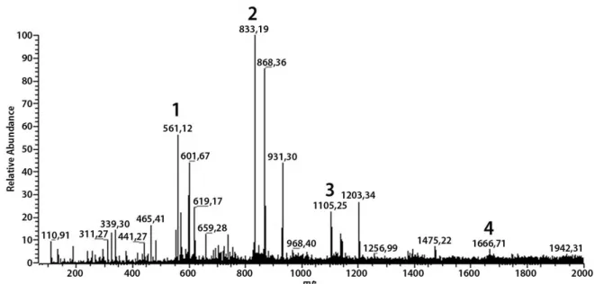
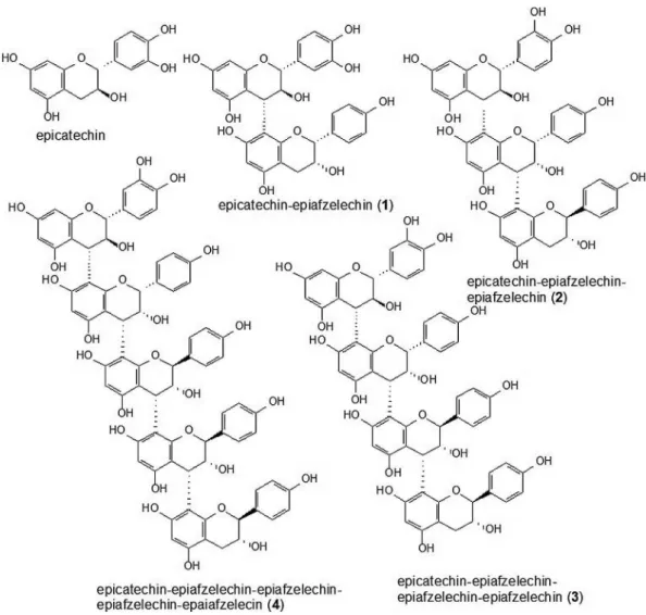
Five phenolic compounds were identified by mass spectrometry fingerprinting: (Epi) free chatechin and other four condensed tannins (Epichatechin-Epiafzelechin ((Epi) Cat-(Epi)Afz), compound 1; Epichatechin-Epiafzelechin-Epiafzelechin ((Epi)Cat-(Epi)Afz-(Epi)Afz), compound 2;
Epichatechin-Epiafzelechin-Epiafzelechin-Epiafzelechin ((Epi)Cat-(Epi)Afz-(Epi)Afz-(Epi)Afz), compound 3; and Epichatechin-Epiafzelechin-Epiafzelechin-Epiafzelechin (Epi)Cat-(Epi)Afz-(Epi)Afz-(Epi)Afz-(Epi)Cat, compound
4)(Figures 1 and 2).
The total phenolic compounds and flavonoids in EE
(643.43 mgGAE g-1 and 11.53 mgQE g-1, respectively) were
higher than those found in EA (187.21 mgGAE g-1 and 8.60
mgQE g-1, respectively) (Table 1). EE exhibited a higher
antioxidant potential with lower EC50 (23 μg mL-1), when
compared to the standard rutin and ascorbic acid (30 and
35 μg mL-1, respectively), as well as when compared to EA
(91 μg mL-1).
Hepatic SOD activity was not modified by the treatments (Figure 3A). However, PCM caused a significant decrease in CAT activity (22.8%). The extracts or rutin had no effect on the hepatic CAT activity levels. But rutin, EE and EA attenuated the decrease in CAT activity produced by PCM (Figure 3B). GST, an important enzyme in the detoxification of xenobiotics, showed a significant reduction (30.2%) of its activity in the PCM group in comparison to the control group (Figure 3C).
GSH, an important non-enzymatic antioxidant, decreased significantly by 22.8% in the PCM group when compared to the control group, and increased significantly by 35% in the PCM + EE group when compared to the PCM group (Table 2). The ascorbic acid level decreased by 16.2% in the PCM group when compared with the control group. However, the animals treated with PCM + EE or PCM + EA exhibited a significant increase of 18.2% and 26.1%, respectively when compared to the PCM group (Table 2). There was a significant
Figure2. Structure of the compounds identified by ESI-IT-MS in crude ethanolic extract from Copaifera multijuga stem bark.
Table 1. Analysis of total phenols, total flavonoids and effective concentration (EC50) of extracts of Copaifera multijuga stem bark. ASA: ascorbic acid; EE: ethanolic extract; EA: ethyl acetate extract; mg GAE/g: mg gallic acid equivalent per gram of extract; mg QE/g: mg of quercetin equivalent per gram of extract; EC50: effective concentration required and the amount of antioxidant to scavenger 50% of the initial DPPH·.
Samples Total phenols (mg GAE/g)
Total flavonoids (mg QE/g)
DPPH. EC
50 (µg/
mL)
EE 643.43 11.53 22.96
EA 187.21 8.60 91.13
Rutin --- --- 30.06
Ascorbic acid
(ASA) --- --- 35.74
Table 2. Effect of ethanolic (EE) and ethyl acetate (EA) extracts of Copaifera multijuga stem bark under oxidative stress induced by paracetamol/ acetaminophen (PCM) in liver tissue. Results are expressed as mean ± SD; n = 8 animals. a p< 0.05 compared with the control group; b p < 0.05 compared with
the PCM group by one way ANOVA followed by a Tuckeytest.
Treatments
GSH (µmol GSH mg
protein-1)
ASCORBIC ACID (µmol ASA g-1
tissue)
CARBONYL (nmol carbonyl
mg protein-1)
Control 164.50 ± 20.01 1.05 ± 0.10 1.70 ± 0.40
Rutin 130.30 ± 23.49 1.05 ± 0.10 2.06 ± 0.49
PCM 127.00 ± 22.31a 0.88 ± 0.10a 2.61 ± 0.49a
PCM + Rutin 145.10 ± 13.38 0.92 ± 0.09 1.98 ± 0.37
PCM + EE 171.40 ± 26.81b 1.04 ± 0.09b 1.46 ± 0.35b
PCM + EA 144.20 ± 28.52 1.11 ± 0.11b 2.07 ± 0.36
EE 134.50 ± 25.16 0.93 ± 0.06 2.31 ± 0.28
increase of 53.5% in protein carbonyls in the PCM group compared to the control group. PCM + EE significantly decreased by 44.1% the damage caused by the PCM (Table 2).
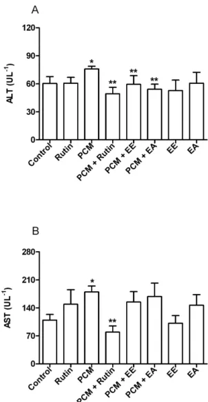
There was a significant increase in ALT in the plasma of animals treated with PCM (25.8%), when compared to the control group. However, the ALT levels in plasma in the PCM + Rutin, PCM + EE and PCM + EA groups decreased significantly (35%, 21.7% and 28.6%, respectively) when compared to the PCM group (Figure 4A). The plasma activity of AST significantly increased by about 65% in the PCM group when compared to the control group, and decreased significantly in the PCM + Rutin group (55.9%) when compared to the PCM group. In contrast to ALT, however, EE and EA did not reverse this change in AST activity (Figure 4B).
Figure3. Effect of ethanolic (EE) and ethyl acetate (EA) extracts of Copaifera multijuga stem bark under oxidative stress induced by paracetamol/ acetaminophen (PCM) in liver tissue: A – SOD; B – CAT; C – GST. Results are expressed as mean ± SD; n = 8 animals. * p < 0.05 compared with the control
group by one way ANOVA followed by a Tuckey test.
Figure4. Effect of ethanolic (EE) and ethyl acetate (EA) extracts of Copaifera multijuga stem bark under oxidative stress induced by paracetamol/ acetaminophen (PCM) in the plasma: A – ALT and B - AST. Results are expressed as mean ± SD; n = 7 animals. * p < 0.05 compared with the control group; ** p < 0.05
CAT activity was not altered in the kidney, but the non-enzymatic antioxidant GSH was significantly reduced by 43.3% in the PCM group when compared to the control. The positive control group (PCM) had an increase in protein carbonyls (71.3%) when compared to the control group. However, the PCM + EA and PCM + Rutin groups had a significant decrease in the carbonylation of 50.7% and 30.4%, respectively (Table 3).
CAT activity and GSH levels were not altered in the brains of mice exposed to PCM. However, the level of ASA was significantly increased in the EE group (by 42%) when
compared to the control group, showing an effect per se (Table
4). Protein carbonylation increased significantly by 28.5% in the positive control group (PCM) when compared to the control group, while the PCM + EE, PCM + EA and PCM + Rutin groups reduced this parameter (38.7%, 35.7%, 31.5%, respectively) when compared to the PCM group. In the EE group the carbonylation of proteins in the brain tissue was significantly reduced (by 42.3%), when compared to the control group (Table 4).
DISCUSSION
The effects observed in vivo in this study in the EE groups
can be related to the phenolic compounds detected in the EE
extract of C. multijuga, mainly the tannins (Shimoda et al.
2008). The presence of free (Epi) catechin at m/z 289 is in
accordance with Nuengchamnong et al. (2009). Condensed
tannins were also detected, such as dimers (compound 1 at m/z 561.12), the ion found at m/z 833.19 that corresponds to trimer compound 2, and the ion at m/z 1105.25 (which can be attributed to compound 3), all of them in line with
Souza et al. (2008). We suggest that m/z 1666.71 corresponds
to a tannin with higher polymerization (corresponding to compound 4), but no data was found in the literature for this compound, since most of the equipment used for this type of analysis does not detect compounds with track acquisition at 100–1500 m/z.
PCM is a drug that can cause toxicity at high doses by inducing oxidative stress in the liver and kidney. The toxic metabolite of PCM (NAPQI) is conjugated with reduced glutathione, thereby reducing the concentration of GSH. With the depletion of GSH, there is an increase in the formation of nitrogen and oxygen reactive species in the liver
and kidney (Stern et al. 2005; Hinson et al. 2010) thus causing
mitochondrial oxidative stress (Hodgman and Garrard 2012). The use of natural products containing antioxidant compounds, such as phenolic compounds, can effectively protect against the toxicity of the PCM metabolite (Jaeschke
et al. 2011). The ethanolic extract from Copaifera multijuga
stem bark showed higher concentration of total phenols and flavonoids, and significantly higher antioxidant potential than the ethyl acetate extract. This can be explained by the presence of phenolic compounds in the ethanolic extract, such as the tannins epicatechin and epiafzelechin, which were extracted in larger quantities by ethanol. In the chromatographic profile
of the hydroalcoholic extract from Copaifera langsdorffii
leaves at 257 nm, the presence of phenolic compounds
such as Quercetin-3-O-α-L-rhamnopyranoside (quercitrin)
and kaempferol-3-O-α-L-rhamnopyranoside (afzelin) was
Table 3. Effect of ethanolic (EE) and ethyl acetate (EA) extracts of Copaifera multijuga stem bark under oxidative stress induced by paracetamol/ acetaminophen (PCM) in kidney tissue. Results are expressed as mean ± SD; n = 8 animals. a p< 0.05 compared with the control group; b p < 0.05 compared with
the PCM group by one way ANOVA followed by a Tuckeytest.
Treatments
CAT (µmol H2O2 min-1
mg protein-1)
GSH (µmol GSH mg
protein-1)
CARBONYL (nmol carbonyl
mg protein-1)
Control 28.35 ± 4.84 61.45 ± 14.30 3.21 ± 0.76 Rutin 25.22 ± 3.81 45.76 ± 14.83 4.35 ± 0.63 PCM 25.67 ± 3.02 34.86 ± 9.77a 5.50 ± 1.20a
PCM + Rutin 23.45 ± 3.57 48.52 ± 9.48 3.83 ± 0.78b
PCM + EE 25.96 ± 1.43 49.85 ± 8.80 4.88 ± 1.02 PCM + EA 26.22 ± 2.85 52.61 ± 13.50 2.71 ± 0.62b
EE 25.81 ± 1.73 49.44 ± 11.94 3.75 ± 0.82 EA 26.44 ± 2.45 45.42 ± 6.22 4.31 ± 0.63
Table 4. Effect of ethanolic (EE) and ethyl acetate (EA) extracts of Copaifera multijuga stem bark under oxidative stress induced by paracetamol/acetaminophen (PCM) in brain tissue. Results are expressed as mean ± SD; n = 8 animals. a p< 0.05 compared with the control group; b p < 0.05 compared with the PCM group by one way
ANOVA followed by a Tuckeytest.
Treatments CAT
(µmol H2O2 min-1 mg protein-1)
GSH (µmol GSH mg protein-1)
ASCORBIC ACID (µmol ASA g-1)
CARBONYL (nmol carbonyl mg protein-1)
Control 5.91 ± 0.94 87.22 ± 14.06 0.52 ± 0.06 4.32 ± 0.46
Rutin 5.53 ± 0.78 93.88 ± 16.55 0.62 ± 0.06 4.05 ± 0.56
PCM 6.29 ± 0.85 88.54 ± 16.78 0.54 ± 0.08 5.55 ± 0.80a
PCM + Rutin 5.95 ± 1.10 86.30 ± 10.56 0.70 ± 0.10 3.81 ± 0.63b
PCM + EE 6.01 ± 0.68 82.36 ± 9.04 0.66 ± 0.11 3.40 ± 0.80b
PCM + EA 6.27 ± 1.15 78.88 ± 10.76 0.64 ± 0.10 3.57 ± 0.63b
EE 6.51 ± 0.92 80.24 ± 10.26 0.74 ± 0.06a 3.20 ± 0.76a
observed (Alves et al. 2013). In the chromatographic study of
the ethanolic extract from C. langsdorffii pulp, the presence of
epicatechingallate, catechin, epicatechin and other phenolic
compounds was observed (Batista et al. 2016).
The toxic metabolite NAPQI is inactivated by
GST-mediated conjugation with GSH (Mohar et al. 2014). GST is
an important enzyme in the detoxification of xenobiotics, and showed lower activity in the group exposed to acute poisoning by PCM, indicating that PCM induced an inactivation of GST. The inhibition of GST, in association with a depletion of GSH, certainly contributed to worsening the hepatic damage caused by PCM. It is notable that the EE restored the GSH in higher levels than rutin, indicating a potential
mechanism of protection of C. multijuga EE extract against
PCM, which is possibly associated with its high concentration of total phenolics.
In some situations the organism may respond to the generation of free radicals by increasing the synthesis of antioxidant enzymes (Bianchi and Antunes 1999). However, in our study the enzymatic antioxidant defense system in hepatic tissue was reduced by PCM. The treatments with extracts and rutin (antioxidant control) did not change these parameters. The decrease in CAT, which was in accordance
with literature (Olaleye and Rocha 2008; Olaleye et al.
2014; Shanmugam et al. 2013), can increase the H2O2 levels.
In this context, this substance, together with the reactive NAPQI, can intensify the hepatic damage caused by PCM (Olaleye and Rocha 2008), promoting a covalent binding of NAPQI to cellular target proteins, starting the initial
phase of PCM toxicity (Jaeschke et al. 2011). The increase
in the levels of protein carbonylation observed in our study can also be associated with these changes. On the other hand, the SOD enzyme, which acts on the conversion of the
superoxide radical (O2•) into hydrogen peroxide (H
2O2), was
not modified by PCM. Animals treated with 300 mg kg-1
of PCM had MnSOD levels restored after 6 hours of injury
(Agarwal et al. 2011). However, in another study total SOD
activity was reduced by PCM (Olaleye and Rocha 2008). Many studies suggest that PCM is hepatotoxic when
administered at high doses (Hinson et al. 2010; Hodgman et
al. 2012; Kisaoglu et al. 2014) and some liver enzymes were
analysed in order to confirm this damage. The hepatotoxic damage of PCM was confirmed by the increase of ALT and AST in plasma, which was also observed in other studies
using PCM in the same dose (250 mg kg-1) (Olaleye and
Rocha 2008) and higher doses [Rosa et al. 2012 (600 mg
kg-1); Shanmugam et al. 2013 (800 mg kg-1); Kisaoglu et al.
2014 (1 g kg-1); Olaleye et al. 2014 (2 g kg-1); and Verma et
al. 2013 (3 g kg-1)]. EE, EA and rutin were able to reverse
the damage caused by PCM on ALT, an enzyme found in the cytoplasm of hepatocytes (Nelson and Cox 2011), suggesting that they may act as hepatoprotectives. Although the extracts
were not effective on AST, an important enzyme located in mitochondria (Nelson and Cox 2011), it is possible that the extracts did not have a pronounced antioxidant activity, which could be sufficient to protect the mitochondrial membrane.
In order to evaluate the protective effects of extracts against PCM-induced oxidative stress, we reported here whether EE and EA extracts had hepatotoxic properties. There was no evidence of any change in aminotransferase activity in the extract groups when compared to the control group. In a study about copaiba oleo-resin with a dose of
0.63 mL kg-1, there was a decrease in serum levels of ALT
and AST (Nuguchi et al. 2002). It can thus be concluded
that both the oleo-resin (at 0.63 mL kg-1) and the stem bark
extract (EE and EA at 250 mg kg-1) are likely not hepatotoxic
in rodents.
NAPQI can reach a high enough concentration in the kidney to react with the nucleophilic cellular constituents (Nelson and Pearson 1990), causing proximal tubular necrosis (Oshima-Franco and Franco 2003). Glutathione detoxifies
NAPQI both in the liver and kidneys (Mudge et al. 1978).
The increase in protein carbonylation in renal tissue in the PCM group may have occurred due to depletion of GSH, triggering cell damage. However, EA and rutin attenuated the increase in protein carbonylation induced by PCM,
indicating a protective effect of the C. multijuga bark extract
against PCM overdosage.
Similarly to renal tissue, CAT activity and GSH were not altered in the brain tissue, yet there was an increase in protein carbonyls, though much lower when compared to liver and kidney tissues), where the toxic metabolite caused major damage by possibly reaching higher concentrations. All treatments were able to reduce the increase in protein carbonyls caused by PCM in the brain.
Non-enzymatic antioxidants are exogenous agents responsible for the inhibition and reduction of injuries caused by free radicals in cells, and include ascorbic acid,
α-tocopherol, β-carotene, polyphenols and flavonoids
(Ighodaro and Akinloye 2017). Ascorbic acid has a significant antioxidant action in the extracellular fluids, and contributes
to the regeneration of α-tocopherol from tocoferoxil radical,
preserving the antioxidant capacity of biological membranes, as well as blocking off chain reactions and lipid peroxidation (Augusto 2006; Bianchi and Antunes 1999). The increasing effect of the extracts on the low level of ascorbic acid (ASA) in
liver tissue depleated in the PCM group suggests that Copaifera
multijuga can either stimulate the synthesis of this important non-enzymatic antioxidant or act as an antioxidant in mice. Although PCM did not cause any changes in ASA levels in
brain tissue, EE per se had an important role in increasing
CONCLUSIONS
Paracetamol overdosage caused liver damage in mice, as evidenced by the increase of ALT and AST in plasma. Oxidative stress caused by PCM was due to the depletion of GSH, reduced activity of CAT and GST and reduction in ASA. These events caused an increase in protein carbonylation, particularly in the hepatic tissue. The kidney was also affected because there was an increase in protein carbonylation and a depletion of GSH. Brain tissue damage occured due to the increase in protein carbonylation. The ethanolic (EE) and ethyl
acetate (EA) crude stem bark extracts of Copaiba multijuga
did not cause hepatotoxic and nephrotoxic effects and reduced several of the toxic effects caused by PCM overdosage. The EE had a generally better effect in most analyses when compared to EA and rutin, probably because of its higher concentration of phenolic compounds, such as the tannins epicatechin and epiafzelechin. Our results should stimulate further studies on
the safety of C. multijuga EE and EA in experimental models,
to determine their potential use as therapeutic agents for PCM overdosage in humans.
ACKNOWLEDGMENTS
The authors express their gratitude to Coordenação de Aperfeiçoamento de Pessoal de Nível Superior (CAPES) and Fundação de Amparo a Pesquisa do Estado de Mato Grosso (FAPEMAT) for granting the scholarships to D. L. Pereira and A. P. S. Cunha, respectively. We would also like to thank Ms. Ivani Kuntz Gonçalves, from Universidade Federal de Viçosa, and Dr. Haroldo Cavalcante de Lima, from the Instituto de Pesquisa Jardim Botânico, Rio de Janeiro for assisting in botanical identification. Dayana Schimidt Moore translated the manuscript.
REFERENCES
Agarwal, R.; MacMillan-Crow, L.A.; Rafferty, T.M.; Saba, H.; Roberts, D.W.; Fifer, E. K.; James, L.P.; Hinson, J.A. 2011. Acetaminophen-Induced Hepatotoxicity in Mice Occurs with Inhibition of Activity and Nitration of Mitochondrial
Manganese Superoxide Dismutase. Journal of Pharmacology and
Experimental Therapeutics, 337: 110–116.
Alves, J.M.; Munari, C.C.; Monteiro Neto, M.A.B.; Furtado, R.A.; Senedese, J.M.; Bastos, J.K.; Tavares, D.C. 2013. In vivo protective effect of Copaifera langsdorffii hydroalcoholic extract
on micronuclei induction by doxorubicin. Journal Applied
Toxicology, 33: 854–860.
Augusto, O. 2006. Radicais livres, bons, maus e naturais. 1.ed. Oficina de Texto, São Paulo, 120p.
Basu, S.; Haldar, N.; Bhattacharya, S.; Biswas, S.; Biswas, M. 2014.
Hepatoprotective activity of Litchi chinensis leaves against
paracetamolinduced liver damage in rats. Middle East Journal
Scientific Research, 7: 292- 296.
Batista, A.G.; Ferrari, A.S.; Cunha, D.C.; Silva, J.K.; Cazarin,C.B.B.; Correa, L.C.; Prado, M.A.; Carvalho-Silva, L.B.; Esteves,
E.A.; Maróstica Júnior, M.R. 2016. Polyphenols, antioxidants, and antimutagenic effects of Copaifera langsdorffii fruit. Food
Chemistry, 197: 1153–1159.
Bianchi, M.L.P.; Antunes, L.M.G. 1999. Free radicals and the main dietary antioxidants. Revista Nutrição, 12: 123-130.
Bradford, M.M. 1976. A rapid and sensitive method for the quantification of microgram quantities of protein utilizing the principle of protein-dye binding. Analytical Biochemistry, 72: 248-254.
Brasil. 2011. Sistema Nacional de informações tóxico farmacológicas - SINITOX. (http://www.fiocruz.br/sinitox/cgi/cgilua.exe/sys/ start.htm?sid=386). Accessed on 13 Dic 2014.
Buege, J.A.; Aust, S.D. 1978. Microsomal lipid peroxidation. Methods
in Enzymology, 52: 302-309.
Carvalho, J.C.T.; Cascon, V.; Possebon, L.S.; Morimoto, M.S.S.; Cardoso, L.G.V.; Kaplan, M.A.C.; Gilbert, B. 2005. Topical
antiinflamatory and analgesic activities of Copaifera duckei
Dwyer. Phytotherapy Research, 19: 946-950.
Costa, J.A.S. 2018. Copaiferain Flora do Brasil 2020 em construção. Jardim Botânico do Rio de Janeiro. (http://floradobrasil.jbrj.gov. br/reflora/floradobrasil/FB82967). Accessed on 02 Aug 2018. Degáspari, C.H.; Waszczynskyj, N. 2004. Propriedades antioxidantes
de compostos fenólicos. Revista Visão Acadêmica, 5: 33-40.
Ghosh, A.; Sil, P.C. 2007. Anti-oxidative effect of a protein from
Cajanus indicus L. against acetaminophen-induced
hepato-nephrotoxicity. Journal of Biochemistry and Molecular Biology, 40: 1039-1049.
Gomes, N.M.; Rezende, C.M.; Fontes, S.P.; Matheus, M.E.; Fernandes, P.D. 2007. Antinociceptive activity of Amazonian
Copaiba oils. Journal of Ethnopharmacology, 109: 486-492.
Gomes, N.M.; Rezende, C.M.; Fontes, S.P.; Hovell, A.M.C.; Landgraf, R.G.; Matheus, M.E.; Pinto, A.C.; Fernandes, P.D.
2008. Antineoplasic activity of Copaifera multijuga oil and
fractions against ascitic and solid Ehrlich tumor. Journal of
Ethnopharmacology, 119: 179-184.
Gomes, N.M.; Rezende, C.M.; Fontes, S.P.; Matheus, M.E.; Pinto, A.C.; Fernandes, P.D. 2010. Characterization of the antinociceptive and anti-inflammatory activities of
fractions obtained from Copaifera multijuga Hayne. Journal of
Ethnopharmacology, 128: 177-183.
Habig, W.H.; Pabst, M.J.; Jacoby, W.B. 1974. Glutathione S-transferase, the first enzymatic step in mercapturic acid
formation. Journal of Biological Chemistry, 249: 7130-7139.
Hinson, J.A.; Roberts, D.W.; James, L.P. 2010. Mechanisms of
acetaminophen-induced liver necrosis. Handbook of Experimental
Pharmacology, 196: 369-405.
Hodgman, M.J.; Garrard, A.R. 2012. A Review of Acetaminophen Poisoning. Critical Care Clinics, 28: 499-516.
Hort, M. A.; Dalbó, S.; Brighente, I.M.C.; Pizzolatti, M.G.; Pedrosa, R.C.; Ribeiro do Valle, R.M. 2008. Antioxidant and hepatoprotective effects of Cyathea phalerata Mart. (Cyatheaceae).
Basic & Clinical Pharmacology & Toxicology (Print), 103: 17-24.
antioxidant defence grid. Alexandria Journal of Medicine:doi. org/10.1016/j.ajme.2017.09.001
Jaeschke, H.; McGil, M. R.; Williams, C.D.; Ramachandran, A. 2011. Current issues with acetaminophen hepatotoxicity. A clinically relevant model to test the efficacy of natural products.
Life Sciences, 88: 737-745.
Karthivashan, G.; Arulselvan, P.; Tan, S.W.; Fakurazi, S. 2015. The molecular mechanism underlying the hepatoprotective potential of Moringa oleifera leaves extract against acetaminophen induced hepatotoxicity in mice. Journal of Functional Foods, 17:115–126. Kisaoglu, A.; Ozogul, B.; Turan, M.I.; Yilmaz, I.; Demiryilmaz,
I.; Atamanalp, S.S.; Bakan, E.; Suleyman, H. 2014. Damage
induced by paracetamol compared with N-acetylcysteine. Journal
of the Chinese Medical Association, 77: 463-468.
Leandro, L.M.; Vargas, F.S.; Barbosa, P.C.S.; Neves, J.K.O.; Silva, J.A.; Veiga-Junior, V.F. 2012. Chemistry and biological activities
of terpenoids from Copaiba (Copaifera spp.) oleoresins.
Molecules, 17: 3866-3889.
Lima, S.R.M.; Veiga Junior, V.F.; Christo, H.B.; Pinto, A.C.;
Fernandes, P.D. 2003. In vivo and in vitro studies on the
anticancer activity of Copaifera multifuga Hayne and its fractions.
Phytotherapy Research, 17: 1048-1053.
Malone, M.H. 1983. The pharmacological evaluation of natural products - general and specific approachs to screening ethnopharmaceuticals. Journal of Ethnopharmacology, 8, 127-147. Manimaran, A.; Sarkar, S.N.; Sankar, P. 2010. Repeated preexposure or coexposure to arsenic differentially alters acetaminophen-induced oxidative stress in rat kidney. Environmental Toxicology, 26: 250-259.
Mesquita, L.M.S.; Colpo, K.D.; da Rocha, C.Q.; Gatte-Picchi, D.; Tangerina, M.M.P.; Zachello-Nunes, B.; Martins, M.B.G.; Vilegas, W. 2017. Anatomical differentiation and metabolomic profiling: A tool in the diagnostic characterization of some
medicinal Plantago species. Brazilian Journal of Botany, 40:
801-810.
Misra, H.P.; Fridovich, I. 1972. The role of superoxide anion in the auto-oxidation of epinephrine and a simple assay for Superoxide
dismutase. Journal of Biological Chemistry, 247: 3170-3175.
Mohar, I.; Stamper, B.D.; Rademacher, P.M.; White, C.C.; Nelson, S.D.; Kavanagh, T.J. 2014. Acetaminophen-induced liver damage in mice is associated with gender-specific adduction of
peroxiredoxin-6. Redox Biology, 2: 377-387.
Mudge, G.H.; Gemborys, M.W.; Ruggin, G.G. 1978. Covalent binding of metabolites of acetaminophen to kidney protein and depletion of renal glutathione. Journal of Pharmacology and
Experimental Therapeutics, 206: 218-26.
Nelson, D.L.; Cox, M.M. 2011.Princípios da Bioquímica de
Lehninger.5 ed. Artmed, Porto Alegre, 1273p.
Nelson, D.P.; Kiesow, L.A. 1972. Enthalphy of decomposition of hydrogen peroxide by catalase at 25 °C (with molar extinction coefficients of H2O2 solution in the UV). Analytical Biochemistry, 49: 474-478.
Nelson, S.D.; Pearson, P.G. 1990. Covalent and non-covalent interactions in acute lethal cell injury caused by chemicals.
Annual Review of Pharmacology and Toxicology, 30: 169-195.
Noguchi, A.; dos Reis, J.M.C.; Dias, C.S.; Epaminondas, W.A.; Azevedo, P.S.R.; Brito, M.V.H. 2002. Níveis séricos de aminotransferases, bilirrubinas e gama-glutamil transpeptidase após a admininstração de óleo de copaíba em ratos. Acta Cirúrgica
Brasileira, 17: 130-134
Nuengchamnong, N.; Krittasilp, K.; Ingkaninan, K. 2009. Rapid screening and identification of antioxidants in aqueous extracts
of Houttuynia cordata using LC–ESI–MS coupled with DPPH
assay. Food Chemistry, 117: 750–756.
Olaleye, M.T.; Amobonye, A.E.; Komolafe, K.; Akinmoladun, A.C. 2014. Protective effects of Parinari curatellifolia flavonoids
against acetaminophen-induced hepatic necrosis in rats. Saudi
Journal of Biological Sciences, 21: 486-492.
Olaleye, M.T.; Rocha, B.T.J. 2008. Acetaminophen-induced liver damage in mice: Effects of some medicinal plants on the oxidative defense system. Experimental and Toxicologic Pathology, 59: 319-327.
Oshima-Franco, Y.; Franco, L.M. 2003. Biotransformação: importância e toxicidade. Saúde em Revista, 5: 69-76. Ozcelik, E.; Uslu, S.; Erkasap, N.; Karimi, H. 2014. Protective
effect of chitosan treatment against acetaminophen-induced hepatotoxicity. Kaohsiung Journal of Medical Sciences, 30: 286-290.
Paiva, L.A.F.; Gurgel, L.A.; Campos, A.R.; Silveira, E.R.; Rao, V.S.N. 2004. Attenuation of ischemia/reperfusion-induced intestinal injury by oleo-resin from Copaifera langsdorffii in rats. Life
Sciences, 75: 1979-1987.
Paiva, L.A.F.; Rao, V.S.N.; Gramosa, N.V.; Silveira, E.R. 1998.
Gastroprotective effect of Copaifera langsdorffii oil-resin
on experimental gastric ulcer models in rats. Journal of
Ethnopharmacology, 62: 73–78.
Perlman, A.I.; Lebow, D.G.; Raphael, K.; Ali, A.; Simmons, L.A. 2013. A point-of-sale communications campaign to provide consumers safety information on drug-dietary supplement
interactions: A pilot study. Health Communication, 28: 729-39.
Pieri, F.A.; Mussi, M.C.; Moreira, M.A.S. 2009. Óleo de copaíba
(Copaifera sp.): histórico, extração, aplicações industriais e
propriedades medicinais. Revista Brasileira de Plantas Medicinais,
11:465-472.
Pisoschi, A.M.; Pop, A. 2015. The role of antioxidants in the
chemistry of oxidative stress: A review. European Journal of
Medicinal Chemistry, 97: 55-74.
Roe, J.H. 1954. Chemical determination of ascorbic, dehydroascorbic,
and diketogulonic acids. In: Glick, D. (Ed). Methods of
Biochemical Analysis. v.1. Interscience, New York, p.115-139.
Rosa, E.J.F.; Silva, M.H.; Carvalho, N.R.; Bridi, J.C.; Rocha, J.B.T.; Carbajo-Pescador, S.; Mauriz, J.L.; González-Gallego, J.; Soares, F.A. 2012. Reduction of Acute Hepatic damage Induced by Acetaminophen after Treatment with Diphenyl Diselenide in Mice. Toxicologic Pathology, 40: 605-613.
Sedlack, J.; Lindsay, R.H. 1968. Estimation of total, protein-bound, and nonprotein sulfhydryl groups in tissue with Ellman’s reagent.
Analytical Biochemistry, 25: 192-205.
Shanmugam, G.; Ayyavu, M.; Rao, D.M.; Devarajan, T.;
umbellate against acetaminophen induced oxidative stress and liver damage in rat. Journal of Pharmacy Research, 6: 342-345. Shimoda, H.; Tanaka, J.; Kikuchi, M.; Fukuda, T.; Ito, H.;
Hatano, T.; Yoshida, T. 2008. Walnut polyphenols prevent liver damage induced by carbon tetrachloride and D-galactosamine: Hepatoprotective hydrolysable tannins in the kernel pellicles
of walnut. Journal of Agricultural and Food Chemistry, 56:
4444–4449.
Sousa, C.M.M.; Silva, H.R.; Vieira Jr., G.M.; Ayres, M.C.C.; Costa, C.L.S.; Araújo, D.S.; et al. 2007. Fenóis totais e atividade
antioxidante de cinco plantas medicinais. Química Nova, 30:
351-355.
Souza, L.M.; Cipriani, T.R.; Iacomini, M.; Gorin, P.A.J.; Sassaki, G.L. 2008. HPLC/ESI-MS and NMR analysis of flavonoids and tannins in bioactive extract from leaves of Maytenusilicifolia,
Journal of Pharmaceutical and Biomedical Analysis, 47: 59–67.
Stern, S.T.; Bruno, M.K.; Hennig, G.E.; Horton, R.A.; Roberts, J.C.; Cohen, S.D. 2005. Contribution of acetaminophen-cysteine to acetaminophen nephrotoxicity in CD-1 mice: I. Enhancement of acetaminophen nephrotoxicity by acetaminophen-cysteine.
Toxicology and Applied Pharmacology, 202: 151–159.
Vásquez, S. P. F.; Mendonça, M. S.; Noda, S. N. 2014. Etnobotânica de plantas medicinais em comunidades ribeirinhas do Município de Manacapuru, Amazonas, Brasil. Acta Amazonica, 44: 457 - 472.
Veiga Jr., V. F.; Pinto, A. C. 2002. O gênero Copaifera L. Química
Nova, 25: 273-286.
Veiga Junior, V.F.; Rosas, E.C.; Carvalho, M.V.; Henriques, M.G.M.O.; Pinto, A.C. 2007. Chemical composition and
anti-inflammatory activity of copaiba oils from Copaifera
cearensis Huber ex Ducke, Copaifera reticulata Ducke and
Copaifera multijuga Hayne. A comparative study. Journal of
Ethnopharmacology, 112: 248-254.
Verma, P. K.; Raina, R.; Sultana, M.; Prawez, S.; Jamwal, N. 2013.
Hepatoprotective mechanisms of Ageratum conyzoides L. on
oxidative damage induced by acetaminophen in Wistar rats.
Free Radicals and Antioxidants, 3: 73-76.
Woisky, R.G.; Salatino, A. 1998. Analysis of própolis: Some
parameters and procedures for chemical quality control. Journal
of Apicultural Research 37: 99-105.
Yan, L.J.; Traber, M.G.; Packer, L. 1995. Spectrophotometric method for determination of carbonyls in oxidatively modified
apolipoprotein B of human low-density lipoproteins. Analytical
Biochemistry, 228: 349-351.
RECEIVED: 08/12/2017 ACCEPTED: 02/08/2018 ASSOCIATE EDITOR: Jorge David