*Correspondence: A. M. Saraiva. Laboratório de Análises Microbiológicos, Departamento de Ciências Farmacêuticas, Universidade Federal de Pernambuco 50740-521 - Recife - PE, Brazil. E-mail: [email protected]
A
vol. 48, n. 1, jan./mar., 2012
Antimicrobial activity and bioautographic study of
antistaphylococcal components from
Caesalpinia pyramidalis
Tull.
Antonio Marcos Saraiva
1*, Cristiane Lopes Saraiva
1, Admário Marques Gonçalves
1,
Rogério Ribeiro Soares
1, Fabrício de Oliveira Mendes
1, Risonildo Pereira Cordeiro
1,
Haroudo Satiro Xavier
2, Maria Nelly Caetano Pisciottano
11Microbiological Analysis Laboratory, Pharmaceutical Science Department, Federal University of Pernambuco, 2Pharmacognosy Laboratory, Pharmaceutical Science Department, Federal University of Pernambuco
The antimicrobial activity of dry methanol and ethyl acetate extracts for the leaves, bark of the stem, peel of the root, lower, fruit and seed of Caesalpinia pyramidalis Tull. (catingueira) was performed
against seventeen isolates of Staphylococcus aureus MRSA multiresistant strains, which included two
isolates of S. aureus MSSA and two ATCC strains. The antimicrobial activity was tested by the agar
diffusion method and the Minimum Inhibitory Concentration (MIC) was determined. The dry methanol extract of the root showed good antimicrobial activity with a MIC of less than 0.5 mg.mL-1. The dry ethyl acetate extracts exhibited lower antimicrobial activity, which might be explained by solubility problems and less diffusion in the agar medium. Results of the bioautographies also conirmed inhibition halos corresponding to the active substances present in the leaves, as well as in the lower of C. pyramidalis.
The phytochemical study of the leaves, bark of the stem, peel of the root, lower and fruit of extracts from C. pyramidalis conirmed the presence of a number of known antimicrobial agents including
ursolic acid, quercetin, catechin, ellagic acid, sitosterol, lavonoids, proanthocyanidins and gallic acid.
Uniterms:Caesalpinia pyramidalis/antimicrobial activity. Caesalpinia pyramidalis/bioautographic study.Caesalpinia pyramidalis/antistaphylococcal components. Staphylococcus aureus/multiresistant. Brazilian Epidemic Clone.
A determinação da atividade antimicrobiana dos extratos metanólicos e em acetato de etila da folha, casca do caule, casca da raiz, lor, fruto e semente de Caesalpinia pyramidalis Tull. foi realizada frente a dezessete isolados de Staphylococcus aureus MRSA multirresistentes, dois isolados de S. aureus
MSSA e duas cepas padrão, pelas técnicas de poço/difusão em ágar e determinação das CMI pelo método de diluição em agar/multiinoculador de Stears. O extrato metanólico de casca da raiz indicou uma boa atividade, com CMI inferior a 0.5 mg.mL-1. Os extratos secos por extração em acetato de etila apresentaram menor atividade que se poderia explicar por problemas de solubilidade e menor difusão no meio de cultura em ágar. Resultados das bioautograias conirmaram zonas de inibição correspondente às substâncias ativas presente na folha, como também na lor da C. pyramidalis. No estudo itoquímico
das folhas, casca da caule, casca da raiz, lor e fruto dos extratos de C. pyramidalis evidenciou-se a presença de vários constituintes com reconhecida atividade antimicrobiana, entre estes o ácido ursólico, quercetina, catequina, ácido elágico, sitosterol, lavonóides, proantocianidinas e ácido gálico. Entre todos os metabólitos citados, somente o ultimo não observamos, por CCD, na casca da raiz de C. pyramidalis.
Unitermos: Caesalpinia pyramidalis/atividade antimicrobiana. Caesalpinia pyramidalis/estudo bioautográico. Caesalpinia pyramidalis/componentes antiestailocócicos. Staphylococcus aureus/
INTRODUCTION
The importance of nosocomial infection caused by Staphylococcus aureus, especially by methicillin resis-tant S. aureus (MRSA) is well known for its frequency,
morbidity, mortality and principally for its dificulty to
treat (Nascimento et al., 2000). The strains of MRSA are
resistant to all β-lactamics, macrolides, tetracycline, ami
-noglycosides, while the two glycopeptides (vancomycin
and teicoplanin) remain the only alternatives for clinical treatment against infections of MRSA multiresistant S. aureus (Shibata et al., 2005). However, S. aureus with intermediate susceptibility to vancomycin (VISA) or S. aureus glycopeptides (GISA) has recently been identiied in different countries. Subsequently, strains of S. aureus resistant to vancomycin (VRSA) have also emerged, pos-sessing a different mechanism to those of VISA strains (Nishi et al., 2004).
Studies conducted in Brazil have described a clone of methicillin resistant Staphylococcus aureus (Brazilian Epidemic Clone-BEC, ST247-SCCmecIIIA) that is
dis-seminated and predominates within hospitals throughout the country. It was also observed that this clone has spread
to other countries in South America (Argentine, Chile, Colombia, Peru, Ecuador, Uruguay), besides Europe (Por-tugal, Italy and the Czech Republic) and Asia (Miranda et al., 2007; Sola et al., 2002; Rodríguez-Noriega et al.,
2010). In light of this evidence, the need to identify new
antimicrobial agents is clear.
Caesalpinia pyramidalis Tull., Leguminous family (Fabaceae) is a tree found in the Northeastern region of
Brazil, popularly known as “catingueira”, “pau-de-por
-co” and “mussitaiba”. In folk medicine, its lowers, seeds,
leaves and bark of the stem are used for the treatment of catarrhal infections, diarrheas and dysentery, besides
being endowed with antipyretic and diuretic properties
(Mendes et al., 2000; Braga, 1960; Rêgo Júnior et al., 2011).
A large number of metabolites have been isolated from `catingueira`, such as: phenylpropanoids, lupeol,
β-sitosterol, bioflavonoids (agastiflavone, amentofla
-vone, sequoialavone and podocarpuslavone), chalcone,
kaempferol, apigenin, lignane, stigamasterol and methyl gallate (Mendes et al., 2000; Bahia et al., 2010; Novais et al., 2003). Crude ethyl acetate from the leaves and roots of C. pyramidalis Tull. tested against strains of S. aureus and Escherichia coli by the agar disc diffusion method
showed inhibition halos in the order of 10 mm for the S.
aureus strains.
MATERIAL AND METHOD
Collection and identification
The plant was collected in the town of Carnaubeira
da Penha, in the hinterland of Pernambuco (State), at a
latitude of 08°19’09”, longitude of 38º44’41” and altitude of 446 meters (MME, 2005), between the months of March and June, 2004. Samples were identiied by employees of
the Herbarium of the Empresa Pernambucana de Pesquisa Agropecuária (IPA), Dr. Rita de Cássia Pereira, and
de-posited with voucher nº 70.008.
Preparation of extracts
For determination of the antibacterial activity of C. pyramidalis, different fresh material parts of the plant
including leaves, bark of the stem, peel of the root, lower, seeds and fruit were triturated, weighed and submitted to
three successive extractions by the process of infusion,
with medium intervals of 72 hours for each solvent. The
order of solvents used was n-hexane, followed by ethylic
acetate and lastly methanol. The extracts thus obtained
were filtered and solvents evaporated at 40 ºC under reduced pressure. These were subsequently weighed and
their output calculated.
The dry extracts obtained from the methanol
extrac-tion were reintroduced into water/DMSO (1:1 v/v) (Saka
-gami et al., 2005) at a concentration of 100 mg.mL-1. The
dry extracts obtained from the ethyl acetate and n-hexane
extractions were also reintroduced in tween 80/water
(4.8:0.2 v/v) at a concentration of 100 mg mL-1.
Bacteria strains
A total of twenty-one Staphylococcus aureus strains,
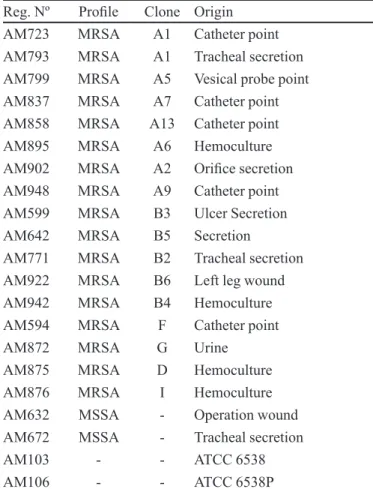
comprising nineteen clinical isolates and two standard strains, were used in the study (Table I). These consisted of
eight multiresistant S. aureus MRSA of the Brazilian
epi-demic clone (BEC), ive isolates of the pediatric epiepi-demic clone, four isolates of the sporadic clones and two MSSA strains which were only resistant to the antibiotics penicil -lin, erythromycin and gentamycin. The multiresistant S. aureus MRSA and S. aureus MSSA were strains from the Microbiological Analysis Laboratory collection, derived
from a study on the susceptibility/resistance proile of S. aureus in Recife-Pernambuco state(Cordeiro, 2004).
Preparation of inoculates
TABLE I - List of Staphylococcus aureus strains assayed
Reg. Nº Proile Clone Origin
AM723 MRSA A1 Catheter point
AM793 MRSA A1 Tracheal secretion
AM799 MRSA A5 Vesical probe point
AM837 MRSA A7 Catheter point
AM858 MRSA A13 Catheter point
AM895 MRSA A6 Hemoculture
AM902 MRSA A2 Oriice secretion
AM948 MRSA A9 Catheter point
AM599 MRSA B3 Ulcer Secretion
AM642 MRSA B5 Secretion
AM771 MRSA B2 Tracheal secretion
AM922 MRSA B6 Left leg wound
AM942 MRSA B4 Hemoculture
AM594 MRSA F Catheter point
AM872 MRSA G Urine
AM875 MRSA D Hemoculture
AM876 MRSA I Hemoculture
AM632 MSSA - Operation wound
AM672 MSSA - Tracheal secretion
AM103 - - ATCC 6538
AM106 - - ATCC 6538P
Reg. Nº: Register Number; AM: Microbiological analysis laboratory collection – Pharmaceutical Science Department
–UFPE; ATCC: American type culture collection; Clone A:
Brazilian epidemic clone (BEC); Clone I, D, G, F: Sporadic
clone; Clone B: Pediatric clone; MRSA: Methicillin resistant
Staphylococcus aureus; MSSA: Methicillin sensitive
Staphylococcus aureus
colonies culture of S. aureus in Mueller-Hinton agar and suspended in sterile physiological solute, comparing
the turbidity with the 0.5 tube of the McFarland scale
(108 UFC mL-1) (CLSI, 2003).
Agar well diffusion method
The inoculum were applied to the surface of the Mueller-Hinton agar and after perforation of the wells
(perforator 6 mm in diameter), 100 µL of the extracts (concentrations of 100 mg .mL-1 and 50 mg mL-1),
stan-dard antibiotic (300 µg mL-1) and of the control solution
(DMSO at 50% (v/v) or tween 80 at 4% (v/v)) were added to each well. After incubation at 37 ºC ± 1 for 24 hours, the diameter of the inhibition halos were measured and results evaluated according to the following scale: inhibition
halos of 9 mm - inactive; 9-12 mm - somewhat active;
13-18 mm - active; 13-18mm - very active (Alves et al., 2000).
Agar dilution method – Determination of Minimum Inhibitory Concentration (MIC)
The minimum inhibitory concentration for the ex-tracts (basic solution of 20 mg.mL-1), dilutions in water
with concentrations of 312.5 µg.mL-1 to 20.103 µg.mL-1,
had been prepared and incorporated to Mueller-Hinton
agar (1:9), thus inal concentrations of 31.5 µg.mL-1 to
2000 µg.mL-1 of the extracts remained. The standard
anti-biotics used were tetracycline and oxacillin (Sigma), with
concentrations as indicated by the CLSI (2003) norms.
The twenty-one inoculates were distributed asepti
-cally in the Stears multi-inoculator, and then deposited on
the surface of the culture medium, and incubated at 36 ºC ± for 24 hours.
Duplicate controls were performed at the beginning
and end of the inoculation control process. Control of the
diluents was also done (tween 80 at 4% and DMSO at
50%).
The results were interpreted comparatively to the strain controls and determined by the irst plate whose concentration inhibited growth.
Phytochemical analysis
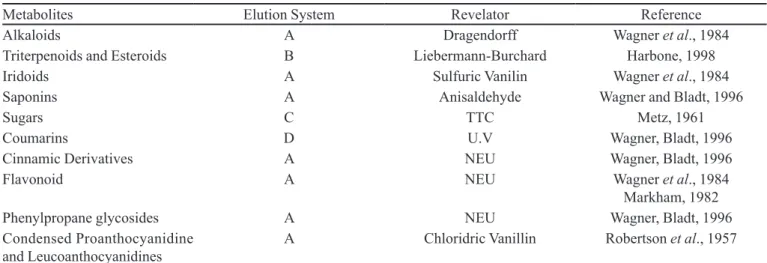
Thin layer chromatography (TLC) with silica-gel GF°254 (Merck) was used. The methanol or ethyl acetate
extracts of the leaves and lower of Caesalpinia
pyramida-lis at the concentration of 20 mg mL-1 were applied using
an analytic capillary (15 µL). The mobile phase for the
methanol extracts was the chromatographic system (1):
AcOEt/MeOH/H2O (81:11:08), while for the correspond
-ing ethyl acetate the system (2): AcOEt/HCOOH/AcOH/
H2O (100:2:2:2 and 100:3:3:3) was employed. TLC was
performed in duplicate, one being used as a chromato-graphic reference and the other for bioautography.
In the phytochemical study, standards of gallic acid, ellagic acid, quercetin, kaempferol, catechin, ursolic acid,
β-sitosterol, β-amyrin, pilocarpine, iridoids, and glucose were used.
The visualization of TLC was carried out under UV light (254 and 366 nm), as shown in Table II.
Bioautographic technique
The developed TLC according to system (1) was
surface, melted Mueller-Hinton agar (MH) was added,
inoculated with a saline suspension of S. aureus ATCC
6538 (bacterial suspension at 108 UFC/mL) and
homoge-neously distributed over the TLC plate. After solidiication
of inoculated MH, it was left for 30 minutes at the sur
-rounding ambient temperature of 25 ºC for predifusion of the active components. Subsequently, it was incubated for 24 to 36 hours ±1 ºC. After this period the bioautography was revealed with a solution of 2,3,5-triphenyltetrazolium
chloride (TTC) at 2.5 mg mL-1 and incubated for a further
4 hours.
The presence of an inhibition zone indicated the existence of active components (Pessini et al., 2003).
RESULTS AND DISCUSSION
Antimicrobial activity
The results of methanol and ethyl acetate extracts from C. pyramidalis against four clinical isolates and one standard strain (AM103) of S. aureus are given in
Table III. The hexane extracts showed no activity. The
diameters of the inhibition halos expressed in millimeters are expressed in the form of a table for each extract and each bacterium.
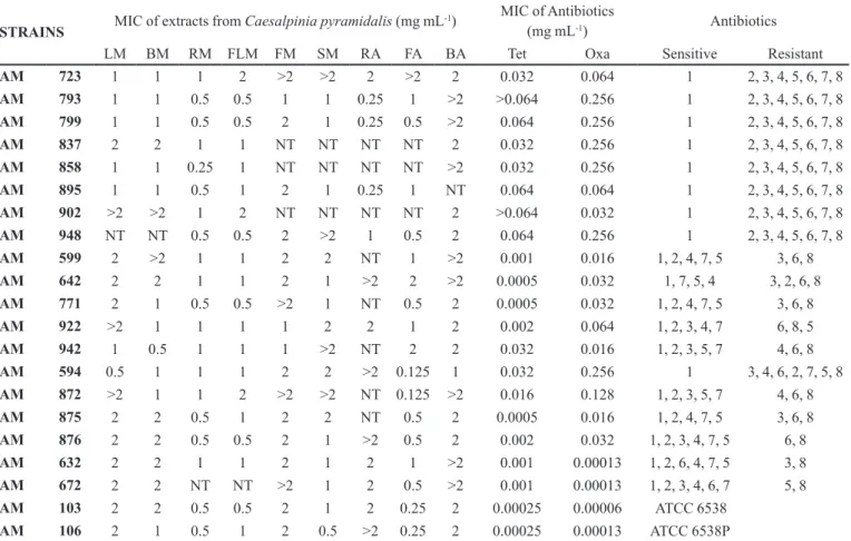
The MIC values of the assayed extracts are shown
separately, according to type of S. aureus MRSA multi-resistant clone. Table IV summarizes the results of eight Brazilian epidemic clones, five pediatric clones, four
sporadic multiresistant clones, two strains of S. aureus
MSSA, and inally two standard strains.
TABLE II - Chromatographic systems used for phytochemical screening of Caesalpinia pyramidalis Tull.
Metabolites Elution System Revelator Reference
Alkaloids A Dragendorff Wagner et al., 1984
Triterpenoids and Esteroids B Liebermann-Burchard Harbone, 1998
Iridoids A Sulfuric Vanilin Wagner et al., 1984
Saponins A Anisaldehyde Wagner and Bladt, 1996
Sugars C TTC Metz, 1961
Coumarins D U.V Wagner, Bladt, 1996
Cinnamic Derivatives A NEU Wagner, Bladt, 1996
Flavonoid A NEU Wagner et al., 1984
Markham, 1982
Phenylpropane glycosides A NEU Wagner, Bladt, 1996
Condensed Proanthocyanidine and Leucoanthocyanidines
A Chloridric Vanillin Robertson et al., 1957
A: EtOAc-HCOOH-AcOH-H2O (100:11:11:26 v/v); B: EtOAc-HCOOH-AcOH-H2O (100:0.5:0.5:0.5 v/v); C: n-BuOH-Me2CO-
Buffer Phosphate pH = 5.0 (40:50:10 v/v); D: Et2O-toluene-AcOH 10% (50:50:50 v/v); NEU: 2-Amino-ethyl-diphenyl borinate,
TTC: Triphenyl Tetrazolium Chloride.
TABLE III - Antimicrobial activity of Caesalpinia pyramidalis
against S. aureus
Extracts mg/Well S. aureus(AM) halo diameter in mm
594 723 922 942 103
LM 10 mg 18 20 20 21 20
5 mg 15 15 16 18 16
BM 10 mg 20 18 18 19 21
5 mg 17 15 16 16 19
RM 10 mg 19 16 17 17 20
5 mg 19 16 17 17 20
FLM 10 mg 20 22 22 22 23
5 mg 17 18 18 19 21
FM 10 mg 20 20 19 21 23
5 mg 18 18 16 19 20
SM 10 mg 17 16 19 17 20
5 mg 15 14 16 16 18
LA 10 mg - - - - 12
5 mg - - - -
-BA 10 mg 16 15 15 16 18
5 mg 14 13 12 12 14
RA 10 mg 18 18 20 19 20
5 mg 16 17 16 17 18
FA 10 mg 14 13 12 18 18
5 mg 12 - - 15
-TT 30 mcg 11 12 27 12 28
L: (leaves); FL (lower); B (bark of stem); R (peel of root);
F (fruit); S (seeds); M (methanol); A (ethyl acetate) TT:
Tetracycline; AM: Microbiological analysis laboratory
TABLE IV - Minimum Inhibitory Concentration (MIC) of extracts from Caesalpinia pyramidalis against Staphylococcus aureus
strains
STRAINS MIC of extracts fromCaesalpiniapyramidalis(mg mL
-1) MIC of Antibiotics
(mg mL-1) Antibiotics LM BM RM FLM FM SM RA FA BA Tet Oxa Sensitive Resistant
AM 723 1 1 1 2 >2 >2 2 >2 2 0.032 0.064 1 2, 3, 4, 5, 6, 7, 8
AM 793 1 1 0.5 0.5 1 1 0.25 1 >2 >0.064 0.256 1 2, 3, 4, 5, 6, 7, 8
AM 799 1 1 0.5 0.5 2 1 0.25 0.5 >2 0.064 0.256 1 2, 3, 4, 5, 6, 7, 8
AM 837 2 2 1 1 NT NT NT NT 2 0.032 0.256 1 2, 3, 4, 5, 6, 7, 8
AM 858 1 1 0.25 1 NT NT NT NT >2 0.032 0.256 1 2, 3, 4, 5, 6, 7, 8
AM 895 1 1 0.5 1 2 1 0.25 1 NT 0.064 0.064 1 2, 3, 4, 5, 6, 7, 8
AM 902 >2 >2 1 2 NT NT NT NT 2 >0.064 0.032 1 2, 3, 4, 5, 6, 7, 8
AM 948 NT NT 0.5 0.5 2 >2 1 0.5 2 0.064 0.256 1 2, 3, 4, 5, 6, 7, 8
AM 599 2 >2 1 1 2 2 NT 1 >2 0.001 0.016 1, 2, 4, 7, 5 3, 6, 8
AM 642 2 2 1 1 2 1 >2 2 >2 0.0005 0.032 1, 7, 5, 4 3, 2, 6, 8
AM 771 2 1 0.5 0.5 >2 1 NT 0.5 2 0.0005 0.032 1, 2, 4, 7, 5 3, 6, 8
AM 922 >2 1 1 1 1 2 2 1 2 0.002 0.064 1, 2, 3, 4, 7 6, 8, 5
AM 942 1 0.5 1 1 1 >2 NT 2 2 0.032 0.016 1, 2, 3, 5, 7 4, 6, 8
AM 594 0.5 1 1 1 2 2 >2 0.125 1 0.032 0.256 1 3, 4, 6, 2, 7, 5, 8
AM 872 >2 1 1 2 >2 >2 NT 0.125 >2 0.016 0.128 1, 2, 3, 5, 7 4, 6, 8
AM 875 2 2 0.5 1 2 2 NT 0.5 2 0.0005 0.016 1, 2, 4, 7, 5 3, 6, 8
AM 876 2 2 0.5 0.5 2 1 >2 0.5 2 0.002 0.032 1, 2, 3, 4, 7, 5 6, 8
AM 632 2 2 1 1 2 1 2 1 >2 0.001 0.00013 1, 2, 6, 4, 7, 5 3, 8
AM 672 2 2 NT NT >2 1 2 0.5 >2 0.001 0.00013 1, 2, 3, 4, 6, 7 5, 8
AM 103 2 2 0.5 0.5 2 1 2 0.25 2 0.00025 0.00006 ATCC 6538
AM 106 2 1 0.5 1 2 0.5 >2 0.25 2 0.00025 0.00013 ATCC 6538P
NT: Not tested; L: (leaves); FL (lower); B (bark of stem); R (peel of root); F (fruit); S (seeds); M (methanol); A (ethyl acetate).; (1) – vancomycin; (2) – ciproloxacin; (3) – erythromycin; (4) –tetracycline (Tet); (5) – gentamicin; (6) – oxacillin (Oxa); (7) – sulfamethoxazole/trimethoprim; (8) – penicillin.; AM: Microbiological analysis laboratory collection – Pharmaceutical Science Department –UFPE
Bioautography
The bioautographic results for the leaves and lower
extracted in methanol and ethyl acetate, and peel of the root extracted in ethyl acetate,from C. pyramidalis are listed in Table V.
TABLE V - Retention factor values related to inhibition halos of extracts of Caesalpinia pyramidalis obtained by bioautographic technique
Chromatograph Systems Extracts Retention Factor (Rfs)
AcOEt/MeOH/H2O (81:11:08) LM 0.55 0.84 0.94
FLM 0.19 0.29 0.48
AcOEt/HCOOH/AcOH/H2O (100:2:2:2 and 100:3:3:3) LA 0.51 0.62 0.80 0.94
FLA 0.62
RA 0.26 0.39 0.58 0.68
L: (leaves); FL (Flower); R (Peel of Root); M (Methanol); A (Ethyl Acetate); AcOEt: Ethyl acetate, MeOH: Methanol, HCOOH:
Formic Acid, AcOH: Acetic Acid.
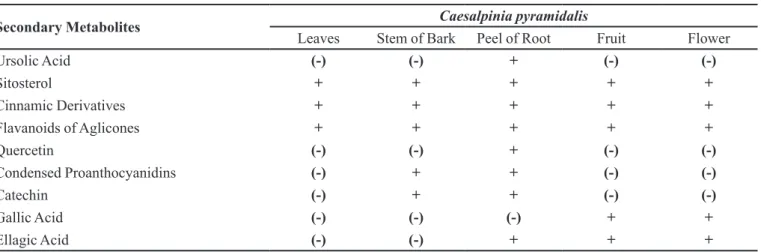
Phytochemical study
The secondary metabolites found in the extracts of the parts studied from C. pyramidalis with well-known
antimicrobial activity are shown in Table VI.
-TABLE VI - Secondary metabolites from Caesalpinia pyramidalis with well-known antimicrobial activity
Secondary Metabolites Caesalpinia pyramidalis
Leaves Stem of Bark Peel of Root Fruit Flower
Ursolic Acid (-) (-) + (-) (-)
Sitosterol + + + + +
Cinnamic Derivatives + + + + +
Flavanoids of Aglicones + + + + +
Quercetin (-) (-) + (-) (-)
Condensed Proanthocyanidins (-) + + (-) (-)
Catechin (-) + + (-) (-)
Gallic Acid (-) (-) (-) + +
Ellagic Acid (-) (-) + + +
+: Metabolite Present; (-): Metabolite Absent
tion of antimicrobial activity, the agar well diffusion tech -nique, despite using larger volumes (100 µL) (Caetano et al., 2002) compared to disks (10 µL) (Voravuthikunchai
and Kitppipit, 2005), has the advantage of allowing the
use of adjuvant to improve the solubility of the extract
constituents and to permit radial as well as superficial
diffusions, conditions resulting in better inhibition halos.
With regard to the ive Staphylococcus aureus strains studied, four represent the three types of MRSA multire-sistant clones and one the standard strain.
In Table III, the extract of the flower methanol (FLM) showed the largest inhibition halos for the five tested strains, with values in the order of 22 mm, at the highest concentration (10 mg/well), corresponding to the very active classiication (Alves et al., 2000), and halos in
the order of 16 mm for concentrations of 5 mg per well , with extracts of leaves, peel of the root, fruit and seed also
having good activity.
In general, extracts from retrieval in ethyl acetate
showed smaller inhibition halos compared with those ex
-tracted from methanol. This fact can be due to the polarity
characteristics of its constituents which resulted in lower
diffusion in aqueous environs and therefore produced
smaller inhibition halos despite having substances with
antimicrobial activity (Lenette et al., 1987).
Observing Table III of the corresponding extracts (leaves, bark of the stem, peel of the root, fruit and seed) in ethyl acetate, the extract of peel of the root produced
inhibition halos in the order of 20 mm, while the extract of the leaves showed no activity except against the standard
strain (AM103). Experimentally, this difference may be explained by the fact that the dry extract of peel of the
root is in the form of powder and dissolved very well in tween 80/water 4% whereas extract from the leaves had a
pasty consistency and was not well dissolved, hindering
its solubility.
The inhibition zone produced by the standard
anti-biotic, tetracycline, conirmed the resistance phenotype
of the clone strains, i.e. Staphylococcus aureus MRSA AM594, S. aureus AM723 and S. aureus AM942, resistant to tetracycline had inhibition halos in the order of 13 mm
or less while the strains S. aureus MRSA AM922 and S.
aureus AM103 sensitive to tetracycline had inhibition halos in the order of 26 mm.
Finally, the two diluents, DMSO at 50% and tween
80 at 4%, exhibited no inhibition.
The presented data are mean values of two deter
-minations.
On the other hand, the presence of active substances
in the leaves as well as in the lower for both types of ex
-tracts was evidenced in the results of the bioautographies
(Table V).
The bioautography technique permits the detection of inhibition halos that reveal the presence of active sub-stances in the TLC (Pessini et al., 2003) against S. aureus ATCC 6538 in the methanolic extracts, one in Rƒ 0.48
for the lower extract and Rƒ 0.55 for the leaves extract. Similarly, for the extracts in ethyl acetate, one showed the presence of inhibition halos in Rƒ 0.62 for the lower
extract, the Rƒ 0.51, Rƒ 0.62, Rƒ 0.80 and Rƒ 0.94 for the leaves extract and the Rƒ 0.26, Rƒ 0.39, Rƒ 0.58, Rƒ 0.68, , Rƒ 0.77 and Rƒ 0.92 for the stem bark extract (Table V). The phytochemical investigation evidenced the presence of substances such as flavonoids (flavonoid aglycones), sitosterol and cinnamic derivatives for all the extracts of C. pyramidalis tested. The presence of gallic
polyphenols (phenolic acid) in the fruit, and ursolic acid and quercetin from the bark of the stem and peel of the root
(root and bark of the stem) thus conirming data from the
literature (Bahia et al., 2010; Bahia et al., 2005).
With reference to MIC (Table IV), analysis of the re-sults reveals that, in the case of S. aureus MRSA Brazilian epidemic clone, that some of the values are less than 0.5 mg.mL-1 for the methanol extracts as well as ethyl acetate
extracts of the peel of the root.
Similar results were found for the methanol extract of the lower.
The MIC values for the ive pediatric clones were again low for the extracts of the peel of the root, being in
the order of 0.25 mg.mL-1 for both methanol and ethyl
acetate extracts.
Also, in Table IV featuring MIC for the four
spo-radic clones together with the susceptible S. aureus
MSSA strains and standard strains, the extracts of the
peel of the root in ethyl acetate had some values as low as
0.125 mg mL-1, even less than those of the MSSA strains
(0.5 mg mL-1) and standard strains (0.25 mg mL-1).
The MIC values of tetracycline and oxacillin con-firmed the phenotype resistance and MRSA or MSSA character of isolates of S. aureus strains, respectively.
In relation to the DMSO diluents at 50% and tween 80 at 4%, these showed no inhibition against any of the twenty-one strains, conirming earlier data for DMSO by
Sakagami et al. (2005).
CONCLUSION
The extracts from C. pyramidalis showed good antimicrobial activity against the S. aureus multiresistant
strains. Also, the bioautography technique allowed visual -izing six inhibition halos for the extracts in ethyl acetate
from stem bark and three from the leaves, which allows
us to conclude that there are at least six plus three active compounds, respectively.
Further experiments aimed at the isolation and
struc-tural identiication of the active compounds are needed, as well as more pharmacological data to validate the popular
use of the extracts from the C. pyramidalis. such as data determining its safety and toxicity.
ACKNOWLEDGEMENTS
We extend our thanks to Prof. Dr. Agnes M. S. Figueiredo – Laboratory of Molecular Biology of Bacteria, Institute of Microbiology – UFRJ, Rio de Janeiro – RJ, Brazil.
REFERENCES
ALVES, T.M.A.; SILVA, A.F.; BRANDÃO, M.; GRAND, T.S.M.; SMÂNIA, E.F.A.; SMÂNIA, J.R.A.; ZANI, C.L.
Biological screening of brazilian medicinal plants. Mem.
Inst. Oswaldo Cruz, v.95, n.3, p.367-373, 2000.
BAHIA, M.V.; DAVID, J.P.; DAVID, J.M. Occurrence of
bilavones in leaves of Caesalpinia pyramidalis specimens.
Quim. Nova, v.33, n.6, p.1297-300, 2010.
BAHIA, M.V.; SANTOS, J.B.; DAVID, J.P.; DAVID,
J.M. Biflavonoids and other phenolics of Caesalpinia
pyramidalis (Fabaceae). J. Braz. Chem. Soc., v.16, n.6b, p.1402-1405, 2005.
BRAGA, R. Plantas do nordeste – especialmente do Ceará. 4.ed.
Natal: Editora universitária da UFRN,1960. 540 p.
CAETANO, N.; SARAIVA, A.; PEREIRA, R.; CARVALHO, D.; PIMENTEL, M.C.B.; MAIA, M.B.S. Determinação de atividade antimicrobiana de extratos de plantas de uso
popular como antiinflamatório. Rev. Bras. Farmacogn.,
v.12, suppl.1, p.132-135, 2002.
CLINICAL LABORATORY STANDARDS INSTITUTE.
CLSI. Performance standards for antimicrobial disk
susceptibility tests. Approved standard. 8.ed. Wayne:
NCCLS, 2003. 58 p. (NCCLS document M2-A8). Available
at: <www.sbac.org.br/pt/pdfs/biblioteca/clsi_OPASM7_
A6.pdf>. Accessed on: 21 jul. 2011.
CORDEIRO, R.P. Peril de sensibilidade/resistência de cepas de
Staphylococcus aureus. MRSA do Hospital das Clínicas da
Universidade Federal de Pernambuco, Recife. Recife, 2004. 55 p. [Dissertation of Master degree in Pharmaceutical Science. Federal University of Pernambuco].
HARBONE J.B. Phytochemical methods. 3.ed. London:
Chapman & Hall, 1998. 320 p.
LENETTE, E.H.; BALOWS, A.; HAUSCLER W.J.; SHADOMY,
H.J. Manual de microbiologia clinica. 4.ed. Buenos Aires:
Editorial medica panamericana, 1987. 1407 p.
MINISTÉRIO DE MINAS E ENERGIA. MME. Projeto cadastro de fontes de abastecimento por água subterrânea - diagnóstico do Município de Carnaubeira da Penha, 2005.
14 p. Available at: <http://www.cprm.gov.br/rehi/atlas/
MARKHAN, K.R. Techniques of flavonoid identification. London: Academic Press, 1982. 113 p.
MENDES, C.C.; BAHIA, M.V.; DAVID, J.M.; DAVID, J.P.
Constituents of Caesalpinia pyramidalis. Fitoterapia, v.71, n.2, p.205-207, 2000.
METZ, H. Thin-layer chromatography for rapid assays of
enzymatic steroid transformations, Naturwissenschaften,
v.48, n.17, p.569-570, 1961.
MIRANDA, O.P.; SILVA-CARVALHO, M.C.; RIBEIRO, A.; PORTELA, F.; CORDEIRO, R.P.; CAETANO, N.; VIDAL, C.F.L.; FIGUEIREDO, A.M.S. Emergency in Brazil
of methicillin-resistant Staphylococcus aureus isolates
carrying SCCmecIV that realated genetically to the USA800
clone. Clin. Microb. Inf., v.13, p.1165-1172, 2007.
NASCIMENTO, G.G.F.; LOCATELLI, J.; FREITAS, P.C.;
SILVA, G.L.Antibacterial activity of plant extracts and
phytochemicals on antibiotic-resistant bactéria. Braz. J.
Microbiol., v.31, n.4, p.247-256, 2000.
N I S H I , H . ; K O M AT S U WA , H . ; F U J I WA R A , T. ; MCLALLUM, N.; SUGAI, M. Reduced content of lysyl-phosphatidyglycerol in the cytoplasmic membrane affects susceptibility to moenomycin, as well as vancomycin,
gentamicin, and antimicrobial peptides. in Staphylococcus
aureus.Antimic. Agents. Chemother., v.48, n.12, p.4800-4007, 2004.
NOVAIS, T.S.; COSTA, J.F.D.; DAVID, J.P.L.; DAVID, J.M.; QUEIROZ, L.P.; FRANÇA, F.; GIULIETTI, A.M.; SOARES, M.B.P.; SANTOS, R.R.. Atividade antibacteriana
em alguns extratos de vegetais do semi-árido brasileiro. Rev.
Bras. Farmacogn., v.13, suppl.2, p.5-8, 2003.
OLIVEIRA, G.A.; FARIA, J.B.; LEVY, C.E.; MAMIZUKA,
E.M.Characterization of the brazilian endemic clone of
methicillin-resistant Staphylococcus aureus (MRSA) from
hospitals throughout Brazil. Braz. J. Infect. Dis., v.5, n.4,
p.163-170, 2001.
PESSINI, G.L.; HOLETZ, F.D.; SANCHES, N.R.; CORTEZ, D.A.; DIAS FILHO, B.P.; NAKAMURA, C.V. Avaliação da atividade antibacteriana e antifúngica de extratos de plantas
utilizadas na medicina popular. Rev. Bras. Farmacogn.,
v.13, suppl.1, p.21-24, 2003.
RÊGO JÚNIOR, N.O.; FERNANDEZ, L.G.; CASTRO, R.D.; SILVA, L.C.; GUALBERTO, S.A.; PEREIRA, M.L.A.; SILVA, M.V. Bioactive compounds and antioxidant activity
of crude extracts of brushwood vegetable species. Braz. J.
Food Technol., v.14, n.1, p.50-57, 2011.
ROBERTSON, E.A.H.; CARTWRIGHT, R.A.; OLDSCHOOL, M.M. Phenolic substances of manufactured tea. I. Fractionation and paper chromatography of water-soluble
substances. J. Sci. Food Agr., v.8, n.2, p.72-80, 1957.
RODRÍGUEZ-NORIEGA, E.; SEAS, C. The changing pattern
of methicillin-resistant Staphylococcus aureus clones in
Latin America: implications for clinical practice in the
region. Braz. J. Infect. Dis., v.14, supl.2, p.87-96, 2010.
SAKAGAMI, Y.; LINUMA, M.; PIYASEMA, K.G.N.P.; DHARMARATNE, H.R.W. Antibacterial activity of
α-mangostin agins vancomycin resistant Enterococci
(VRE) and synergism with antibiotics. Phytomedicine, v.2,
n.3, p.203-208, 2005.
SHIBATA, H.; KONDOK, K.; KATSUYAMA, R.; KAWAZOE, K.; SATO, Y.; MURAKAM, K.; TAKAISHI, Y.; ARAKAKI,
N.; HIGUTI, T.Alkyl gallates, intensieiers of β-lactam
susceptibility in methicillin-resistant Staphylococcus
aureus.Antimicrob. Agents Chemother., v.49, n.2, p.549-555, 2005.
SOLA, C.; GRIBAUDO, G.; VINDEL, A.; PATRITO, L.;
BOCCO, J.L.Identiication of a novel methicillin-resistant
Staphylococcus aureus Epidemic Clone in Córdoba,
Argentina, Involved in Nosocomial Infections. J. Clin.
Microbiol., v.40, n.4, p.1427-1435, 2002.
VORAVUTHIKUNCHAI, S.P.; KITPPIPIT, L . Activity of medicinal plant extracts against hospital isolates
of methicillin-resistant Staphylcoccus aureus. Clin.
Microbiol. Infect., v.11, n.5, p.493-512, 2005.
WAGNER, H.; BLADT, S.; ZGAINSKI, E.M. Drogenanalyse.
Berlin: Springer, Verlag, 1984. 321 p.
WAGNER, H.; BLADT, S. Plant drug analysis – A thin layer
chromatography atlas. 2.ed. Munich: Springer. 1996. 384 p.
Received for publication on 06th July 2011