RESUMO.- [Atividade antimicrobiana, toxicidade agu-da e crônica do óleo essencial de Lippia origanoides.] Atualmente nota-se um aumento do interesse pelas plan-tas medicinais, fruto da grande procura por terapias alter-nativas. Neste trabalho foi avaliada a atividade
antimicro-biana e a toxicidade do óleo essencial da Lippia origanoides (alecrim-pimenta). O óleo essencial de alecrim-pimenta foi obtido por arraste com vapor d’água e seus constituintes
foram identificados por cromatografia acoplada a espec -trofotômetro de massa (GC/MS). Entre os 15 compostos
identificados os mais abundantes foram o carvacrol (29%), o-cimeno (25,57%) e metil timol éter (11,50%). Os óleos
foram submetidos a ensaios antimicrobianos para deter-minação da CIM e da CBM. Os resultados mostraram que
a dose de 120μl/mL de qualquer um dos óleos testados foi eficiente em inibir o crescimento dos micro-organis -mos Escherichia coli (ATCC 25922), Staphylococcus aureus
(ATCC 25923) e Salmonella cholerasuis (ATTCC 10708).
Os efeitos tóxicos agudos e crônicos foram verificados em animais segundo método de classes - Toxicidade aguda oral (dose fixas) - OECD 420. As concentrações de 30, 60
Antimicrobial activity and acute and chronic toxicity of the
essential oil of
Lippia origanoides
1Viviane A. Andrade2, Anna C. Almeida3, Dayane S. Souza3, Keila G.F. Colen3, Auricélio A. Macêdo4, Ernane R. Martins3, Francine S.A. Fonseca3 and Renato L. Santos4*
ABSTRACT.- Andrade V.A., Almeida A.C., Souza D.S., Colen K.G.F., Macêdo A.A., Martins E.R., Fonseca F.S.A. & Santos R.L. 2014.Antimicrobial activity and acute and chronic toxicity of Lippia origanoides.Pesquisa Veterinária Brasileira 34(12):1153-1161. Laboratório de Pa -tologia Molecular, Escola de Veterinária, Universidade Federal de Minas Gerais, Av. Antônio
Carlos 6627, Belo Horizonte, MG 31270-000, Brazil. E-mail: [email protected]
Currently, there is a growing interest in medicinal plants, because of an increased demand for alternate therapies. In this study, the antimicrobial activity and toxicity of the essential oil
of Lippia origanoides (L. origanoides) were investigated. The essential oil of L. origanoides was
extracted by steam-dragging distillation and its constituents were identified by chromatogra
-phy coupled with mass spectrometry. Among the 15 compounds identified, the most abundant were carvacrol (29.00%), o-cymene (25.57%), and thymol methyl ether (11.50%). The essen
-tial oil was studied in antimicrobial assays to determine the MIC and MBC. The results indica
-ted that a concentration of 120μL/mL of oil was sufficient to inhibit the growth of the following microorganisms: Escherichia coli (ATCC 25922), Staphylococcus aureus (ATCC 25923) and Sal-monella cholerasuis (ATCC 10708). Acute and chronic toxic effects of orally administered oil
were investigated in Wistar rats by using standard methods. Doses of 30, 60 and 120mg/kg of the essential oil did not induce significant changes in weight, behavior or hematological and biochemical parameters in the animals. There were no signs of any histopathological changes to the liver, kidneys or heart of the treated rats, suggesting that Lippia origanoides oil is
non--toxic after oral administration in acute or chronic toxicity studies. The results obtained in this study show that the essential oil of L. origanoides has a high safety margin, with no detectable
toxic effects in rats treated with doses to 120mg/kg. In addition, L. origanoides oil
demons-trated potent antimicrobial activity against S. aureus, E. coli and S. cholerasuis. Based on these
findings, this essential oil may have practical application as a veterinary antimicrobial.
INDEX TERMS: Lippia origanoides, essential oil, antimicrobial, acute toxicity, chronic toxicity.
1 Received on April 29, 2014.
Accepted for publication on July 21, 2014.
2 Pós-doutoranda do Instituto Educacional Santo Agostinho, Faculdade
de Saúde e Desenvolvimento Humanos, Universidade Federal de Minas Gerais (UFMG), Campus Montes Claros, Av. Osmane Barbosa 937, JK, Mon -tes Claros, MG 394004-006, Brazil.
3 Instituto de Ciências Agrárias, UFMG, Campus Montes Claros, Avenida
Universitária 1000, Montes Claros, MG 39404-547. E-mail: annachristina [email protected]
4 Laboratório de Patologia Molecular, Escola de Veterinária, UFMG, Av.
e 120 mg/kg de óleo essencial não induziram alterações significativas no peso, no comportamento dos animais e
nem nos parâmetros hematológicos e bioquímicos.
Tam-bém não houve presença de alterações histopatológicas no fígado, rins e coração sugerindo que o óleo de alecrim
--pimenta é atóxico após administração oral em condições
agudas ou crônicas. Os resultados obtidos neste trabalho levam a concluir que o óleo essencial de alecrim-pimenta possui uma margem elevada de segurança, com efeitos
tó-xicos inexistentes além de apresentar atividade antimicro
-biana eficaz contra os micro-organimos S. aureus, E. coli e S. cholerasuis. Sua utilização na medicina veterinária deve ser considerada como uma grande viabilidade econômica e sustentável.
TERMOS DE INDEXAÇÃO: Lippia origanoides, alecrim-pimenta, óleo essencial, antimicrobiano, toxicidade aguda, toxicidade crô-nica.
INTRODUCTION
Owing to the current widespread and frequent use of syn -thetic antimicrobials, there has been an increase in the re-sistance of microorganisms to these compounds. Because
of this problem, there is an increasing need to find new products with antimicrobial activity, including alkaloids and terpenes, which are present in some vegetables. Me -dicinal plants and plant-derived essential oils have proven
inhibitory and antiseptic activity, and may have potential for application in the field of public health, industry and
treatment of animals. There is increasing interest in
medi-cinal plants as well as their active ingredients, which may
possess antibiotic properties.
Brazil has the richest plant diversity in the world, with approximately 22% of the planet’s biodiversity and more than 55,000 catalogued species of plants, among an esti
-mated total of 350,000 to 550,000 species (Gatyas & Sava
-ge 2010). The National Program for Medicinal Plants and Phytotherapics published the National List of Medicinal Plants of Interest to the unified health system of Brazil (Sis
-tema Único de Saúde or SUS) in January 2009. In this list, medicinal plants that can potentially produce products of
interest to the SUS are listed. Lippia origanoides as among
the species listed (Brasil 2006).
Lippia origanoides Cham. (Verbanaceae) is a wild shrub
native to the semiarid regions of Brazil. Its leaves are fre
-quently used in phytotherapy. L. origanoides contains 4.5%
of an essential oil incredibly rich in thymol, one of the main active ingredients extracted by steam-dragging distillation (Matos 2002). Some other substances in L. origanoides,
such as flavonoids and quinones, may contribute to the ac
-tion of the main active components (Matos 2002).
The antimicrobial effect of L. origanoides essential oil has been tested against the Staphylococcus aureus and Es-cherichia coli isolated from artisanal Minas cheese
produ-ced in Brazil (Castro et al. 2011). The results showed the bactericidal activity of the essential oil of L. origanoides,
suggesting the possibility of its use as an alternative anti -bacterial agent. Other studies assessed the antimicrobial
and antiseptic activity of L. origanoides essential oil on
25 strains of bacteria (coagulase-negative Staphylococcus, coagulase-positive Staphylococcus, esculin-positive Strep-tococcus, esculin-negative StrepStrep-tococcus, and Mannheimia haemolytica) isolated from sheep with mastitis in the north
region of Minas Gerais (Castro et al. 2011). The antimicro
-bial activity of the L. origanoides extract was also tested on isolates of S. aureus in which inhibition of microbial growth
was shown (Oliveira et al. 2006a, 2006b).
In addition, the toxicity of L. origanoides essential oil has been assessed. The investigators in these studies did
not observe any acute or subchronic toxicity after oral ad
-ministration (Fontenelle et al. 2007). In another acute toxi
-city study, this essential oil was shown to have some toxic
effects in mice after peritoneal administration (Almeida et
al. 2010).
Results with L. origanoides oil found in the literature are
varied. This may be due to differences in the parts of the
plant used, differences in the methods used for extraction,
differences in inoculation methods, as well as differences in the analysis of results. There were also physiological varia
-tions between plants cultivated and grown under different environmental conditions, which may have contributed to
differences in the composition of the extracts and the expe-rimental differences found in the literature.
Therefore, further research on L. origanoides oil is nee-ded to determine the potential of this medicinal plant as a
phytotherapeutic product for use in veterinary medicine. So, the aim of this study was to assess the phytochemical composition, the antimicrobial activity, and the toxicity of
the essential oil of L. origanoides.
MATERIALS AND METHODS
Plant collection. Lippia origanoides plants were collected from the reserve at the Institute of Agricultural Sciences (ICA) at
Federal University of Minas Gerais (UFMG). The collection areas were recorded with the aid of a global positioning system receiver.
Aerial parts of L. origanoides plants were collected in the morning
and stored in polyethylene bags, protected from moisture, light, heat, insects and contamination by microorganisms. After collec
-tion, the material was stabilized and dried at the Laboratory of Medicinal Plants at ICA/UFMG in an oven with forced air circula
-tion and a ventila-tion system and controlled and constant tempe
-rature of 60 ± 2°C (Falkenberget al. 2007).
Essential oils extraction and analysis. The L. origanoides
essential oil was obtained using the technique steam-dragging distillation in a pilot distiller (Linax®, model D20, SP, Brazil). After
3 h of extraction, the oil was separated from the hydrolyte by li
-quid-liquid partitioning, removed with a micropipette and stored in sterile amber glass vials under refrigeration (4-8°C). Analyses were carried out on three different samples of L. origanoides oil
based on the month in which the plants were collected: oil 1 was extracted from plants collected in December 2011, oil 2 was ex
-tracted from plants collected in March 2012 and oil 3 was extrac
-ted from plants collec-ted in May 2012.
The chromatographic analyses were carried out using a gas chromatography system, Agilent Technologies (GC 7890A), cou
-pled with a mass spectrometer detector (MS 5975C) (GC-MS). A HP-5 MS capillary column was used (Agilent Technologies, sta
split/splitless injector was maintained at 220°C. The temperature gradient was set from 50°C (2 min) up to 240°C (1 min) at a rate of 3°C/min. The sample volume introduced was 1μL in the injection mode with division of flow and a split ratio of 1:10, using a Combi PAL injector. The mass spectrometer was operated with electron ionization at 70 e V and a quadruple mass analyzer, operated in scan mode (monitoring) between 29 and 550 (m/z). The samples were diluted and injected into the GC-MS and the constituents were identified using a computer program provided by the ma -nufacturer.
Antimicrobial activity assay. The microbial sensitivity test of L. origanoides essential oil was performed using the disc-diffu
-sion method adapted from Pinto et al. (2003), for three different microorganisms Escherichia coli (ATCC 25922), Staphylococcus aureus (ATCC 25923) and S. cholerasuis (ATCC 10708). Sterile
filter paper discs (Whatman nº1, 6mm diameter) were impreg
-nated with 10µL of oil at concentrations of 30µL/mL, 60µL/mL and 120µL/mL. The inoculum of microorganisms was prepared and standardized according to the 0.5 McFarland standards which correspond to 106 colony-forming units. The halos were read and
measured in millimeters after 24h of incubation at 37°C by using a pachymeter (Mitunox Instrumentos, SP/Brazil). The tests were
carried out in duplicate.
The minimum inhibitory concentration (MIC) and the mini
-mum bactericidal concentration (MBC) were defined according to recommendations by Rios et al. (1988) as the lowest concen
-trations of oil preventing visible growth of the microorganism subculture. For the MIC test, a solution was prepared with sterile BHI (Brain Heart Infusion) broth. This solution was prepared with 40µL of Tween 80 and the essential oil diluted to give a concen
-tration of 120µL/mL of L. origanoides. From this solution,
macro-dilution was performed to obtain concentrations of 60 and 30µL/ mL of L. origanoides oil. The test was carried out in duplicate for
each of the three samples of oil used and the tubes were incubated at 37°C for 24h in an incubator. The MBC test was carried out as described above, by using an aliquot from the tube contents of the MIC tests that showed no turbidity after 24h of incubation, and were streaked on petri dishes containing TSA agar medium. The plates were incubated at 37°C for 24h.
Acute oral toxicity assay. The experimental procedures
using animals described in this methodology were approved by
the Ethics Committee for Animal Experimentation at UFMG,
un-der Protocol number 034/2008.The method for assessing acute toxicity established by Organization for Economic Co-operation and Development (OECD) (Guideline 420 2001) is a gradual pro -cess using groups of animals of the same sex per step.
It was defined by testing the concentrations close to the do
-ses used in the microbiological tests. A single dose of 0 (group 1 - control), 30 (group 2), 60 (group 3) or 120mg/kg (group 4) L. origanoides oil was administered by gavage to five fasted Wistar rats. After treatment, all animals remained in the experimental
bioterium at UFMG where they were kept under standard condi
-tions with unrestricted access to food and water, a circadian cycle of light for 12h and dark for 12h, and a controlled temperature of 22 ± 2°C.
Systematic behavioral observations (hippocratic screening) were made to provide a general measure of the effects of the test substance on the conscious state, general disposition, activity, co
-ordination, reflexes, and effects on the central autonomic nervous systems (Malone & Robichaud 1962). Parameters (general activi
-ty, vocal fremitus, irritabili-ty, response to touch, tail squeeze res
-ponse, contortion, posterior train position, straightening reflex, body tonus, grab strength, ataxia, auricular reflex, corneal reflex, tremors, convulsions, hypnosis, anesthesia, lacrimation, urina
-tion, defeca-tion, piloerec-tion, hypothermia, respira-tion, cyanosis,
hyperemia, death) were assessed at 15 min, 30 min, 1h, 2h, 4h and 8h after administration and then daily for 14 days.
The animals were weighed on alternate days to monitor wei
-ght gain. Samples of about 4mL of blood, collected by cardiac puncture were stored in two types of tubes, one with HB antico
-agulant (Laborlab®) to determine the hematological parameters
and the other, without anticoagulant, to obtain the serum to as
-sess the biochemical parameters. The hematological analyses were carried out in a specialized laboratory, service provider. The surviving animals were sedated with 80mg of ketamine +15mg of xylazine per kg, injected intraperitoneally, and were considered to be anesthetized by immobility of the body, but with normal res
-piratory frequency and amplitude. The animals were sacrificed by cervical dislocation. Macroscopic and histopathological analy
-ses of the liver, heart, and lung were performed and these organs were weighed to determine the absolute weights. Relative organ weights were also calculated (relative to body weight).
Chronic oral toxicity test. The study on chronic toxicity of L. origanoides oil was carried out based on the OECD fixed dose pro
-tocol 452/2001 using single dose level of 120mg/kg (since signs of toxicity were not expected at lower doses), and fewer animals were used. The test animals were kept under the same conditions as previously described for the acute toxicity tests. To investiga
-te the chronic toxicity of the L. origanoides essential oil, after 30
days of oral administration, hematological, histopathological and biochemical (serum) parameters were assessed as described by Fontenelle et al. (2007).
The animals were separated into four groups (n=5 per group) and treated with the essential oil of L. origanoides or saline
solu-tion (control) orally by gavage. Blood samples were collected on day 0 (one day before administering the essential oil or vehicle) and on days 15 and 30.The serum concentrations of urea, crea -tinine, glutamic oxaloacetic transaminase (GOT or AST) and
glu-tamic pyruvic transaminase (GPT or ALT) were determined. The blood samples taken on days 0 and 30 were used to determine erythrocyte, and leukocyte counts, hemoglobin, hematocrit and for biochemical analyses. The blood was stored in two types of tu
-bes, as described above. The hematological analyses were perfor
-med in an accredited laboratory. The values obtained were com
-pared within and between groups. At the end of the experimental period (30 days) histopathological analyses of the heart, liver and kidneys were performed.
The animals were observed for 30 days, with the animal beha
-vior observed daily, assessing the following parameters: locomo
-tion, behavior, respira-tion, fur, and skin alterations, eyes, tremors, salivation, diarrhea, lethargy, drowsiness, number of deaths and causes of death. The data were recorded on a monitoring form for each animal. The animals were weighed on alternate days.
For the histopathological analysis, sections of the heart, liver
and kidneys were collected and fixed by immersing in a 10% bu
-ffered formaldehyde solution for 24h, followed by dehydration
in increasing concentrations of alcohol, diaphonized in xylol and
soaked in paraffin. The sections were cut to 5μm thickness and
stained with hematoxylinand eosin (HE) for histopathological
analysis. Any damage was assessed according to intensity and was
given a score for each tissue: 0= no damage, 1= discrete damage,
2= moderate damage, and 3= severe damage.
The results were analyzed using analysis of variance (ANOVA) followed by a post-hoc test. When appropriate, the Student t-test with the Bonferroni adjustment was used for pairwise compari
-sons. A value of p<0.05 was used to indicate a statistically signi
RESULTS
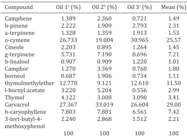
The content of sample of essential oil of Lippia origanoides
were analyzed using GC-MS is described in Table 1. Fifteen compounds were identified with o-cymene, thymol methyl
ether and carvacrol being the compounds present in the hi-ghest concentrations in all three oil samples (Table 1). The
average concentration of these compounds was 29.0% for carvacrol, which was the compound present in the highest amount in all the oils, followed by o-cymene (25.6%) and thymol methyl ether (11.5%).
The antimicrobial activity of the L. origanoides essential
oils was tested against the microorganisms Staphylococcus aureus, Ehcerichia coli, and S. cholerasuis. The disk-diffu
-sion test results are shown in Figure 1. A concentration of 60μL/mL of L. origanoides (oil number 1) had a significant
inhibitory effect on the growth of E. coli, while only a con
-centration of 120μL/mL of oils number 2 and 3 inhibited the growth of this microorganism with a significant diffe
-rence according to the Bonferroni test and two-way ANOVA
(Fig.1A). In the case of S. aureus, only a concentration of
120μL/mL of oil 2 significantly inhibited microbial gro
-wth (Fig.1B). Oils 1 and 3 did not form a halo larger than
those observed for the control groups (Fig.1B). For Salmo-nella cholerasuis all the tested concentrations (30, 60 and
120μL/mL) of all oils tested (1, 2 and 3) significantly inhi
-bited the growth of this microorganism, suggesting that a low concentration of. L. origanoides oil can inhibit the
gro-wth of Salmonella cholerasuis (Fig.1C).
The results of the MIC and MBC tests indicate that 120 μL/mL of all of the oils tested inhibited microbial growth. A concentration of 60 μL/mL inhibited the growth of E. coli
with all three oils and of S. aureus with oil two. A concen
-tration of 120µL/mL is enough to cause death of the micro -organisms.
With regard to acute oral toxicity, the animals did not show any changes in locomotion, behavior, respiration, skin, or fur. The animals were stables throughout the entire treatment. There was no significant difference in weight of the animals during the experiment (Fig.2A). Macroscopic analyses of tissues showed that no organs (heart, liver, kid
-ney) from the groups treated with L. origanoides showed
Fig.1. Sensitivity test of three batches of Lippia origanoides oil against different microorganisms. (A) Esch-erichia coli, (B) Staphylococcus aureus, and (C) Salmonella spp. Oil 1 extracted in December 2011. Oil
2 extracted in May 2012. Oil 3 extracted in March 2012. Asterisk (*) indicates a significant difference between treatments (p<0.01).
Table 1. Chemical composition of the essential oils in
Lippia origanoides
Compound Oil 1a (%) Oil 2b (%) Oil 3c (%) Mean (%)
Camphene 1.389 2.360 0.721 1.49 b-pinene 2.222 1.909 2.793 2.31 a-terpinene 1.328 1.359 1.913 1.53 o-cymene 26.733 19.004 30.965 25.57
Cineole 2.203 0.895 1.264 1.45
g-terpinene 5.731 7.190 8.696 7.21 b-linalool 0.907 0.909 1.220 1.01
Camphor 1.270 3.369 0.760 1.80
borneol 0.687 1.906 0.734 1.11
thymolmethylether 12.778 9.121 12.610 11.50 l-bornyl acetate 3.220 5.204 0.556 2.99
Thymol 4.122 3.008 3.090 3.41
Carvacrol 27.367 33.019 26.604 29.00 b-caryophyllene 7.803 7.881 6.561 7.42 3-tert-butyl-4- 2.240 2.868 1.512 2.21 methoxyphenol
100 100 100 100
a Oil extracted in December 2011, b Oil extracted in May 2012, c Oil
extrac-ted in March 2012.
Fig.2. Change in weight of animals and organs. (A) Weight of animals in control group and those treated
with different doses of Lippia origanoides essential oil. The values represent the weights measured in
rats over 14 days after a single dose treatment. (B) Weight of organs expressed as percentage (%) total
weight of animals. Experimental group with n=5 animals. Asterisk (*) indicates a significant difference
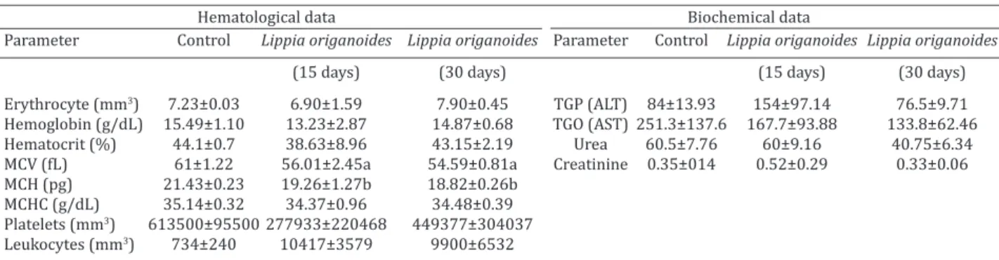
There was a significant change in mean corpuscular volu
-me (MCV), which is the index that helps determine the size of erythrocytes in the diagnosis of anemia and in mean cor
-puscular hemoglobin (MCH) which represents the weight of hemoglobin in the erythrocyte (Table 3).
Plasma levels of AST, ALT, urea and creatinine were me
-asured after 15 or 30 days of dosing with the L. origanoides
oil. These values did not differ significantly when compa
-red to the control group suggesting that there was no he -patotoxic or nephrotoxic effects on the animals (Table 3).
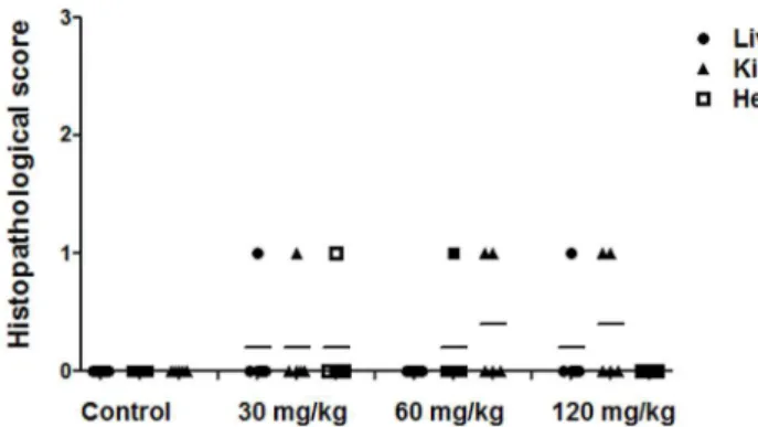
Histopathological analyses of the liver, heart, and kid
-neys did not show significant abnormalities (Fig.5A). Toge -ther, these data suggest that the L. origanoides essential oil
does not have toxic effects when administered at a concen
-tration of 120mg/kg for a period of 30 days.
DISCUSSION
The essential oils of medicinal plants are typically a com
-plex mixture of chemical compounds (primarily terpenes) which can act individually, additively or synergistically to improve health. As well as being used as therapeutic
agents, bioactive substances isolated from the essential
oils of plants may be used as prototype molecules for new synthetic compounds and as tools in physiological studies.
Fig.4. Change in animals and organs weight. (A) Weight of animals in control group and those treated with 120 mg/
kg of Lippia origanoides essential oil. The values represent the weights measured in rats over 30 days of treat -ment. (B) Weight of organs expressed as percentage (%) total weight of animals. Experimental group with n= 5
animals (p<0.001).
any morphological changes and their weights also did not significantly differ between the treatments and the control (Fig.2B).
The hematological parameters of animals treated with
L. origanoides oil in the acute toxicity study were within
the reference limits. There were no significant differences in the full hematological analysis (leukocytes, red cells, eo
-sinophils, lymphocytes and basophils, hemoglobin, hema
-tocrits and platelets). Biochemical analysis of blood taken from animals treated with L. origanoides oil showed that
there was an alteration only in the enzyme, glutamic oxalo
-acetic transaminase (AST) in animals treated with 60 mg/ kg of L. origanoides oil, indicating a possible hepatic
altera-tion in the animals of this group (Table 2).
Histopathological studies on the liver, heart, and kidneys were performed to determine the presence of pathological alterations caused by the L. origanoides oil. The results
in-dicate that there was no dose-dependent histopathological
alteration in the organs from the treated groups compared
with the control (Fig.3).
During the study assessing the chronic toxicity of L. ori-ganoides oil, there were no changes in behavior or body
weight of the animals throughout the experiment (Fig.4A). There were no significant changes in the weight of the or
-gans (expressed as percentage of total body weight) in tre
-ated animals when compared to the control group (Fig.4B).
The hematological and biochemical parameters
measu-red to assess the chronic toxicity are described in Table 3.
Fig.3. Histopathological analysis of the acute toxicity of Lippia origanoides oil. Animals were treated with 30, 60 or 120 mg/
kg administered orally for 14 days. Assessment score used for analysis: 0 = no damage, 1 = discrete damage, 2 = moderate damage, 3 = severe damage. The statistical analysis performed was the non-parametric Kruskal-Wallis test with Dunn’s mul
-tiple comparison test (p<0.0005).
Table 2. Biochemical parameters of Wistar rats treated with a single oral dose of Lippia origanoides essential oils Parameter Control 30mg/kg 60mg/kg 120mg/kg TGP (ALT) 98.66±12.73 77. 6±3.81 82±6.81 75.4±8.47 TGO (AST) 103.66±46.27 185±42.78 248.75±83.97a 196.2±11.15
Thymol and carvacrol are monoterpenoid phenols bio
-synthesized in plants from g-terpinene and p-cymene (Ba
-ser & Demirci 2007, Lopez et al. 2011). Therefore, these precursors are always present in essential oils that contain thymol and carvacrol. In addition, other biosynthesized intermediates may also be present, such as terpinen-4-ol,
cumin alcohol, and p-cymen-8-o l (Baser & Demirci 2007).
Previous studies have shown that the essential oil of na -tive specimens of L. origanoides from the North East of
Bra-zil is composed of approximately 60% thymol (Fontenelle et al. 2007). Carvacrol, an optical isomer of thymol, is also
found in the essential oil of Origanum vulgare (Tian & Lai
2006) making up more than 92% of its essential oils. In this study, lower concentrations of thymol and carva
-crol were found in the L. origanoides oil compared to
con-centrations described in the literature. However, the effects against the tested microorganisms showed that, even in lower concentrations, o-thymol and carvacrol could inhibit microbial growth (Fig.1).
Also in this study, a much higher concentration of o
--cymene was observed (approx.25%, Table 1). Although
this compound is a biological precursor of carvacrol, no
bactericidal activity has been described for this compound
o-cymene. However, studies on p-cymene, an isomer of o
--cymene, indicate that while it is not effectively bactericidal when used alone, but when combined with carvacrol, it is
quite effective against Bacillus cereus in vitro (Cimanga et
al. 2002). This effect may be because p-cymene becomes in
-corporated in the lipid bilayer of Bacillus cereus, facilitating the transport of carvacrol through the plasma membrane
(Cimanga et al. 2002). It is possible that the observation of
bactericidal effect of Lippia origanoides oil in this study was
due o-cymene, which was found in high concentrations in this preparation, may have acted in combination with thy
-mol and carvacrol inducing inhibition growth of microor -ganism (Fig.1).
The variation in the proportions of the active
compoun-ds between oils used in different studies may be explained based in period of collect of plants. Its know that time of the year when the aerial parts were collected to extract the oils can influence the concentrations of compounds. Fac
-tors such as temperature, relative humidity, sun and wind exposure may directly influence the chemical composition of essential oils (Simões & Spitzer 2004). The quantities of chemical components in the essential oils may vary ac
-cording to the stage of development of the plant (Simões & Spitzer 2004) and different period of the year (Santos et al. 2009). Plants are richer in essential oils when the wea
-ther is stable, warm, sunny which are the best conditions for harvesting. There was no significant difference in the relative quantity of compounds in the three batches of oils used in this study, despite having been collected at different times of the year (Table 1).
Morais (2009) suggested that genetic factors, age of the plant and climatic and environmental factors, as well as the period of the year the plants were collected, may cause significant alterations in the production of secondary me
-tabolites influencing the composition of essential oils and thereby the oil’s antimicrobial effect. According to Costa et al. (2011), the essential oil of L. origanoides inhibited the
growth of S. aureus and E. coli with inhibition halos of 8.2
and 23.2 mm respectively at a concentration of 160µL/mL. Klancnik et al. (2010) tested different essential oils
against different bacteria and concluded that despite
diffe-Fig.5. Histopathological analysis of the chronic toxicity of Lippia origanoides oil. Histopathological results of animals treated
with 120 mg/kg for 30 days. Assessment score used for analy
-sis: 0 = no damage, 1 = discrete damage, 2 = moderate dam
-age, 3 = severe damage. The statistical analysis performed was the non-parametric Kruskal-Wallis test with Dunn’s multiple comparison test (p<0.0005).
Table 3. Hematological and biochemical parameters of Wistar rats treated with oral doses of 120 mg/kg of Lippia origanoides essential oils for 30 days
Hematological data Biochemical data
Parameter Control Lippia origanoides Lippia origanoides Parameter Control Lippia origanoides Lippia origanoides
(15 days) (30 days) (15 days) (30 days)
Erythrocyte (mm3) 7.23±0.03 6.90±1.59 7.90±0.45 TGP (ALT) 84±13.93 154±97.14 76.5±9.71
Hemoglobin (g/dL) 15.49±1.10 13.23±2.87 14.87±0.68 TGO (AST) 251.3±137.6 167.7±93.88 133.8±62.46 Hematocrit (%) 44.1±0.7 38.63±8.96 43.15±2.19 Urea 60.5±7.76 60±9.16 40.75±6.34 MCV (fL) 61±1.22 56.01±2.45a 54.59±0.81a Creatinine 0.35±014 0.52±0.29 0.33±0.06 MCH (pg) 21.43±0.23 19.26±1.27b 18.82±0.26b
MCHC (g/dL) 35.14±0.32 34.37±0.96 34.48±0.39 Platelets (mm3) 613500±95500 277933±220468 449377±304037
Leukocytes (mm3) 734±240 10417±3579 9900±6532
rent mechanisms of action, in general, essential oils have greater effectiveness against gram-positive bacteria.
Klanc-nik et al. (2010) present data that indicate that the main reason for differences in bacterial susceptibility may be due to the external membrane that covers the cell wall of gram-negative bacteria, which limits the diffusion of com
-pounds through the lipopolysaccharide cell wall. Other au
-thors have speculated whether differences in sensitivity of
essential oils described in the literature are also associated
with differences in methodology, including the method of extraction (Othman et al. 2011, Radulovic et al. 2013). Also, antimicrobial activity may be dependent on the site of ac
-tion in the cell may potentiate these differences in activity (Negi 2012).
The results obtained here indicate that the
concentra-tion of 120µl/mL of L. origanoides oil inhibited the growth of the tested microorganisms. This concentration proved to be effective in all tests, suggesting that this is an effective concentration for antimicrobial use. Our results are in
agre-ement with data in the literature, which show the bacterici
-dal activity of the L. origanoides essential oil against various
microorganisms (Veras et al. 2014) and suggests that this concentration is most efficient in oils obtained from plants collected between the months of December and May. The
results obtained in other studies using the essential oil of L. origanoides extracted from plants from the North of Minas
Gerais also concur with the optimal concentration found in this study (Castro et al. 2011, Costa et al. 2011, Pinho et al. 2012).
The acute toxicity test data indicate that Wistar rats tre
-ated with L. origanoides oil showed normal behavior and
physiology in all treatment groups. The lack of organ da
-mage in the histopathological analyses suggests that there were no toxic effects from the L. origanoides oil. The
he-matological parameters were within the reference limits, suggesting that there were no significant effects of the L. origanoides oil treatments when compared to the control.
Histopathological studies of the liver, heart, and kidneys showed no significant dose-dependent histopathological
alterations in the treated groups compared to the control (Fig.3). These suggest that oral administration of L. origa-noide soil for 14 consecutive days, at the doses used in this
study was not toxic.
Studies in literature about effect of acute toxicity of L. origanoides essential oil are scarce but was demonstrated that Lippia sidoides extract, administrated by intraperito
-neal way, has a DL50 of 1329,17mg/kg that was considered of high toxicity (Almeida et al. 2010). This result is different from showed in this work, but is important highlight that methods and protocols was distinct of those presented in this study.
Our studies are consistent and can be confirmed by se
-rum biochemical analyses that showed no alteration in the serum levels of the enzyme, glutamic oxaloacetic transami
-nase (AST). In only one of the dose groups (60mg/kg L. ori-ganoides oil) there was alteration that may indicate hepa
-tic toxicity in the animals of this group (Table 2). However, other analyses performed to investigate hepatic changes
do not suggest that this isolated result is due to treatment
with L. origanoides oil. Since this enzyme, AST, is found in
high concentrations in the cytoplasm and mitochondria of hepatocytes, skeletal and cardiac muscles, and is also found in the kidney, pancreas and erythrocytes, damage to any of these organs could increase the levels of this enzyme in the blood. Because there is no laboratory technique to deter
-mine the origin of AST found in the blood, damage to any
of these organs is a possible source of increased serum AST
(Lehninger et al. 2005).
In this study, no damage to any of these organs, kidney, liver and heart, was observed that could explain the alte
-ration in AST in the 60mg/kg group. These organs were chosen because they are the common target organs of toxic agents. These data are in agreement with data in the lite
-rature, where other studies have shown no cell damage in these tissues in tests assessing the toxicity of L. origanoides
and/or its components. Studies with Phyllanthus emblica
Linn. plants demonstrated that alterations in weigh organs
is frequently but this alterations not causes change in ma
-croscopic or mi-croscopic changes were detected in the in
-ternal organs or tissues in any of the treatment rats (Jaijoy et al. 2010).
The results of the chronic toxicity test showed no evi
-dence of chronic toxicity under the conditions in this study. During the study, no changes in behavior or body weight were observed in the treated animals compared to control animals (Fig.4A). The animals were weighed on alternate days and no weight loss was recorded during treatment. There was also no significant alteration in heart, kidney, or lung organ weight (expressed as percentage of total body weight) observed in the treated animals when compared to the control group (Fig.4B).
The hematological and biochemical parameters
measu-red to assess the chronic toxicity are described in Table 3. A significant decrease was observed in the mean corpuscular volume (MCV) which is the index that helps to determine the size of erythrocytes. A decrease was also observed in mean corpuscular hemoglobin (MCH) which represents the amount of hemoglobin in the erythrocytes (Table 3). These parameters are below the levels for the control animals and beyond the reference range for Wistar rats (Dantas et al. 2006, Castello-Branco et al. 2011) and may indicate some interference in erythropoiesis.
No alterations were seen in the serum measurements of AST, ALT, urea and creatinine after 15 or 30 days of do
-sing with L. origanoides oil. This suggests that there was no toxic effect of oral L. origanoides oil in these rats (Table 3).
The histopathological analyses of the liver, heart and kid
-neys did not show any significant abnormalities (Fig. 5). As AST and ALT are considered sensitive indicator of hepatic
damage and into the limits can provide a qualitative
evalu-ation of damage degree suffering by hepatocytes (Martin et al. 1981) this result suggest that no alterations in liver was observed after treatment with L. organoides oil. The
use of this oil cannot be hepatotoxic, when used in doses
tested.
Few toxicity studies on L. origanoides oil or the
com-pounds thymol and carvacrol are sparse have been repor
in agreement with the results described by Fontenelle et al. (2007) and Almeida et al. (2010) despite a difference in methodology.
A recent study by Ferraz et al. (2013) showed the po
-tential activity of thymol, present in plants of the genus Li -ppia, in tumor cells, indicating other important prospects
for medicinal use. Cleff et al. (2008) described the lack of toxicity of repeated doses of Ocimum gratissimum
(admi-nistered orally and intravaginally in rats) which like L. ori-ganoides, has considerable quantities of thymol and carva
-crol. However, Cabello et al. (2014) who analyzed the effect of thymol and carvacrol in human CACO-2 cells, observed
dose and time dependent effects including lipid degene-ration, mitochondrial damage, nucleolar segregation and apoptosis.
Together these data suggest that the essential oil of L. origanoides does not cause any significant toxic effects
when administered at a concentration of 120mg/kg under the conditions of this study.
Using natural products derived from plants is becoming
increasingly common worldwide and research to identify the beneficial effects and the possible adverse effects of their use, is becoming increasingly important, especially studies that take into account the effects of these products
at the molecular level.
CONCLUSIONS
The essential oil of Lippia origanoides has potential as
an antimicrobial, according to the sensitivity tests in the present study.
This extract showed no acute or chronic toxicity in rats, as confirmed by no change in the body weight of the treated animals compared to controls and no significant changes in
the levels of metabolic in serum.
A detailed assessment of hematological, biochemical and histopathological effects of L. origanoides oil did not
indicate any significant changes in physiological functions or organ function in animals treated with L. origanoides oil.
Based on these findings, this essential oil may have practical application as a veterinary antimicrobial. Further exploration of this medicinal plant for veterinary phytothe
-rapy is warranted.
Acknowledgements.- We would like to thank everyone who contributed to this study with financial support from the agencies FAPEMIG (Fundação de Amparo à Pesquisa do Estado de Minas Gerais, Brazil), CNPq (Conse -lho Nacional de Desenvolvimento Científico e Tecnológico, Brazil), CAPES (Coordenação de Aperfeiçoamento de Pessoal de Nível Superior, Brazil), and Pró-Reitoria de Pesquisa of UFMG (PRPq/UFMG).
REFERENCES
Almeida A.C., Sobrinho E.M., Pinho L., Souza P.N.S., Martins E.R., Duarte E.R., Santos H.O., Brandi I.V., Cangussu A.S. & Costa J.P.R. 2010. Toxici -dade aguda de extratos hidroalcoólicos de folhas de alecrim-pimenta, barbatimão, aroeira e farelo da casca de pequi administrados intrape-ritonealmente. Ciência Rural 40(1). Disponível em <http://www.scielo. br/pdf/cr/v40n1/a415cr1346.pdf> Access in March 2014.
Baser K.H.C. & Demirci F. 2007. Chemistry of essential oils, p.43-86. In: Berger R.G. (Ed.), Flavors and Fragrances: chemistry, bioprospecting and sustainability. Springer, Heidelberg.
Brasil 2006. Decreto nº 5813, de 22 de junho de 2006. Aprova a Política Nacional de Plantas Medicinais e Fitoterápicos e dá outras providências. Agência Nacional de Vigilância Sanitária, Ministério da Saúde, Brasília, DF. Disponível em <http://bvsms.saude.gov.br/bvs/publicacoes/politi -ca_nacional_fitoterapicos.pdf>Acessado em junho de 2013.
Cabello M.R.L., Praena D.G., Pichardo Moreno F.J., Bermúdez J.M., Aucejo S. & Cameán A.M. 2014. Cytotoxicity and morphological effects induced by carvacrol and thymol on the human cell line Caco-2. Food Chem. Toxicol. 64:281-290.
Castello-Branco A.C.S., Diniz M.F.F.M., Almeida R.N., Santos H.B., Oliveira K.M., Ramalho J.A. & Dantas J.G. 2011. Biochemical and hematologi-cal parameters of Wistar rat and Swiss mice in the Professor Thomas George Animal Laboratory. Revta Bras. Ciênc. Saúde 15:209-214. Castro C.E., Ribeiro J.M., Diniz T.T., Almeida A.C., Ferreira L.C., Martins E.R.
& Duarte E.R. 2011. Antimicrobial activity of Lippia sidoides Cham. (Ver-benaceae) essential oil against Staphylococcus aureus and Escherichia coli. Revta Bras. Pl. Med. 13:293-297.
Cimanga K., Kambu K., Tona L., Apers S., De Bruyne T., Hermans N., Totte J., Pieters L. & Vlietinck A.J. 2002. Correlation between chemical composi -tion and antibacterial activity of essential oils of some aromatic medici-nal plants growing in the Democratic Republic of Congo. J. Ethnophar -macol. 79:213-220.
Cleff M.B., Meinerz A.R., Sallis E.S., Antunes T.A., Mattei A., Rodrigues M.R., Meireles M.C.A. & Braga J.R.M. 2008. Toxicidade pré-clínica em doses repetidas do óleo essencial de Origanum vulgare L. (Orégano) em ratas
Wistar. Lat. Am. J. Pharm. 27:704-709.
Costa J.P.R., Almeida A.C., Martins E.R., Rodrigues M.N., Santos C.A. & Me -nezes I.R. 2011. Atividade antimicrobiana do óleo essencial de alecrim --pimenta e do extrato bruto seco do barbatimão diante de bactérias iso -ladas do leite. Biotemas 24:1-6.
Dantas J.A., Ambiel C.R., Cuman R.K.N., Baroni S., Bersani A. & Ciomar A. 2006. Valores de referência de alguns parâmetros fisiológicos de ratos do Biotério Central da Universidade Estadual de Maringá, Estado do Pa -raná. Acta Scient. Health Sci. 28:165-70.
Falkenberg M.B., Santos R.I. & Simões C.M.O. 2007. Introdução à análise fi -toquímica, p.229-245. In: Simões C.M.O. (Ed.), Farmacognosia da Planta ao Medicamento. 6ª ed. Editora da UFRGS, Porto Alegre/RS, Editora da UFSC, Florianópolis/SC.
Ferraz R.P.C., Bomfin D.S., Carvalho N.C., Soares M.B.P., Silva T.B., Machado W.J., Prata A.P.N., Costa E.V., Moraes V.R.S., Nogueira P.C.L. & Bezerra D.P. 2013. Cytotoxic effect of leaf essential oil of Lippia gracilis Schauer (Ver-benaceae). Phytomedicine 20:615-621.
Fontenelle R.O.S., Morais S.M., Britol E.H.S., Kerntopf M.R., Brilhante R.S.N., Cordeiro R.A., Tome A.R., Queiroz M.G.R., Nascimento N.R.F., Sidrim J.J.C. & Rocha M.F.G. 2007. Chemical composition, toxicological aspects and antifungal activity of essential oil from Lippia sidoides. Cham. J. Antimi-crob. Chemother. 59:934-940.
Gatyas G. & Savage C. 2010. IMS Forecasts Global Pharmaceutical Market Growth of 5-8% annually through 2014: maintains expectations of 4-6% growth in 2010. IMS Health Incorporated, NORWALK, CT. Disponível em <http://www.imshealth.com/portal/site/imshealth> Accessed in Jan. 2014.
Jaijoy K., Soonthornchareonnon N., Lertprasertsuke N., Panthong A. & Sireeratawong S. 2010. Acute and chronic oral toxicity of standardized water extract from the fruit of Phyllanthus emblica Linn. Int. J. Appl. Res.
Nat. Prod. 3(1):48-58.
Klančnik A., Piskernik S., Jeršek B. & Možina S.S. 2010. Evaluation of dif -fusion and dilution methods to determine the antibacterial activity of plant extracts. J. Microbiol. Methods 81:121-126.
Lehninger A.L., Nelson D.L. & Cox M.M. 2005. Principles of Biochemistry. 4th ed. Freeman and Company, New York.
Lopez M.A., Stashenko E.E. & Fuentes J.L. 2011.Chemical composition and antigenotoxic properties of Lippia alba essential oils. Genet. Mol. Biol. 34:479-488.
Martin D.W., Mayes P.A., Rodwell Y.W. & Harper S. 1981. Review of Bio -chemistry. Lange Medical, California. 688p.
Matos F.J.A. 2002. Farmácias Vivas. 4ª ed. EUFC, Fortaleza.
Morais L.A.S. 2009. Influência dos fatores abióticos na composição quími -ca dos óleos essenciais. Horticult. Bras. 27:4050-4063.
Negi P.S. 2012. Plant extracts for the control of bacterial growth: effica -cy, stability and safety issues for food application.Int. J. Food Microbiol. 156:7-17.
Pinho L., Souza P.N.S., Macedo Sobrinho E., Almeida A.C. & Martins E.R. 2012. Atividade antimicrobiana de extratos hidroalcoólicos das folhas de alecrim-pimenta, aroeira, barbatimão, ervabaleeira e do farelo da casca de pequi. Ciência Rural 42:326-331.
OECD 2001. Test no.420: Acute Oral Toxicity: acute toxic class method. OECD Guidelines for the Testing of Chemicals, Health Effects, 17 Dez. 2001.
Oliveira F.P., Lima E.O., Siqueira Júnior J.P., Souza E.L., Santos B.H.C. & Bar -reto H.M. 2006a. Effectiveness of Lippia sidoides Cham. (Verbenaceae) essential oil in inhibiting the growth of Staphylococcus aureus strains isolated from clinical material. Revta Bras. Farmacognosia 16:510-516. Oliveira P.F., Lima O.E. & Júnior S.P.J. 2006b. Effectiveness of Lippia sidoides
Cham. (Verbenaceae) essential oil inhibiting the growth of Staphylococ-cus aureus strains isolated from clinical material. Braz. J. Pharmacogno -sy 16:510-516.
Othman M., Loh H.S., Wiart C., Khoo T.J., Lim K.H. & Ting K.N. 2011. Optimal methods for evaluating ntimicrobial activities from plant extracts. J. Mi-crobiol. Methods 84:161-166.
Pinto T.J.A., Kaneko T.M. & Ohara M.T. 2003. Controle Biológico de Quali-dade de Pprodutos Farmacêuticos, Correlatos e Cosméticos. 2ª ed. Athe -neu, São Paulo.
Radulović N.S., Mladenović M.Z., Blagojević P.D., Stojanović-Radić Z.Z., Ilic --Tomic T., Senerovic L. & Nikodinovic-Runic J. 2013. Toxic essential oils. III. Identification and biological activity of new allylmethoxyphenyl es -ters from a Chamomile species (Anthemis segetalis Ten.). Food Chem. Toxicol. 62:554-565.
Rios J.L., Recio M.C. & Villar A. 1988. Screeening methods for natural prod -ucts with antimicrobial activity: review of the literature. J. Etnopharma-col. 23:127-149.
Santos A., Paduan R.H., Gazin Z.C., D’Oliveira J.E., Pérsio S., Cortez D.A.G., & Cortez L.E.R. 2009. Determinação do rendimento e atividade antimicro -biana do óleo essencial de Cymbopogon citratus (DC.) Stapf em função
de sazonalidade e consorciamento. Revta Bras. Farmacognosia, 19:436-441.
Simões C.M.O. & Spitzer V. 2004. Óleos voláteis, p.467-496. In: Simões C.M.O., Schenkel E.P., Gosmann G., Mello J.C.P., Mentz L.A. & Petrovick R. (Eds), Farmacognosia: da planta ao medicamento. 5ªed. UFRGS, Porto Alegre.
Tian H. & Lai D.M. 2006. Analysis on the volatile oil in Origanum vulgare.
Zhongyaocai, 29:920-921.
Veras H.N.H., Rodrigues F.F.G., Botelho M.A., Menezes I.R.A., Coutinho H.D.M. & Costa G.M. 2014. Antimicrobial effect of Lippia sidoides and thymol on Enterococcus faecalis biofilm of the bacterium isolated from