Full paper published online: May 30, 2010 ISSN 1678-9199.
American cutaneous leishmaniasis in dogs from an endemic urban area in Cianorte municipality, Paraná State, Brazil
Cerino DA (1), Veloso J (2), Perles TF (3),Zanzarini PD (4), Lonardoni MVC (4),
Silveira TGV (4)
(1) Graduate Program in Health Sciences, State University of Maringá, Maringá, Paraná State, Brazil; (2) Cianorte City Health Office, Cianorte, Paraná State, Brazil; (3) Introductory Science Program, Department of Clinical Analysis, State University of Maringá, Maringá, Paraná State, Brazil; (4) Department of Clinical Analysis, State University of Maringá, Maringá, Paraná State, Brazil.
ABSTRACT: American cutaneous leishmaniasis (ACL) was investigated in dogs from an urban endemic area in Cianorte, Paraná state, Brazil. Of 169 studied dogs, none presented suspected ACL lesions. Eleven animals (6.6%) had anti-Leishmania braziliensis antibodies (titers ≥ 40) detected by the immunofluorescent antibody test (IFAT) while four (2.4%) showed L. braziliensis-complex DNA by the polymerase chain reaction (PCR). Although no associations were found between IFAT or PCR results and age, sex, origin, free-roaming animals or length of residence at the address, the majority of IFAT- or PCR-positive dogs were from the urban area of the city and were allowed to roam freely beyond their neighborhood. The presence of anti-Leishmania braziliensis antibodies and L. braziliensis-complex DNA in dogs from this urban area near a native-forest park indicates the importance of following up on these dogs to confirm the ACL diagnosis.
KEY WORDS: Leishmania braziliensis, fluorescent antibody technique, polymerase chain reaction, urban park.
CONFLICTS OF INTEREST: There is no conflict.
FINANCIAL SOURCE: The National Council for Scientific and Technological Development – CNPq (grant n.40.0227/99-1) and Araucária Foundation, Brazil.
CORRESPONDENCE TO:
INTRODUCTION
American cutaneous leishmaniasis (ACL) is a widespread zoonosis from the
southern United States to northern Argentina (1). In Brazil, it comprises a serious
public-health problem due to its high incidence rate and broad geographic distribution
(2). From 1980 to 2005, 98.7% of all ACL cases in the southern region of Brazil
occurred in the state of Paraná (3). In northwest Paraná, ACL cases are mostly
provoked by L. (V.) braziliensis (4-7). Images from orbital remote sensing of this
region reveal a close relationship between ACL and areas of modified native forest,
small river or lakeside forests or their remnants (8).
The life cycle of parasites of the genus Leishmania depends on vertebrate (wild
mammals) and invertebrate (sandflies) hosts (9, 10). Until the 1940s, ACL was
considered a wild animal zoonosis that occasionally affected humans in contact with
forests. In recent decades, the literature has reported changes in the epidemiology of
the disease (8). Recently, this infection has been occurring in highly populated rural
areas and in urban peripheries. Normally, remnants of native forests surround such
areas, where enzootic foci persist (8). The adaptation of sandflies and wild
reservoirs, such as synanthropic rodents, to densely populated areas is evident and
favors the transmission of this infection in peridomiciles and domiciles of rural and
urban areas (11, 12). In endemic areas, besides humans, domestic animals –
especially dogs and horses – are frequently affected by the disease (4, 13-16). The
concomitance of dog and human infection cases in the same environment suggests
that dogs may play a role in maintaining ACL transmission in domiciliary
environments (17, 18).
Cianorte city, in northwest Paraná, presents one of the highest incidences in the
state. In 2005, it was 35.4 per 100,000 inhabitants, with a large concentration of
human ACL cases in the urban area (8). The Cianorte urban area is surrounded by a
modified native forest reserve (about 7 km long, called the Green Belt City Park) that
was labeled as “the site where the infection probability is higher”.
The presence of human ACL cases in the urban area, the proximity of houses to the
forest and the possibility that dogs may play a role in the ACL transmission cycle
were the motives for the present study, which aims to investigate Leishmania sp.
MATERIALS AND METHODS Area of Study
Cianorte is situated in the Northwest Mesoregion (23°39’ S; 52°38’ W) of Paraná
state in southern Brazil, at an altitude of 530 meters. The city presents 812 km2 total area, of which the urban perimeter occupies approximately 35 km2. Its urban population is 53,735 inhabitants, while the rural is 8,401 (19).
The study was carried out in the urban area of the city, in nine blocks (# 73, 74, 77,
78, 79, 80, 96, 97 and 98) of the district Zone 3, in the vicinity of the Manduí part of
the Green Belt Park, which presents altered native forest vegetation.
Study Design
The study was carried out from February to August 2006. The blocks were chosen
according to their proximity to the forest reserve and because they showed a high
concentration of human ACL cases within the Cianorte urban area from 1993 to 1998
(8).
All residences on these blocks were inspected and all dog owners were informed
about the disease and the study procedures. Those who agreed to participate in the
study gave written authorization, through a free and informed consent form.
Each dog was clinically examined for the presence of suspected ACL lesions and
provided blood samples. Data were collected – including name and address of the
owner; name, age and sex of the dog; length of residence at the current address,
origin, and whether the animal could roam freely beyond the domiciliary area – and
recorded on epidemiological index cards.
The study was submitted to the Committee for Ethical Conduct in the Use of Animals
in Experiments (CEAE) of the State University of Maringá.
Biological Material
An aliquot (1-1.5 mL) of blood was added to an equal volume of ACD solution (25
mM citric acid; 50 mM sodium citrate; 81 mM glucose). The leukocyte layer was
obtained after centrifugation (1,200 g for ten minutes) and stored at –20°C until DNA
Immunofluorescent Antibody Test (IFAT)
IFAT for Leishmania was carried out according to Silveira et al. (20), using
Leishmania (Viannia) braziliensis promastigotes as antigens and anti-dog
immunoglobulin G conjugated with fluorescein (Sigma, Germany). Titers of at least
40 were considered positive results (5). The Imunocruzi® antigen (Biolab, Brazil) was
employed to reveal anti-Trypanosoma cruzi antibodies. Serum obtained from dogs
with ACL (positive microscopy of lesions) was used as positive control, whereas
serum of dogs from non-endemic areas served as the negative control; all samples
were previously analyzed by IFAT.
Polymerase Chain Reaction (PCR)
The phenol-guanidine isothiocyanate method was utilized to extract the DNA from
leukocyte layer samples (21). In brief, the samples were thawed and centrifuged
(3,500 g for 15 minutes) and the sediment washed with PBS. Three-hundred
microliters of phenol-guanidine isothiocyanate solution and 50 μL of chloroform were
added to the sediment. After centrifugation (9,200 g for ten minutes), the supernatant
was added to 300 μL of absolute ethanol and centrifuged (9,200 g for 15 minutes).
The sediment was washed twice with 300 μL of absolute ethanol, dried in a drying
bath (Bioplus IT-2002®, Brazil) at 95oC, rehydrated in 50 μL of TE buffer (10 mM TRIS; 1 mM Na2EDTA.H2O, pH 8.0) and stored at 4°C until use. Negative and
positive controls were, respectively, uninfected dog blood and dog blood with L. (V.)
braziliensis (MHOM/BR/1987/M11272) promastigotes. For each set of samples
submitted to DNA extraction, one positive and one negative extraction control were
employed.
The primers LU-5A (5´-TTT ATT GGT ATG CGA AAC TTC–3´) and LB-3C (5´-CGT
(C/G)CC GAA CCC CGT GTC-3´) were utilized for DNA amplification, as described
by Harris et al. (22), in which a fragment of 146 to 149 base pairs (bp) of RNA gene
multicopy leader sequence of the L. braziliensis complex is amplified. The reaction
medium (25 µL) consisted of 1 µM of each primer (Invitrogen, Brazil), 0.2 mM dNTP
(Invitrogen, USA), 1 U Taq DNA polymerase (Invitrogen, Brazil), 1.5 mM MgCl2,
enzyme buffer and 5 µL of the DNA sample. A positive [2 pg DNA obtained by boiling
L. (V.) braziliensis (MHOM/BR/1987/M11272) promastigotes] and a negative
The amplification was carried out in a Personal Thermocycler® (Biometra, Germany)
at a constant temperature of 95°C for five minutes. Then, 30 cycles were carried out
comprising denaturation (95°C, 1.5 minute), annealing (57°C, 1.5 minute) and
polymerization (72°C, 2 minutes) processes. Subsequently, microtubes were kept at
72°C for ten minutes and then stored at 4°C until use. The amplified samples were
submitted to electrophoresis in 2% agarose gel, containing 0.1 µg/mL of ethyl
bromide, at 10-15 V/cm. The presence of bands was verified in a transilluminator
(MacroVue UV-20®, Hoefer, USA). For every five amplified samples, one positive
and one negative amplification control were amplified.
Data Analysis
The software EpiInfo® (Centers for Disease Control and Prevention, USA), version
3.32, was employed to analyze data, at a significance level of 5%. The associations
between positive IFAT or PCR results and age, sex, length of residence in the area,
ability to roam freely beyond the domiciliary area and the animal origin were
assessed by Fisher’s exact test.
RESULTS
A total of 169 dogs – 106 (62.7%) males and 63 (37.3%) females – from 117 houses
were examined. Their ages ranged from two months to 13 years (mean: 3 years ±1
month); the owners of 13 animals did not know the ages of their dogs.
The lengths of residence of dogs at their current addresses ranged from one month
to 13 years (mean: 2 years ±9 months). Regarding the origin, 91 (53.8%) were from
the urban area of the city, 29 (17.2%) were stray dogs, 17 (10.1%) were from a rural
area, 16 (9.5%) were born in their current house, 11 (6.5%) were from urban areas of
other municipalities and five (2.9%) were of origins unknown to their owners did. With
regard to the ability to roam freely beyond the domiciliary area, 94 dogs (55.6%) were
able to leave their yards and 75 (44.4%) were not.
None of the animals had suspected ACL lesions. IFAT, carried out in 166 dogs, was
positive in 11 (6.6%), all from different houses. Seven of these dogs (63.6%) had
titers of 40, and four (36.4%) had titers of 80. Examination for anti-T. cruzi antibodies
in these 11 dogs showed negative results (titers ≤ 20). PCR, carried out in 167 dogs,
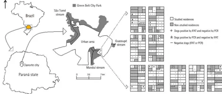
through IFAT. The PCR sensitivity was 0.5 pg. Figure 1 shows residential locations of
dogs with positive serology and PCR.
Figure 1. Location of Cianorte city, Paraná, Brazil. The study area and the analyzed dogs are indicated according to the results of the immunofluorescent antibody test
(IFAT) and polymerase chain reaction (PCR).
No association between the IFAT and PCR results was found (Fisher; p = 0.7554).
Additionally, no relation between IFAT or PCR results and other parameters (age,
sex, origin, habit of roaming freely and length of residence at the address) was found
Table 1. Epidemiological data and results of IFAT and PCR from dogs studied in Cianorte city, Paraná, Brazil
IFAT (n = 166) PCR (n = 167) Positive
(11 – 6.6%) Negative p*
Positive
(4 – 2.4%) Negative p*
Age
≤ 3 years 8 93 1 100
> 3 years 3 62 0.3095 3 63 0.1709
Sex
Male 8 95 2 102
Female 3 60 0.3403 2 61 0.4858
Origin
Urban area 8 109 1 106
Unknown place 3 46 0.5842 3 47 0.0955
Habit of roaming freely
Yes 9 2 3 91
No 84 71 0.0675 1 72 0.4106
Length of residence at the address
≤ 3 years 9 113 2 120
> 3 years 2 42 0.4035 2 43 0.2939
DISCUSSION
The study region in the urban area of Cianorte was considered by Lima et al. (8) a
place where Leishmania infection is highly probable due to the elevated
concentration of human ACL cases diagnosed from 1993 to 1998. In northwest
Paraná, Leishmania (Viannia) braziliensis has been isolated from both human and
canine ACL cases (6, 14). The role of dogs in ACL transmission cycle is still
controversial. It is not completely understood whether the animals participate or if
they are only accidentally infected with the parasite, as are humans (23).
In the present study, no dog showed suspected ACL lesions, but IFAT was positive in
6.6% of them. This test has been used in several studies to detect Leishmania
infection in dogs (24-27). Although serological cross-reactivity in visceral
leishmaniasis is well known, autochthonous cases of visceral leishmaniasis have
never been reported in Paraná (3, 28). Passos et al. (18) found 3.2% positivity in
Paraná state, several studies have demonstrated higher percentages of IFAT
positivity in dogs without lesions that lived in rural areas of endemic regions (5, 7, 15,
29). In these localities, dogs presenting ACL lesions have also been found (14, 15,
29, 30).
The PCR test using LU-5A and LB-3C primers had a sensitivity of 0.5 pg for DNA
from the L. braziliensis complex, close to that found by Harris et al. (22). DNA from
the L. braziliensis complex was detected in 2.4% of analyzed dogs. In other studies
of animals from endemic areas, higher percentages have been found in dogs without
suspected lesions. However, using both different protocols to obtain the DNA and
diverse targets for PCR, Reithinger et al. (31)found 7.1% positivity and Velásquez et
al. (29) 15.4% positivity, after an outbreak of human ACL. In both studies, dogs with
lesions were also discovered. The lower positivity for IFAT and PCR in the present
study may be related to the location from which these dogs originated, an urban
area, and to the fact that none of them had suspected lesions. Follow-up of positive
animals is essential to confirm the ACL diagnosis, as suggested by Madeira et al.
(32).
No association was found between positive IFAT and PCR results. Furthermore, in
other ACL endemic areas, no connection between serology and the presence of
Leishmania DNA has been reported (29, 33). An explanation for this finding is that
dogs had not yet developed an immune response to this infection (33). There was
also no relationship between positive results from these techniques and age, sex,
origin, length of residence or habit of roaming freely beyond the canine domiciliary
area. However, most animals that revealed positive serology (63.6%) and other four
positive PCR dogs (100%) were from either the urban area of the city or had been
found in its streets (strays); nine dogs (81.8%) with positive serology and three
(75.0%) with positive PCR were allowed to roam freely in streets and could enter the
edges of the forest. These data suggest that forest may constitute a possible natural
focus of ACL transmission in the urban area, where these dogs could have contact
with Leishmania (34, 35).
A similar work in Rio de Janeiro showed 74.6% seropositivity in dogs born either in
houses or somewhere else in the study area, and that all serologically positive
animals were allowed to leave their yards, suggesting that they participated in the
The present study demonstrated the presence of anti-Leishmania braziliensis
antibodies and of L. braziliensis-complex DNA in dogs from an urban area of
Cianorte, which indicates the importance of subsequently monitoring the dogs to
confirm the ACL diagnosis. Further studies should be carried out in order to better
understand ACL transmission in the urban area and the role of domestic dogs in its
cycle.
REFERENCES
1. Marzochi MCA. Leishmanioses no Brasil: as leishmanioses tegumentares. J Bras Med.
1992;63(1):82-104.
2. Gontijo B, Carvalho MLR. Leishmaniose tegumentar americana. Rev Soc Bras Med
Trop. 2003;36(1):71-80.
3. Ministério da Saúde. Secretaria de Vigilância em Saúde [Internet]. [place
unknown]: Secretaria de Vigilância em Saúde; 2009 [cited in 2009 Jan 20]. Available
from: http://portal.saude.gov.br/portal/saude/visualizar_texto.cfm?idtxt=25340.
4. Aguilar CM, Rangel EF, Garcia L, Fernandez E, Momen H, Grimaldi Filho G, et al.
Zoonotic cutaneous leishmaniasis due to Leishmania (Viannia) braziliensis
associated with domestic animals in Venezuela and Brazil. Mem Inst Oswaldo Cruz.
1989;84(1):19-28.
5. Silveira TGV, Teodoro U, Lonardoni MVC, Toledo MJO, Bertolini DA, Arraes
SMAA, et al. Investigação sorológica em cães de área endêmica de leishmaniose
tegumentar, no Estado do Paraná, sul do Brasil. Cad Saúde Pública.
1996;12(1):89-93.
6. Silveira TGV, Arraes SMAA, Bertolini DA, Teodoro U, Lonardoni MVC, Roberto
ACBS, et al. Observações sobre o diagnóstico laboratorial e a epidemiologia da
leishmaniose tegumentar no Estado do Paraná, sul do Brasil. Rev Soc Bras Med
Trop. 1999;32(4):413-23.
7. Lonardoni MVC, Silveira TGV, Alves WA, Maia-Elkhoury ANS, Membrive UA,
Membrive NA, et al. Leishmaniose tegumentar americana humana e canina no município
de Mariluz, Estado do Paraná, Brasil. Cad Saúde Pública. 2006;22(12):2713-6.
8. Lima AP, Comunello E, Minelli L, Teodoro U. Distribuição da leishmaniose tegumentar
por imagens de sensoriamento remoto orbital, no Estado do Paraná, Brasil. An Bras
9. Grimaldi JR G, David JR, McMahon-Pratt D. Identification and distribution of New
World Leishmania species characterized by serodeme analysis using monoclonal
antibodies. Am J Trop Med Hyg. 1987;36(2):270-87.
10. Grimaldi JR G, Tesh RB, McMahon-Pratt D. A review of the geographic
distribution and epidemiology of leishmaniasis in the New World. Am J Trop Med
Hyg. 1989;41(6):687-725.
11. Teodoro U, La Salvia Filho V, Lima EM, Spinosa RP, Barbosa OC, Ferreira MEMC, et
al. Observações sobre o comportamento de flebotomíneos em ecótopos florestais e
extraflorestais, em área endêmica de leishmaniose tegumentar americana, no norte do
Estado do Paraná, sul do Brasil. Rev Saúde Públ. 1993;27(4):242-9.
12. Brandão-Filho SP, Brito ME, Carvalho FG, Ishikawa EA, Cupolillo E,
Floeter-Winter L, et al. Wild and synanthropic hosts of Leishmania (Viannia) braziliensis in
the endemic cutaneous leishmaniasis locality of Amaraji, Pernambuco State. Brazil.
Trans R Soc Trop Med Hyg. 2003;97(3):291-6.
13. Aguilar CM, Rangel EF, Deane LM. Cutaneous leishmaniasis is frequent in
equines from an endemic area in Rio de Janeiro, Brazil. Mem Inst Oswaldo Cruz.
1986;81(4):471-2.
14. Lonardoni MVC, Teodoro U, Arraes SMAA, Silveira TGV, Bertolini DA, Ishikawa EAY,
et al. Nota sobre leishmaniose canina no noroeste do Estado do Paraná, sul do Brasil.
Rev Saúde Públ. 1993;27(5):378-9.
15. Zanzarini PD, Santos DR, Santos AR, Oliveira O, Poiani LP, Lonardoni MVC, et
al. Leishmaniose tegumentar americana canina em municípios do norte do Estado
do Paraná, Brasil. Cad Saúde Pública. 2005;21(6);1957-61.
16. Vedovello Filho D, Jorge FA, Lonardoni MVC, Teodoro U, Silveira TGV.
American cutaneous leishmaniasis in horses from endemic areas in the north-central
mesoregion of Paraná state, Brazil. Zoonoses Public Health. 2008;55(3):149-55.
17. Falqueto A, Coura JR, Barros GC, Grimaldi Filho G, Sessa PA, Carias VRD, et al.
Participação do cão no ciclo de transmissão da leishmaniose tegumentar no município de
Viana, Estado do Espírito Santo, Brasil. Mem Inst Oswaldo Cruz. 1986;81(2):155-63.
18. Passos VMA, Andrade AC, Silva ES, Figueiredo EM, Falcão AL. Inquérito canino em
foco recente de leishmaniose tegumentar no município de Sabará, região metropolitana
19. Instituto Brasileiro de Geografia e Estatística. Censo 2000 [Internet]. 10th ed. [place unknown]: Instituto Brasileiro de Geografia e Estatística; 2000 [cited in 2006 oct 30].
Available from: http://www.ibge.gov.br.
20. Silveira TGV, Arraes SMAA, Pereira DS, Lonardoni MVC, Dias MLGG, Ramos M,
et al. Avaliação da reação de imunofluorescência indireta para leishmaniose
tegumentar americana em pacientes da região noroeste do Estado do Paraná,
Brasil. Rev Unimar. 1990;12(1):177-88.
21. Chomczynski P, Sacchi N. Single-step method of RNA isolation by acid
guanidinium thiocyanate-phenol-chloroform extraction. Anal Biochem.
1987;162(1):156-9.
22. Harris E, Kropp G, Belli A, Rodriguez B, Agabian N. Single-step multiplex PCR
assay for characterization of New World Leishmania complex. J Clin Microbiol.
1998;36(7):1989-95.
23. Savani ESMM, Galati EAB, Camargo MCGO, D'Auria SRN, Damaceno JT,
Balduino SA. Inquérito sorológico sobre leishmaniose tegumentar americana em
cães errantes no Estado de São Paulo, Brasil. Rev Saúde Públ. 1999;33(6):629-31.
24. Barros GC, Sessa PA, Mattos EA, Carias VRD, Mayrink W, Alencar JTA, et al.
Foco de leishmaniose tegumentar americana nos municípios de Viana e Cariacica,
estado do Espírito Santo, Brasil. Rev Saúde Públ. 1985;19(2):146-53.
25. Follador I, Araujo C, Cardoso MA, Tavares-Neto J, Barral A, Miranda JC, et al.
Surto de leishmaniose tegumentar americana em Canoa, Santo Amaro, Bahia,
Brasil. Rev Soc Bras Med Trop. 1999;32(5):497-503.
26. Serra CMB, Leal CA, Figueiredo F, Schubach TM, Duarte R, Uchôa CMA, et al.
Leishmaniose tegumentar canina em Morada das Águias (Serra da Tiririca) Maricá, Rio
de Janeiro, Brasil. Cad Saúde Pública. 2003;19(6):1877-80.
27. Santos GPL, Sanavria A, Marzochi MCA, Santos EGOB, Silva VL, Pacheco RS,
et al. Prevalência da infecção canina em áreas endêmicas de leishmaniose
tegumentar americana, do município de Paracambi, Estado do Rio de Janeiro, no
período entre 1992 e 1993. Rev Soc Bras Med Trop. 2005;38(2):161-6.
28. Camargo ME, Rebonato C. Cross-reactivity in fluorescence test for Trypanosoma
and Leishmania antibodies. A simple inhibition procedure to ensure specific results.
29. Velásquez LG, Membrive N, Membrive U, Rodrigues G, Reis N, Lonardoni MVC, et
al. PCR in the investigation of canine American tegumentary leishmaniasis in
northwestern Paraná State, Brazil. Cad Saúde Pública. 2006;22(3):571-8.
30. Massunari GK, Voltarelli EM, Santos DR, Santos AR, Poiani LP, Oliveira O, et al.
A serological and molecular investigation of American cutaneous leishmaniasis in
dogs, three years after an outbreak, in the northwest of Paraná State, Brazil. Cad
Saúde Pública. 2009;25(1):97-104.
31. Reithinger R, Lambson BE, Barker DC, Davies CR. Use of PCR to detect
Leishmania (Viannia) sp. in dog blood and bone marrow. J Clin Microbiol.
2000;38(2):748-51.
32. Madeira MF, Uchôa CMA, Leal CA, Silva RMM, Duarte R, Magalhães CM, et al.
Leishmania (Viannia) braziliensis em cães naturalmente infectados. Rev Soc Bras
Med Trop. 2003:36(5):551-5.
33. Reithinger R, Espinoza JC, Coutenay O, Davies CR. Evaluation of PCR as
diagnostic mass-screening tool to detect Leishmania (Viannia) spp. in domestic dogs
(Canis familiaris). J Clin Microbiol. 2003;41(4):1486-93.
34. Camargo LB, Langoni H. Impact of leishmaniasis on public health. J Venom Anim
Toxins incl Trop Dis. 2006;12(4):527-48.
35. Voltarelli EM, Arraes SMAA, Perles TF, Lonardoni MVC, Teodoro U, Silveira
TGV. Serological survey for Leishmania sp. infection in wild animals from the
municipality of Maringá, Paraná state, Brazil. J Venom Anim Toxins incl Trop Dis.