Vasco Sequeira Oliveira
Role of Wnt signaling in heart disease
M in ho |2 01 0 Va sc o Se qu ei ra O liv ei ra R o le o f W n t si g n a lin g in h e a rt d is e a se
Master Thesis - Molecular Genetics
Vasco Sequeira Oliveira
Escola de Ciências
Role of Wnt signaling in heart disease
Work performed under both supervision of
Professor Sónia Pinho
and co-supervision of
Professor Adelino Leite Moreira
from the Faculty of Medicine of Porto
and co-supervision of
Professor Maria João Sousa
This master thesis dissertation would not have been possible without the guidance and the help of several individuals, who directly or indirectly contributed and extended their valuable assistance and knowledge, and to whom I express my deepest gratitude:
First and foremost, my utmost gratitude to Professor Adelino Leite Moreira for kindly accepting me at the Physiology Department of the Faculty of Medicine of Porto. At the same time, I cannot forget to mention all the encouragement and motivation thus entrusted to me, for which I am exceedingly grateful.
To my supervisor, Professor Sónia Pinho, whose support turned possible to hurdle the obstacles in the completion of the work and to whom I also extend my gratitude.
To Professor Maria João Sousa, I would like to express my feelings of appreciation and gratitude by all the support given and for the opportunity to undergo the Molecular Genetics Masters degree of the University of Minho.
I want to share my deepest gratitude to Dr. Inês Falcão Pires, for all the guidance, precious help, assistance, valuable advices and friendship along this time, which I will never forget. To Dr. Patrícia Terra and Dr. Carla Morgado from the Histology Department of the Faculty of Medicine of Porto, I would like to also thank for the scientific cooperation and assistance provided.
To the Physiology Department I also extend my gratitude: to Mr. Armando Jorge, Mrs. Margarida Silva, Mrs. Rosa Gonçalves, Professor Carmen Brás Silva, Dr. André Lourenço, Dr. Nádia Gonçalves, Mrs. Marta Oliveira and Dr. Daniel Gonçalves for all the kindness and prompt availability; a special note should be given to Dr. Antónia Teles and Mrs. Francelina Melo, for constantly displaying their help, confidence and sincerity towards me and my work.
I am also thankful to many of my friends there: to Inês Boal “Sequeira”, Daniela Silva, Dulce Fontoura, Marta Tavares, Carolina Rocha, Manuel Pinto, Luís Mendonça, Rui Cerqueira, Francisco Nóvoa and José Pedro Pinto, for all the joyful moments, constant
advices, which I will never forget.
I am also much indebted to Filipa Paiva, for all the kindness, patience and prompt help, which were very fruitful for the shaping of this thesis.
To my best friends, Daniela, Ivo, Katarina and Mousas, my deepest and sincere gratitude for putting up with me for so long, you are my backbone.
Last but certainly not least, I would like to show and express my enormous gratitude, devotion, honor and love to the most important people in my life: my parents and grandparents who deserve special mention for their tireless support and guidance. Thanks for instructing me the fundamental and basic truths of life and for all the caring and dedication ever since I was a child.
Words fail me to convey my deep gratitude to my brothers, Valtinho and Viti, whose persistence, assurance and unfailing support, have lightened my way.
Heart failure has a major social-economic impact in our society. Despite major advances in the understanding of this pathology, the mechanisms of its development, as well as its pathophysiology, remain unclear. Therefore, it is our priority to clarify how extra- and intracellular factors are able to modulate heart function. Several pathways and/or factors had already been associated with different phases of heart failure development namely TGF-β, IGF, calcineurin, several GPCRs, MAPK, Akt and GSK-3. More recently, several studies started shedding some light on a putative role of Wnt signaling in heart failure development.
Wnt signaling is a major regulator of cell-fate specification during development, proliferation, survival, migration and adhesion. Several diseases including cancer, diabetes, osteoporosis and psychiatric disorders are the result of deregulation of canonical Wnt signaling, due to either genetic alterations or changes in the levels of its effectors. The role of canonical Wnt signaling in heart development is well established and it has been shown to be biphasic, in the sense that its activation is initially required for the commitment of cells to a cardiac lineage and in its inhibition, cardiogenesis is triggered. In heart failure development, a possible role for Wnt signaling has only recently been reported, yet, its results are contradictory. Nonetheless, it was not addressed a possible role exerted by extracellular modulators and receptors of the Wnt pathway. Because of its role in the development of other diseases, and since its extracellular and membrane effectors are regarded as potential targets of pharmacological intervention in the treatment of such pathologies, it became imperative the understanding of Wnt signaling regulation in heart disease and how these interventions would affect heart function. Taking these facts into account, our first goal was to perform a detailed gene expression analysis of different Wnt ligands, receptors and co-receptors, during heart disease development in a type 1 diabetes mellitus rat model. Since in other contexts, Wnt signaling interacts with other pathways known to present a role in the development of diabetic heart disease, such as PPARs and FOXO proteins, we also checked their expression levels. With this approach we aimed starting to unveil a possible role for Wnt signaling in heart disease development as well as possible interactions with other pathways, known to be important of this pathology.
A insuficiência cardíaca apresenta um impacto socioeconómico grande na nossa sociedade. Apesar de grandes avanços na compreensão desta patologia, os mecanismos do seu desenvolvimento, assim como a sua fisiopatologia, permanecem obscuros. De tal forma, é nossa prioridade o esclarecimento de como factores extra- e intracelulares são capazes de modular a função cardíaca. Diversas vias e/ou factores já foram associados a diferentes fases do desenvolvimento de insuficiência cardíaca, nomeadamente TGF-β, IGF, calcineurina, várias GPCRs, MAPK, Akt e GSK-3. Mais recentemente, vários estudos sugerem/apontam um potencial papel da via dos Wnts, no desenvolvimento de insuficiência cardíaca.
A via das Wnts é um importante regulador do desenvolvimento, proliferação, sobrevivência e adesão celulares. Várias doenças como cancro, diabetes, osteoporose e disfunções psiquiátricas, são o resultado da desregulação da via canónica das Wnts, devido a alterações genéticas ou alterações a nível celular dos seus factores.
A sua função no desenvolvimento cardíaco é bem conhecida e revelou-se bifásica, já que, inicialmente, a sua activação é necessária para diferenciação numa linhagem cardíaca e posteriormente, a sua inibição activa a cardiogénese. Vários estudos sugerem um potencial envolvimento da via das Wnts na insuficiência cardíaca, no entanto, os seus resultados são contraditórios. Assim, não foi possível identificar o papel desempenhado por moduladores extracelulares e receptores desta via. Devido ao seu papel no evoluir de outras doenças, e porque os seus receptores são potenciais alvos de intervenções farmacológicas no tratamento de tais patologias, tornou-se indispensável o conhecimento da via das Wnts na doença cardíaca e como essas intervenções poderão afectar o coração. Assim, o nosso primeiro objectivo passou por realizar uma análise à expressão genética dos vários ligandos, receptores e co-receptores, durante o desenvolvimento da doença cardíaca num modelo de rato com diabetes tipo 1. Dado que em outros contextos a via das Wnts interagir com outras vias conhecidas por deterem um papel no desenvolvimento da cardiomiopatia diabética, tais como PPARs e FOXOs, também analisamos os seus níveis de expressão. Com esta abordagem, pretendemos revelar o potencial papel da via das Wnts na fisiopatologia da doença cardíaca, assim como, possíveis interacções com outras vias relevantes e associadas a esta patologia.
1 | WNT SIGNALING ... 19
1.1 Wnt/β-catenin signaling (Canonical Wnt pathway) ... 19
1.2 Wnt/β-catenin independent pathway (Non-canonical Wnt signaling) ... 21
2 | CANONICAL WNT SIGNALING: EXTRACELLULAR AND MEMBRANE PLAYERS ... 22
2.1 Wnt ligands ... 22
2.2 Wnt receptors ... 22
2.2.1 Fzd receptors ... 22
2.2.2 LRP5/6 receptors ... 23
2.3 Unusual Wnt receptors: Drl/Ryk and Ror2 ... 24
3 | OTHER EXTRACELLULAR PLAYERS: ANTAGONISTS AND AGONISTS ... 24
3.1 The antagonists ... 24
3.2 The agonists ... 25
4 | INTRACELLULAR PLAYERS ... 25
4.1 Dishevelled ... 25
4.2 β-catenin structure ... 26
4.3 The ‘scaffold’ destruction complex of β-catenin: GSK-3, CK1, Axin and APC ... 27
5 | CANONICAL WNT SIGNALING ACTIVATION ... 29
6 | NUCLEAR EVENTS ... 32
7 | THE ROLE OF WNT SIGNALING:EMBRYONIC AND ADULT HEART ... 33
7.1 Embryonic heart ... 33
7.2 Adult heart ... 33
8 | DIABETES MELLITUS AND HEART DISEASE ... 35
9 | THE ROUTE FROM DIABETES MELLITUS TO OXIDATIVE STRESS AND VICE-VERSA/BACK ... 36
9.1 ‘Forkhead box, subclass O’ (FOXO) ... 37
9.1.1 FOXO members regulation: Post-translational modifications (PTMs) ... 38
9.1.2 FOXOs nuclear exclusion/negative regulation ... 38
9.1.3 FOXOs nuclear import/positive regulation ... 39
9.2 FOXO and canonical Wnt signaling: β-catenin, the Libra ... 40
10 | LIPID HOMEOSTASIS ... 41
10.1 Peroxisome Proliferator-Activated Receptors (PPARs) ... 42
10.2 PPARs in the heart ... 43
10.3 PPARs convergence with canonical Wnt signaling and FOXOs ... 44
12.2 Echocardiography assessment ... 50 12.3 Myocardial function ... 50 12.4 Molecular Studies ... 51 12.5 Statistical analysis ... 53 13 | RESULTS ... 57 13.1 Echocardiographic evaluation ... 57 13.2 General features ... 58 13.3 Myocardial function ... 59 13.4 Gene expression ... 61 14 | DISCUSSION ... 81
14.1 Effects of diabetes mellitus: myocardial structure ... 81
14.2 Gene expression ... 82
15 | CONCLUSIONS ... 93
16 | SUPPLEMENTS ... 97
APC Adenomatus Polyposis Coli LV Left Ventricle
AU Arbitrary units MAK Metastasis-associated kinase
BCL9 B-cell lymphoma 9 MAPK Mitogen-activated protein kinase BNP Type-B natriuretic peptide MMTV Mouse mammary tumors virus
BSA Body surface area MSC Mesenchymal Stem Cell
CAMKII Ca2+/calmodulin-dependent protein kinase II
MST1 Ste20-like kinases
CBP CREB binding protein n Number of experimental units CDK2 Cyclin-dependent kinase 2 NCoR Nuclear receptor corepressor CK Casein kinase NFAT Nuclear factor of activated T Cells CPCs Cardiac Progenitor Cells NHR Nuclear hormone receptor CREB cAMP response element-binding NPC Nuclear pore complex
CRM1 Chromosomal region maintenance 1 PAR-1 Protease-activated receptor 1 CSCs Cardiac Stem Cells PCP Planar cell polarity
CtBP C-terminal binding protein PtdIns Phosphatidylinositol
CTRL Control PI3K PtdIns-3-kinase
DKK1 Dickkopf 1 PI4KIIa PtdIns-4-kinase type II
DM Diabetes mellitus PIP5KI PtdIns-4-phosphate 5-kinase type I
Drl Derailed PIP2 PtdIns(4,5)-biphosphate
Dvl Dishevelled PKA cAMP-dependent protein kinase
DYRK1 Dual-specificity
tyrosine-phosphorylated and regulated kinase 1
PKB/Akt Protein kinase B
ECs Endothelial Cells PKC Protein kinase C
EF Ejection Fraction PLC Phospholipase C
ERK Extracellular-signal-regulated kinase PPARs Peroxisome proliferator-activated receptors
ET-1 Endothelin-1 PRMTs Protein arginine methyltransferases FOXO Forkhead box, subclass O PTMs Post-translational modifications
FS Fractional shortening Pygo Pygopus
FZD Frizzled p300 CREB binding protein-associated
factor GAPDH Glyceraldehyde-3-phosphate
dehydrogenase
RGS Regulators of G proteins signaling GLUT4 Glucose transporter 4 ROS Reactive Oxygen Species
GPCRs G protein-coupled receptors Rspo R-spondin
GRK G protein-coupled receptor kinases RXR Retinoid X receptor
Gro Groucho SCs Stem Cells
GSK-3 Glycogen synthase 3 SCR-1 Steroid receptor co-activator-1 HR Heart Rate sFRPs Secreted frizzled-related proteins IGF Insulin growth factor SGK Serum- and glucocorticoid-inducible
kinase
IL-1 Interleukin-1 SMCs Smooth muscle cells
IKK IkappaB kinase SMRT Silencing mediator of retinoid, thyroid hormone receptors IP Intraperitoneal SCFβ-TrCP Skp1/Cul1/F-boxβ-TrCP
JNK C-jun kinase SEM Standard error of the mean
LDL Low-density lipoprotein Stbm Strabismus LEF Lymphoid enhancer-binding factor STZ Streptozotocin LRP LDL-receptor related protein TCF T Cell factor
protein Xpo1 Exportin 1
IMAGE 1|OVERVIEW OF CANONICAL WNT SIGNALING. ... 20
IMAGE 2|DVL STRUCTURE... 26
IMAGE 3| β-CATENIN DOMAINS STRUCTURE. ... 26
IMAGE 4|AXIN STRUCTURE. ... 28
IMAGE 5|APC DOMAINS STRUCTURE... 28
IMAGE 6|MODEL OF WNT RECEPTOR ACTIVATION:INITIATION AND AMPLIFICATION. ... 30
IMAGE 7|MODEL OF WNT RECEPTOR ACTIVATION:SIGNALSOME. ... 31
IMAGE 8|MODEL OF WNT RECEPTOR ACTIVATION:RECEPTOR ENDOCYTOSIS. ... 31
IMAGE 9| FOXOs STRUCTURE. ... 37
IMAGE 10| β-CATENIN, THE LIBRA. ... 40
TABLE 1|SPECIFIC PCR PRIMER PAIRS USED IN THE WORK. ... 51
TABLE 2|SPECIFIC PCR PRIMER PAIRS USED IN THE WORK. ... 53
TABLE 3|DOPPLER ECHOCARDIOGRAPHIC MEASUREMENTS... 57
TABLE 4|GENERAL FEATURES OF CTRL AND DM ANIMALS. ... 58
TABLE 5|COMPARISON BETWEEN GROUPS OF THE MEAN RELATIVE EXPRESSION FOR BNP AND ET-1 GENES. ... 62
TABLE 6|COMPARISON BETWEEN GROUPS OF THE MEAN RELATIVE EXPRESSION OF THE HIGHEST EXPRESSED WNT GENES. ... 63
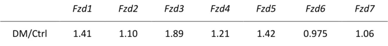
TABLE 7|COMPARISON BETWEEN GROUPS OF THE MEAN RELATIVE EXPRESSION FOR FZD GENES. ... 63
TABLE8|COMPARISON BETWEEN GROUPS OF THE MEAN RELATIVE EXPRESSION FOR FZD AND LRP GENES. ... 65
TABLE 9|COMPARISON BETWEEN GROUPS OF THE MEAN RELATIVE EXPRESSION FOR WNT AND UNRELATED WNT GENES.. ... 66
TABLE 10|COMPARISON BETWEEN GROUPS OF THE MEAN RELATIVE EXPRESSION FOR FZD, LRP6 AND UNUSUAL RECEPTOR GENES.. ... 67
TABLE 11|COMPARISON BETWEEN GROUPS OF THE MEAN RELATIVE EXPRESSION FOR GATA4,AXIN2 AND CYCLIND1 GENES. ... 67
TABLE 12|COMPARISON BETWEEN GROUPS OF THE MEAN RELATIVE EXPRESSION FOR KINASES AND LRP6 GENES. . 70
TABLE 13|COMPARISON BETWEEN GROUPS OF THE MEAN RELATIVE EXPRESSION FOR PPAR GENES. ... 70
TABLE 14|COMPARISON BETWEEN GROUPS OF THE MEAN RELATIVE EXPRESSION FOR Foxo GENES.. ... 73
TABLE 15|COMPARISON BETWEEN GROUPS OF THE MEAN RELATIVE EXPRESSION FOR GSK-3β, ET-1, GATA4 AND BNP GENES AT FOUR- AND SIX-WEEKS OF TREATMENT.. ... 73
TABLE 16|COMPARISON BETWEEN GROUPS OF THE MEAN RELATIVE EXPRESSION FOR FZD3, FZD5, WNT2B, WNT11 AND LRP6 GENES AT FOUR- AND SIX-WEEKS OF TREATMENT. ... 76
TABLE 17|COMPARISON BETWEEN GROUPS OF THE MEAN RELATIVE EXPRESSION FOR PPARγ, Foxo3 AND Foxo4 GENES AT FOUR- AND SIX-WEEKS OF TREATMENT. ... 77
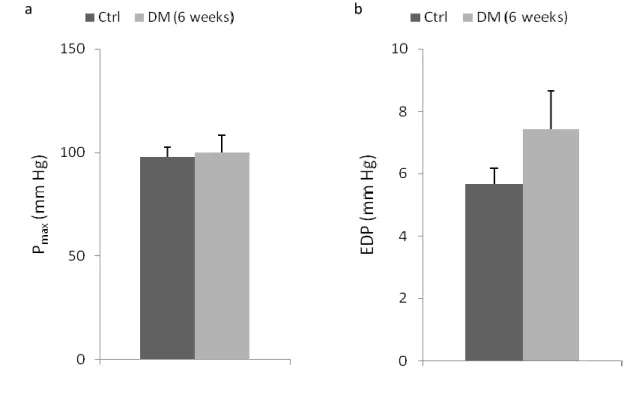
FIGURE 1|BASELINE HEMODYNAMIC ASSESSMENT OF LEFT VENTRICLE FUNCTION.. ... 59
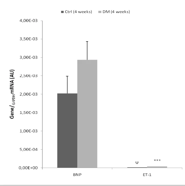
FIGURE 2|BASELINE HEMODYNAMIC ASSESSMENT OF LEFT VENTRICLE FUNCTION. ... 60 FIGURE 3|EXPRESSION OF BNP AND ET-1 GENES, IN LEFT VENTRICLE HEART SAMPLES OF FOUR-WEEK ANIMALS,
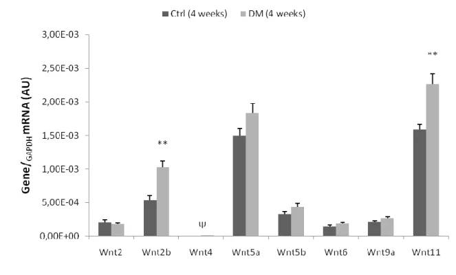
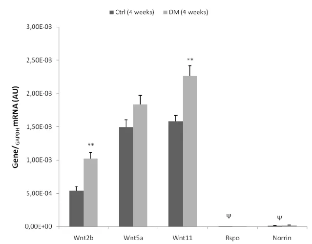
COLLECTED AFTER TREATMENT. ... 61 FIGURE 4|EXPRESSION OF WNT GENES, IN LEFT VENTRICLE HEART SAMPLES OF FOUR-WEEK ANIMALS, COLLECTED
AFTER TREATMENT... 62
FIGURE 5|EXPRESSION OF FZD GENES, IN LEFT VENTRICLE HEART SAMPLES OF FOUR-WEEK ANIMALS, COLLECTED AFTER TREATMENT... 64 FIGURE 6|EXPRESSION OF RELEVANT FZD AND LRP GENES, IN LEFT VENTRICLE HEART SAMPLES OF FOUR-WEEK
ANIMALS, COLLECTED AFTER TREATMENT... 64
FIGURE 7|EXPRESSION OF RELEVANT WNT AND UNRELATED WNT GENES, IN LEFT VENTRICLE HEART SAMPLES OF FOUR-WEEK ANIMALS, COLLECTED AFTER TREATMENT. ... 65 FIGURE 8|EXPRESSION OF FZD, LRP6 AND UNUSUAL RECEPTOR GENES, IN LEFT VENTRICLE HEART SAMPLES OF FOUR
-WEEK ANIMALS, COLLECTED AFTER TREATMENT. ... 66 FIGURE 9|EXPRESSION OF GATA4,AXIN2 AND CYCLIND1 GENES, IN LEFT VENTRICLE HEART SAMPLES OF FOUR-WEEK
ANIMALS, COLLECTED AFTER TREATMENT... 68 FIGURE 10|EXPRESSION OF RELEVANT KINASES AND LRP6 GENES, IN LEFT VENTRICLE HEART SAMPLES OF FOUR-WEEK ANIMALS, COLLECTED AFTER TREATMENT... 69 FIGURE 11|EXPRESSION OF PPAR GENES, IN LEFT VENTRICLE HEART SAMPLES OF FOUR-WEEK ANIMALS, COLLECTED
AFTER TREATMENT... 71
FIGURE 12|EXPRESSION OF Foxo GENES, IN LEFT VENTRICLE HEART SAMPLES OF FOUR-WEEK ANIMALS, COLLECTED AFTER TREATMENT... 72 FIGURE 13|EXPRESSION OF GSK-3β, ET-1, GATA4 AND BNP GENES, IN LEFT VENTRICLE HEART SAMPLES OF SIX-WEEK
ANIMALS, COLLECTED AFTER TREATMENT... 74
FIGURE 14|EXPRESSION OF FZD3, FZD5, WNT2B, WNT11 AND LRP6 GENES, IN LEFT VENTRICLE HEART SAMPLES OF SIX-WEEK ANIMALS, COLLECTED AFTER TREATMENT.. ... 75 FIGURE 15|EXPRESSION OF PPARγ, Foxo3 AND Foxo4 GENES, IN LEFT VENTRICLE HEART SAMPLES OF SIX-WEEK
I
NTRODUCTION
"Learning never exhausts the mind.” Leonardo da Vinci
1 | W
NT SIGNALINGWnt signaling is known to play a critical role in a vast array of biological processes, by which regulate cell proliferation, polarity establishment, migration, cell fate differentiation and stem cell self-renewal [1-2]. As a consequence of their participation in a multitude of cellular events, mutations in the Wnt signaling pathway are usually involved in diseases, such as cancer, premature osteoporosis, diabetes and cardiovascular diseases
[1-2]
.
The Wnt pathway is categorically divided in three branches, taking mostly into account, whether they require or not the transcription factor β-catenin: the canonical or Wnt/β-catenin signaling [3], the Planar Cell Polarity (PCP) [4] and the Wnt/Ca2+ pathways [5]. Of the three, canonical Wnt signaling is the best understood and acts by regulating the amount of the transcriptional regulator β-catenin, whereas the other two act independent of β-catenin and are termed non-canonical or β-catenin-independent pathways [2-5].
1.1 Wnt/β-catenin signaling (Canonical Wnt pathway)
β-catenin, a homolog of the fly Armadillo, is a key component of the cadherin cell adhesion system and the canonical Wnt signaling [6-7]. In the absence of a Wnt ligand, cytoplasmic β-catenin levels are continuously being kept low, because free β-catenin is being targeted to a proteasome-mediated degradation and only membrane β-catenin complexed with the cadherin cell adhesion system is protected from degradation. β-catenin degradation is achieved through the formation of a rigorous complex, composed of the scaffolding protein Axin, the glycogen synthase kinase 3 (GSK-3), the tumor suppressor Adenomatus Polyposis Coli gene product (APC) and Casein Kinase 1 (CK1) [1, 3,
8]
. This protein complex promotes β-catenin sequential phosphorylation exerted by CK1 and GSK-3, with subsequent recognition and binding to SCFβ-TrCP, an E3 ubiquitin-protein ligase, leading to its ubiquitylation and further proteasomal degradation by the 26S proteasome [9-10]. Removal of cytoplasmic β-catenin prevents it from entering the nucleus and binding to the T cell factor (TCF) and lymphoid enhancer-binding factor (LEF) proteins, which is being repressed by the Groucho (Gro) family of transcription repressors [2]. Thereby, inhibiting the role of β-catenin as a transcriptional co-activator of target genes.
Conversely, upon reception of Wnt ligands, β-catenin levels increase. This is mostly due to stabilization against proteolysis of uncomplexed β-catenin in the cell and is independent of the cadherin cell adhesion system. Wnt ligands are known to interact with cell surface receptors, such as Frizzled (Fzd) receptors and its co-receptor [low-density lipoprotein (LDL)-receptor-related protein] (LRP), leading to the formation of a ternary Fzd-Wnt-LRP complex [1, 3]. Consequently, this complex by the action of the Fzd receptor, activates and recruits the Dishevelled (Dvl) cytoplasmic phosphoproteins, resulting in LRP phosphorylation and activation, with following Axin recruitment to the plasma membrane
[2-3]
. This results in the disruption of the degradation complex, thence blocking cytoplasmic β-catenin degradation. Stabilized cytoplasmic β-catenin translocates into the nucleus, where displaces Groucho from the co-activators TCF/LEF, activating Wnt target genes [2-3]. Taken together these observations, is noteworthy that instead the traditional cascade of phosphorylation/dephosphorylation events or the production of intracellular second messenger proteins, cells constantly synthesize and degrade β-catenin, unless canonical Wnt pathway is initiated [10].
Image 1 | Overview of canonical Wnt signaling.
a) In the absence of Wnt ligands, cytoplasmic β-catenin forms a complex with Axin, APC, GSK-3 and CK1,
leading to its phosphorylation, further recognition by the E3 ubiquitin ligase β-TrCP and targeting to proteasomal degradation. Wnt target genes are repressed by TCF/LEF-Groucho association.
b) In the presence of Wnt ligands, a trimeric complex consisted by Fzd-Wnt-LRP is formed. Heteromeric
G-proteins and Dvl G-proteins are activated, leading to the recruitment of Axin to LRPs. This disrupts the degradation complex, subsequent β-catenin stabilization and nuclear translocation, where displaces Groucho from TCF/LEF, activating Wnt target genes.
1.2 Wnt/β-catenin independent pathway (Non-canonical Wnt signaling)
The existence of alternative Wnt signaling pathways, termed non-canonical Wnt pathways, is being supported by accumulating evidences over the last decade. Nonetheless, the precise molecular details are still to be unraveled. Several proteins have been reported to be involved in non-canonical Wnt signal transduction pathways namely Fzd receptors, the transmembrane protein strabismus (Stbm), phospholipase C (PLC), protein kinase C (PKC), Ca2+/calmodulin-dependent protein kinase II (CAMKII), c-jun kinase (JNK), Rho family GTPases and Dvl proteins [4-5, 11-12].
Various reports suggested that non-canonical Wnt signaling might be separated into two distinct pathways, including the Planar Cell Polarity (PCP) and the Wnt/Ca2+ pathways, which may aid to explain the different downstream gene profile observed. In the presence of Wnt ligands, PCP signaling is initiated through Fzd and Stbm (activate Dvl proteins) that in turn activate Rho family of GTPases, Rac and RhoaA, stimulating JNK activity with subsequent JNK-mediated transcriptional regulation [13-14].
On the other hand, Wnt/Ca2+ pathways promote intracellular calcium increase due to PLC activation, after G proteins induction (activated by Fzd receptors). Ca2+-sensitive proteins, such as CAMKII and PKC, detect the increased intracellular Ca2+, causing nuclear translocation of nuclear factor of activated T cells (NFAT), a Ca2+-regulated transcription factor [12, 15].
Yet, other models proposed the incorporation of both these pathways into a single non-canonical Wnt pathway or Wnt regulatory network. Of note, is that effectors of the Wnt pathway, such as Fzd and Dvl, appear to function in both canonical and non-canonical pathways, turning the understanding of Wnt signaling an even more complex task, because it is possible that the two could be simultaneously activated and functionally interacting. In addition, another interesting feature about these signaling pathways is the antagonistic regulation observed, where activation of one might even repress activation of the other [16].
2 | C
ANONICAL WNT SIGNALING:
EXTRACELLULAR AND MEMBRANE PLAYERS 2.1 Wnt ligandsIn 1973, Sharma et al. [17] isolated a Drosophila melanogaster mutant gene termed ‘wingless’ (wg; a fly with no wings), which was subsequent related to cause abnormal wing and mesothorax developments [18]. A decade later, Nusse et al. [19] reported a new gene that was responsible for mammary tumors development (mouse mammary tumors virus; MMTV) in mice, called ‘INT-1’ and just three years later, Rijsewijk et al. [20] identified INT-1 as the mammalian homologue of Drosophila gene wingless. The name ‘WNT’ (wingless-type MMTV integration site family) thus alludes to the original genes wingless and INT-1.
Till date, at least, 19 Wnt ligand members had been reported in mammals, conferring a high degree of complexity and Wnt signaling specificity [21]. Wnts are cysteine-rich secreted proteins (presenting up to 23 or 24 highly conserved cysteine residues) of approximately 350-400 amino acids and highly conserved throughout evolution (conserved in all metazoan animals) [19-20]. These proteins are monomeric and share a Wnt signature motif (C-K-C-H-G-[LIVMT]-S-GS-C) [22]. Active Wnts can also be found expressed combinatorially [23], which appears to activate signaling in a distinct manner.
2.2 Wnt receptors
Two distinct receptor families are critical for the activation of canonical Wnt signaling [1]. The first, belongs to the Fzd class of proteins that are essential for both canonical and non-canonical Wnt signaling, and constitute high affinity Wnt receptors. The second, comprise members of a single transmembrane-spanning protein family recognized as the gene arrow in Drosophila [24] and as LRP5/6 in vertebrates [25], which function specifically as Fzd co-receptors in canonical Wnt signaling.
2.2.1 Fzd receptors
In the mammalian genome, 10 Fzd protein members had been identified to play a central role in Wnt signaling [26]. Interestingly, because Fzds present seven transmembrane-spanning (7TM) domains that evocates of classical G protein-coupled receptors (GPCRs), they comprise a separate class of GPCRs, the “Class Frizzled” [27].
Fzd proteins expose their large N-terminal domain, that contains a cysteine-rich domain, on the extracellular side and to which, it binds Wnt ligands [28]. Besides one N-terminal region, structural analyses of Fzd receptors predicts, three extra- and three intracellular loops, and an intracellular C-terminal domain [29]. The C-terminal domain contains a PDZ (Postsynaptic density 95, Discs large, Zonula occludens-1)-binding domain, by which cytoplasmic proteins interact with the receptor [29-30]. Amino acid sequence analyses showed that the Fzd sequence is highly rich in putative consensus sites for various serine/threonine and tyrosine kinases [29].
2.2.2 LRP5/6 receptors
The low-density lipoprotein (LDL) receptor (LDLR) family consists of cell surface proteins that are involved in receptor-mediated endocytosis of cognate ligands [31-32]. Two of these members, LRP5 and LRP6 comprise a subfamily of the LDLR family, which mediate diverse steps in metabolism and development.
LRP5 and LRP6 are highly homologous proteins that present high co-expression during embryogenesis and adult tissues remodeling [33-34]. It is likely that LRP6 plays a more dominant role during embryogenesis [25], while LRP5 is critical in adult tissue homeostasis [35]. Most of the LRP amino acid sequence is localized extracellular and consists of YWTD (tyrosine, tryptophan, threonine and aspartic acid) domains, EGF (epidermal growth factor)-like domain and LDL repeats [36-37]. Intriguingly, although being a co-receptor for canonical Wnt signaling, LRPs extracellular domain has a poor affinity to Wnt ligands compared to Fzds, yet, bind with high affinity to its antagonists Dickkopf1 (Dkk1) and Sclerostin [38-39]. The YWTD domain is important for LRPs endoplasmic reticulum maturation and membrane trafficking, which requires a specific chaperone molecule called Boca in Drosophila [40] and Mesd in mice [41].
The intracellular domain has proline-, serine- and threonine-rich residues and contains five PPPSP*x+S (‘x’ denotes any amino acid) repetitive motifs [42]. The phosphorylation of these motifs is a requirement for Axin recruitment and subsequent β-catenin stabilization. Upstream of the PPPSP[x]S repeats exist an S/T cluster that is also a target for phosphorylation [43].
Till date, five classes of protein kinases had been identified to phosphorylate LRPs, being divided in two groups. The first, comprises proline-directed kinases which
phosphorylate PPPSP motifs, such as GSK-3 [44], cAMP-dependent protein kinase (PKA) [45], Pftk members [46] and G protein-coupled receptor kinases 5/6 (GRK5/6) [47]. The second contains non-proline-direct kinases like CK1 family members [48] that phosphorylate [x]S sites, the S/T cluster and additional N-terminal regions.
2.3 Unusual Wnt receptors: Drl/Ryk and Ror2
Derailed (Drl), a transmembrane tyrosine kinase receptor from the RYK subfamily, has been shown to be an unusual, yet, essential component of Wnt signaling [49]. Drosophila Wnt5 (Dwnt5), is a regulator of axon guidance in the central nervous system, and embryos lacking Dwnt5, share a similar phenotype to those that lack Drl, that is, they display aberrant neuronal projections across the midline. Drl binds to Dwnt5, through its extracellular Wnt inhibitory factor (WIF) domain, indicating that it is a Dwnt5 receptor in the central nervous system [50]. However, how Drl downstream signals are transduced, remain unclear. Unlike Drl in Drosophila, the mammalian Ryk homolog functions as a co-receptor along with Fzd [51].
Another tyrosine kinase receptor is known to exist, Ror2, which contains a cysteine-rich domain similar to that of Fzd [52]. Upon Wnt5a binding to Ror2, an inhibitory action over canonical Wnt signaling takes place, although paradoxically, Wnt5a can also induce activation of the canonical pathway by directly engaging Fzd4 [53] or Fzd5 [54].
3 | O
THER EXTRACELLULAR PLAYERS:
ANTAGONISTS AND AGONISTS3.1 The antagonists
Wnt antagonists can be categorically divided according to their model of action and to their ability to inhibit canonical Wnt signaling. Some proteins like Dickkopf (Dkk)
[55]
, Wise [56] and Sclerostin [38], which are capable of inhibiting canonical Wnt signaling by binding to the LRP5/6 receptor, are grouped and classified together. On the other hand, those like secreted frizzled-related proteins (sFRPs) [57], Wnt inhibitory factor 1 (WIF1) [58] and Xenopus Cerberus [59], directly bind to and inhibit Wnts and thence, are classified as the sFRP class. In addition, sFRPs are also capable of binding and blocking the access of Wnts to Fzd receptors, thereby, presenting dual inhibitory functions of Wnt signaling [60].
Thus, theoretically, components of the sFRP class will inhibit both canonical and non-canonical pathways, while those binding to LRPs, will specifically inhibit the non-canonical pathway [61].
3.2 The agonists
Norrin and R-spondin (Rspo) are at least two types of proteins that are unrelated to Wnt ligands and activate the Fzd/LRP receptors [62-63]. Norrin is a secreted protein that is mutated in Norrie disease, which is a developmental disorder defined by vascular abnormalities in the eye. Norrin acts by binding with high affinity to Fzd4 and therefore, activates the canonical Wnt signaling in an LRP5/6-dependent manner [62]. The Rspo proteins constitute a novel class of ligands that induce canonical Wnt signaling, by exhibiting synergy with Wnts, Fzds, and LRP6 [63]. Rspo2 genes are often co-expressed in a variety of tissues with Wnts, raising the possibility that Rspo2 proteins display a positive feedback role, in order to reinforce canonical Wnt signaling [63]. In line with this notion, it had already been demonstrated that Rspo proteins are capable of physically interacting with the extracellular domains of LRP6 and Fzd8, therefore activating Wnt target genes
[64].
4 | I
NTRACELLULAR PLAYERS4.1 Dishevelled
Dishevelled (Drosophila: dsh; Mammalian: Dvl) is a ubiquitously expressed cytoplasmic scaffolding protein that is known to interact with activated Fzd receptors. Dsh contains 750 amino acids, displaying high homology with three Dvl homologue genes (DVL1, DVL2 and DVL3) that had been identified in mice and humans [65]. All Dvl family members possess three conserved regions: an N-terminal DIX (Dishevelled/Axin) domain (also found in the terminus of Axin proteins), a central PDZ domain (also found in the C-terminal domain of Fzds) and a C-C-terminal DEP domain, implicated in membrane targeting
[66]
. Additionally, other two conserved regions had been implicated to mediate protein interactions and/or phosphorylation, the basic and the proline-rich regions [67].
Image 2 Image 2 | Dvl structure.
It is interesting to mention that, like Wnts and Fzd receptors, also Dvl is a common player between both canonical and non-canonical Wnt pathways and, like β-catenin, it can shuttle between the cytoplasm and the nucleus, yet, it is unclear how the nuclear localization of Dvl is governed.
4.2 β-catenin structure
β-catenin, a protein of 781 amino acids, is arranged in three distinct regions that share a 71% identical amino acid sequence to the fly Armadillo gene product [68]. It possesses a large central region (residues 141-664) composed of 12 repeats, known as the Armadillo (Arm) repeats, being each arm disposed in a three α-helix configuration. The arm repeats are structurally very similar, however, presenting some irregularities among the whole arm repeats structure [69].
Image 3 | β-catenin domains structure.
Interestingly, the 12 arm repeat structure forms a stiff scaffold for the binding of many factors, such as TCF, Axin and APC [70]. Contrasting, the N- and C-terminal domains are much smaller and much more flexible [10, 68]. Some authors suggested that these two domains would interact with the central arm domain by a fold-back mechanism, regulating the ability of this region to bind to different co-factors [71-72]. Plus, because these two domains are negatively charged, contrasting to the highly positively charged arm domain, is deductible that the three domains interact in a highly and non-specific, dynamic fashion.
4.3 The ‘scaffold’ destruction complex of β-catenin: GSK-3, CK1, Axin and APC
As already stated, cytoplasmic β-catenin downregulation is carried out by the formation of a multiprotein destruction complex that targets β-catenin to proteasomal degradation.
GSK-3 is a ubiquitously expressed constitutively active serine/threonine kinase (GSK-3 active form is dephosphorylated), responsible for cellular substrates phosphorylation and is one of the kinases responsible for β-catenin phosphorylation to subsequent proteasome recognition. This kinase specifically recognizes and phosphorylates critical residues in the N-terminal region of β-catenin (Ser33, Ser37 and Thr41), however, by itself, GSK-3 does not efficiently phosphorylate β-catenin [10]. In fact, Liu et al. [73] demonstrated that β-catenin phosphorylation by GSK-3, is preceded by a “priming” phosphorylation step governed through a member of the CK1 family of kinases, CK1α, also in the N-terminal region of β-catenin (Ser45).
At least, three CK1s have been implicated in the canonical Wnt signaling namely CK1α, CK1ε and CK1γ, being the role of the first isoform the best characterized of this family [10]. The potential role of this kinase is well illustrated in CK1α deletion experiments, where high concentration levels of β-catenin are observed, through inhibition of β-catenin phosphorylation and further cytoplasmic accumulation [73].
At the ‘heart’ of the multiprotein destruction complex lays the scaffolding protein Axin, which plays a critical role in bringing GSK-3, CK1α, and β-catenin together to efficiently promote the phosphorylation reaction [10]. Surprisingly, Axin concentration is extremely low when compared to other components, in frog embryos experiments, suggesting that Axin is a rate limiting factor to the assembly of the destruction complex
[74]
. Moreover, the importance of Axin in β-catenin degradation is highlighted by the increased levels of β-catenin in several human cancers due to mutations in the human AXIN1 gene [75]. In order to potentiate and exert a more dynamic role in the formation of the catenin destruction complex, Axin uses separate domains to interact with GSK-3, β-catenin and CK1α.
A hydrophobic groove in the C-terminal region of GSK-3 binds a central region within the Axin protein, leaving the GSK-3 active site free to phosphorylate β-catenin [76]. C-terminal to the GSK-3 binding site, a short conserved region in Axin, called the
β-catenin-binding domain (CBD), is responsible and sufficient for specific β-catenin-Axin interaction [8, 77]. This interaction occurs in a parallel orientation, where β-catenin through its arm repeats 3 and 4, specifically binds to the Axin-CBD [78]. Thence, the resulting configuration theoretically allows a close proximity between the N-terminal domain of β-catenin with GSK-3, and so, facilitate β-β-catenin N-terminal phosphorylation [78]. On the other hand, the exact binding site for CK1α on Axin is still not known, yet, deletion analysis demonstrated that it binds C-terminal to the β-catenin-binding site [79].
Axin contains two other conserved domains that are suggested to play a role in signal transduction. The RGS (Regulators of G proteins Signaling) domain near its N-terminal region and a C-N-terminal DIX domain [80-81]. Through its RGS domain, Axin contacts to another component of the multiprotein destruction complex, the APC protein.
Image 4 | Axin structure.
APC mutations are found in over 80% of colon cancers, making it the most common event for β-catenin stabilization, during oncogenic development [10]. The central region of APC contains two separated domains, both capable of interacting and binding to β-catenin [10, 82]. The first domain comprises three motifs of 15-amino acid repeats (A, B, C), while the second domain contains seven motifs of 20-amino acid repeats [10]. Three serine-alanine-methionine-proline (SAMP) repeats, intercalated among the 20-amino acid APC repeats, are present in the second domain [82]. These SAMP repeats are responsible for mediating APC-Axin binding, through interaction with the Axin-RGS domain [83].
5 | C
ANONICAL WNT SIGNALING ACTIVATIONBinding of a specific Wnt ligand to a Fzd receptor and its co-receptor LRP5/6, rapidly leads to the reconfiguration and subsequent formation of a ternary complex, composed of Fzd-Wnt-LRP. The ternary complex acts as a platform to ensure LRPs proper cytoplasmic domains phosphorylation, which is a key step in receptor activation [37, 84]. Phosphorylation of the LRPs occurs at five PPPSP[x]S repetitive motifs in the cytoplasmic domain, which creates perfect docking sites for the recruitment of the Axin protein. Because Axin is thought to exist at very low concentrations in cells, its sequestration by LRPs would directly compete with its function in the β-catenin destruction complex.
A dual kinase mechanism responsible for the PPPSP[x]S motifs phosphorylation has been predominantly attributed to GSK-3 and CK1 [42-43]. GSK-3 accounts for the most PPPSP phosphorylation and thus, reveal a positive role for GSK-3 in canonical Wnt signaling, which has been in the shadow of the strong negative role it occupies in the destruction complex [2, 42]. At the same time, CK1 has been identified as the responsible for the [x]S phosphorylation motifs observed in LRPs [42]. Intriguingly, these regulatory steps are similar to the β-catenin phosphorylation events, where cytoplasmic Axin brings into close proximity both kinases to functionally co-operate in LRPs phosphorylation [43-44].
In response to Wnt ligands, Dvl is highly phosphorylated through a Fzd-mediated mechanism and further recruited via Fzds-PDZ domain [85]. Various kinases had been reported to phosphorylate Dvl namely CK1 [86], CK2 [87], protease-activated receptor (PAR-1) [88], PKC [67], and metastasis-associated kinase (MAK) [89].
Dvl phosphoproteins assembly serves to create a platform for Axin-GSK-3-CK1 co-localization and thence, is critical for LRP phosphorylation with subsequent Axin recruitment [44, 48]. However, because it is still unclear how the precise steps of receptors activation occur several models had been proposed.
One model stipulates that because activated Dvl interacts with Axin, its recruitment to the plasma membrane triggers LRP6 phosphorylation by GSK-3 [44, 90]. Interestingly, because Axin is required for LRP6 phosphorylation and that in turn, phosphorylated LRP6 recruits Axin, suggests a positive feedforward loop, where it strongly amplifies phosphorylation of all five PPPSP[x]S motifs. Indeed, Baig-Lewis et al. [91] proposed a two step activation in which, a signal initiation that is coordinated by
Wnt-Fzd-LRP complexes, leads to the translocation of cytoplasmic Axin to the surface membrane, in a Dishevelled-dependent manner. This event would result in a partial inhibition of the destruction complex that would be sufficient to trigger an initial Wnt signal response. However, in order to generate a stronger signaling cascade, active Dvl might promote LRPs phosphorylation, which ultimately leads to cytoplasmic Axin recruitment, with subsequent signaling amplification [91]. Supporting this model, Mao et al. [92] showed that Axin-LRP5 interactions only occur, after Wnt signaling initiation at the surface membrane.
Image 6 | Model of Wnt receptor activation: Initiation and Amplification.
After formation of a ternary complex consisted by Fzd-Wnt-LRP, Dvl recruits Axin that in turn is associated with both GSK-3 and CK1, resulting in the phosphorylation of one or more PPPSP motifs in LRP - Initiation. Presumably, partially phosphorylated LRP might recruit and more efficiently associates with Axin-GSK-3-CK1, promoting more PPPSP motifs phosphorylation – Amplification. Note: Although also associated with Axin-GSK-3, one CK1 is omitted in the picture at the right to the ease of understanding the picture.
Another model proposes the formation of LRPs signalsomes, upon Wnt ligand binding [48]. Signalsomes consist in groups of proteins that cluster together to carry out a specific signaling task. Presumably, these multiprotein complexes comprise some sort of endocytic vesicles that have no common vesicular traffic markers, except for occasional co-localization with caveolin [48, 93].
The role of Wnt ligands is therefore highlighted by their capacity to create a bridging point between LRPs and Fzd, which co-polymerize on a Dvl platform. Bilic et al. [48] suggested that Dvl proteins cluster together with LRP6 and other components of Wnt signaling namely Fzd, Axin and GSK-3β, in a LRP6-signalsome manner. Consequently, this clustering of LRP6 provides an increase amount of local receptors concentration, with their further phosphorylation triggered by CK1γ and subsequent Axin recruitment [48]. Indeed, CK1γ phosphorylation occurs upstream of the PPPSP[x]S repeats in a S/T cluster region [43]. This region after being phosphorylated, creates a perfect docking site for GSK-3, which presumably aids in the LRP6-GSK-3 interactions [94]. Taken together, this supports a
sequential priming model, where it initiates from the S/T cluster and follows C-terminal
[93]
.
Image 7 | Model of Wnt receptor activation: Signalsome.
Signalsome formation through Dvl polymerization with receptors clustering. Dvl oligomerization induces the aggregation of Fzd-Wnt-LRP complexes, resulting in Axin recruitment and further LRP phosphorylation by GSK-3 and CK1. CK1γ potentiates phosphorylation.
Of note, Dvl itself generates cytoplasmic polymers, which can be found as microscopic punctae and that can be recruited to the plasma membrane, upon Wnt signaling activation [95]. This dynamic polymerization facilitates the aggregation of large Dvl-Axin complexes, which is exerted by both Dvl and Axin DIX domains [93, 95]. However contrasting to the general view, other regions of both proteins may also be involved in this interaction. The ability of Dvl polymerization is thus an important feature to signalosome formation, but at the same time, also requires phospholipids and lipid kinases [93].
Image 8 | Model of Wnt receptor activation: Receptor endocytosis.
PIP2-mediated formation, promoted by PPI4KIIα and PIP5KI kinases. PIP5KI binds directly to Dvl and induces
Under Wnt3a stimulation, Dvl induces the formation of phosphatidylinositol 4,5-biphosphate (PtdIns(4,5)P2; PIP2) by sequential regulatory steps of PtdIns-4-kinase type II (PI4KIIa) and PtdIns-4-phosphate 5-kinase type I (PIP5KI). In fact, PI4KIIa was observed to be regulated by both PDZ and DEP domains of Dvl, while the DIX domain was identified to bind and activate PIP5KI [96]. This results in the formation of a ternary complex composed of Dvl-PI4KIIa-PIP5KI, leading to PIP2 formation [96-97], which is required for Wnt3a-induced clustering and phosphorylation of LRP6 [67]. Because PIP2 is well known to induce general receptors endocytosis, it is normal to speculate that LRPs internalization might in fact be a key step in Wnt signaling.
6 | N
UCLEAR EVENTSUpon cytoplasmic stabilization, β-catenin enters the nucleus to further induce a Wnt genetic program, however, shuttles through an unclear mechanism.
Over the years, an emergent body of evidence has shown that the transcriptional activity of β-catenin is modulated by a variety of interacting partners. Despite presenting potent transcriptional activator domains at the N- and C-terminus, β-catenin-DNA interactions are very weak, thus it must depend on interactions with DNA-binding factors to regulate gene expression [98].
TCF/LEF members consist in a subfamily of the HMG-box-containing superfamily of transcription factors that are involved in β-catenin nuclear translocation, with further DNA association [99]. Via their HMG domain, TCF/LEFs ensure the binding to a conserved sequence on DNA, the Wnt-response element (WRE: C/T-C-T-T-T-G-[A/T]-[A/T]), leading to β-catenin-mediated gene transcription [100].
The binding of β-catenin to an N-terminal region on TCF/LEFs assists the assembly of multimeric complexes, consisting in transcriptional activators like CBP/p300 [cAMP response element-binding (CREB) binding protein/CREB binding protein-associated factor]
[101]
and B-cell lymphoma 9 (BCL9) and its nuclear associator Pygopus (Pygo) [102], which are capable of activating target genes.
In the absence of Wnt signaling, TCF represses gene expression through interacting with Groucho (Gro; TLE in human), which is capable of promoting histone deacetylation and chromatin condensation [103]. Thus, TCF/LEFs not only function as transcriptional
activators, but also as transcriptional repressors, because in the absence of β-catenin they assemble complexes with transcriptional co-repressors, such as C-terminal Binding Protein (CtBP) [104] and Gro, forming multimeric transcriptional repressor complexes.
Under Wnt signaling activation, β-catenin physically interacts and displaces Gro from TCF/LEFs, with subsequent recruitment of others transcriptional co-activators [105].
7 | T
HE ROLE OF WNT SIGNALING:
E
MBRYONIC ANDA
DULT HEART7.1 Embryonic heart
Despite its many roles in many cell types, including the development of the embryonic heart, the role of canonical Wnt signaling in the adult and/or disease heart remains still elusive [106-107].
The actual paradigm of embryonic cardiac development ensures that canonical Wnt signaling is initially required for the commitment of a cell to a cardiac lineage [106-108] and that in its inhibition cardiogenesis is triggered [109-110]. Apparently, canonical Wnt signaling acts early during development to enhance cardiac specification, through primarily induction of a special group of cells called, cardiac progenitor cells (CPCs). Nonetheless, in later stages, silencing of canonical Wnt effectors by specific antagonists, promotes specification of cardiac precursors, leading to the formation of the heart cellular content, such as cardiomyocytes, smooth muscle cells (SMCs) and endothelial cells (ECs). As a matter of fact, it has been demonstrated that later activation of canonical Wnt signaling, during cardiac stem cell differentiation, blocks cardiac induction and differentiation [111]. By contrast, non-canonical Wnt signaling activation, as observed by Wnt11, is required for the induction of cardiac tissues [112].
7.2 Adult heart
On the other hand, the role of Wnt signaling in the adult heart and/or disease heart is still undergoing its first steps. It was widely accepted that the heart consisted of a post-mitotic organ with a fixed number of terminal differentiated myocytes, which could not reenter cell cycle [113]. This way, in the absence of cardiac diseases, myocytes would maintain throughout life till the death of the organism. Fortunately, in the last decade
some reports challenged this notion and supported evidences in favor of the regeneration of the young, adult and aged myocardium [114-115]. These results highlighted a novel route of understanding about the growth and aging heart that is attributed to a resident niche of stem cells (SCs) located in the apex, the atria and ventricular myocardium, called cardiac stem cells (CSCs) [116-117]. Thus, CSCs would presumably be responsible for the biology of the heart namely formation of myocytes, SMCs and ECs [118]. This view of the heart as a self-renewing organ, whereby myocytes regenerate throughout the lifespan of the organism, contrasts to the general/old view of the terminal differentiated heart.
Anversa et al. [119] suggested a classification of cardiac immature cells, in the adult heart, into 4 types: 1) CSCs, which give rise to 2) cardiac progenitor cells (CPCs); thereafter, CPCs would differentiate into 3) precursors cells; lately, these precursors cells may originate 4) amplifying cells, resulting in SMCs, ECs and myocytes; expressing the first 3 cell types the molecular markers, c-kit, MDR1 and Sca-1; the second, the third and the fourth express also GATA4, while the last no longer express these markers.
The adult heart can react to distinct stimuli, such as exercise (physiological) or pressure overload (pathological), through cardiac enlargement. Cardiac enlargement is defined by an enlargement of cells, hypertrophy, which in pathological conditions results in irreversible life-threatening heart failure [120]. Several reports identified a role for GSK-3β [121-122] and β-catenin [123-124] in the development of hypertrophic responses. It has been shown that GSK-3β inhibits cardiac hypertrophy [121-122], whereas β-catenin stabilization results indirectly in hypertrophic responses increase [124]. Nonetheless, these authors reported that β-catenin stabilization occurred by a Wnt-independent mechanism (involving Akt/protein kinase B (PKB)) and CyclinD1, a Wnt/β-catenin target gene, did not express. However, contrary to this result, Masuelli et al. [123] reported in hypertrophic cardiomyopathic hearts an accumulation of β-catenin regulated by an increased Wnt expression, with subsequent GSK-3β decrease. In accordance, canonical Wnt signaling had also been reported to be associated to skeletal muscle hypertrophy, reinforcing a possible parallel role in the adult and disease heart [125]. Of interest, it was demonstrated that stabilized β-catenin in isolated cardiomyoctes or in vivo, results in increased myocytes growth, with or even without a hypertrophic stimulus [124, 126]. Nonetheless, this stabilization is thought to occur through Akt activation, rather than a Wnt dependent mechanism.
Although these studies demonstrate that activation of GSK-3β/β-catenin is of great importance for normal myocytes growth, they however do not directly implicate, per se, Wnt signaling activation. At the same time, there is still scarce or no information on the function of several Wnt effectors (ligands, receptors, co-receptors) in heart disease and to that extent the elucidation of such factors has to be defined.
8 | D
IABETES MELLITUS AND HEART DISEASECardiovascular diseases are the leading cause of morbidity and mortality in patients suffering from diabetes mellitus [127]. Diabetes, which represents a deficiency or a resistance to insulin, promotes great alterations at cardiac levels, such as cell hypertrophy, metabolic abnormalities, extracellular matrix alterations, oxidative stress and apoptosis, that may impair myocardial function, thereby contributing to diabetic cardiomyopathy development [128-129]. Diabetic cardiomyopathy has been characterized as a ventricular dysfunction that occurs in diabetic patients, independent of coronary artery diseases or hypertension [130].
It is commonly accepted that diabetic patients present an oxygen toxicity enhancement due to an increase generation of reactive oxygen species (ROS). The excessive release of ROS induces oxidative stress that subsequent leads to abnormal gene expression, impaired signal transduction and cardiomyocytes apoptosis [131-133]. Taking these evidences, it was hypothesized that oxidative damage could alter the structure and function of CPCs that are thought to be recruited during heart disease development, resulting in defects of myocyte formation.
In response to cardiac damage, it has been reported that CPCs are activated by growth factors, leading to enhanced cell division and repair [208-209]. It is now believed that in the first stages of cardiac disease, CSCs and CPCs are recruited and induced to differentiate in order to renew the cardiac tissue content.
Rota et al. [134] reported an altered CPCs function in diabetic animals, which present high levels of ROS. At the same time, because ROS are a considerable threat to cellular constituents/organelles, such as injury to lipids and membranes, proteins and nucleic acids, these authors measured the telomeric length in CPCs and mycotes. They observed a reduced length on both cells suggesting that diabetes simulates myocardial aging and
cellular senescence. In addition, these authors also evaluated the expression of the tumor suppressor proteins, p53 and p16INK4a, and determined an increased expression of both proteins, with subsequent observation of apoptosis and necrosis activation [134].
In addition, Gonzalez et al. [135] observed that chronological aging contributes to the shortening of telomeres in CPCs, which generates progeny that rapidly acquires the senescent phenotype and thence, ventricular dysfunction. However, they have also found that the senescence heart presents fully functional CPCs (containing long telomeres), which right after activation, migrate to the damaged regions, generating a young population of cardiomyocytes, partially reverting the aging myopathy. This could suggest that the aging heart like other organs is the result of stem cells dysfunction. Thence, since the senescent heart contains functional competent CPCs there is at least, a potential to correct cardiac dysfunction and extend myocardium lifespan. Of note, functional CPCs are found after myocardial infarction, yet, these CPCs do not repair the damage [136]. A possible explanation could be the fact that CPCs may not sense the presence of damage around or that their activation, growth and migration could be defective.
Hence, because CPCs seem to be highly affected in cardiac aging and disease, a clear understanding of the CPCs role in both cardiac pathophysiology and physiological aging may expand frontiers and rise new strategies to improve and prolong life.
9 | T
HE ROUTE FROM DIABETES MELLITUS TO OXIDATIVE STRESS AND VICE-
VERSA/
BACKAs already mention, diabetic cardiomyopathy interferes in CPCs regulation, presumably due to ROS toxicity enhancement, which is well associated to the aging heart. This way, diabetic cardiomyopathic patients turned out as useful systems for the study of Wnt signaling pathways and therefore, comprehension of all molecular mechanisms therein involved.
In order to defend itself from stress conditions, such as diabetes, the heart detains a cardioprotective system, which is responsible for the activation of mechanisms that evolve into an initial adaptive hypertrophic response [120]. Nevertheless, despite starting as a natural cellular response to counteract the harmful injuries, the accompanying remodeling of the heart tissue through fibrosis and dilation might progressively result in
irreversible life-threatening heart failure [120]. Hence, the heart detains several cardioprotective systems that aid protecting against its metabolic malfunctioning and impaired molecular mechanisms.
One of such mechanisms of defense consists in the activation of specific transcription factors, of which the subclass of transcription factors family members Forkhead box O (FOXO) proteins, play a central role. FOXOs are able to confer oxidative stress resistance, by trans-activation of antioxidant enzymes (i.e. MnSOD and catalase)
[137-138]
and also regulation of cell cycle arrest [139], apoptosis [140], DNA damage repair events
[141]
and even cell metabolism [142].
9.1 ‘Forkhead box, subclass O’ (FOXO)
The Forkhead family of transcription factors comprise a family of near 100 different members, which share a highly conserved “winged-helix” structural motif that comprises the DNA-binding domain, known as the Forkhead box/Fox box (Fox) [143]. In mammals, the subclass O (‘other’) contains four ‘Forkhead box, subclass O’ (FOXO) family members that are known as FOXO1 (FKHR), FOXO3 (FKHRL1), FOXO4 (AFX/Mllt7) and FOXO6, being all related to the Caenorhabditis elegans ortholog Daf16 and the Drosophila ortholog dFoxo
[143-144]. FOXOs are traditional transcription factors because they contain a DNA-binding
domain located N-terminal and a trans-activation domain in the C-terminal end.
Image 9 | FOXOs structure.
Of the four FOXO members, FOXO6 appears to be more elusive in the developing brain due to a specific temporal and spatial expression [145]. This apparently contrasts with the other three members that possess a ubiquitously expression.
FOXO1, FOXO3 and FOXO4 exhibit a high versatility gene regulation, where post-translational modifications, such as phosphorylation, acetylation, methylation, O-linked glycosylation and ubiquitylation, change FOXOs intracellular localization, turnover and trans-activation properties [146]. This way, FOXO members are capable of regulating a wide range of genes, through association with other transcription activators/factors, such as
β-catenin [147-148], PPAR-α [149], PPAR-γ [150], Androgen receptor (AR) [151], Progesterone receptor (PR) [152], Smad3 and Smad4 [153], among others.
Mutational analysis of FOXO genes have been implicated in embryonic heart development, since mutant forms of its members’ exhibit deficient vascular and cardiac growth [154-155]. Moreover, because FOXOs negatively regulate cell cycle progression, overexpression of its members results in impaired cardiomyocyte proliferation, decreased myocardium thickness and heart size, with subsequent heart failure [156]. Thus, because FOXOs are transcription factors that govern the steady state of cells, it is of great significance the understanding of their transcriptional activity regulation.
9.1.1 FOXO members regulation: Post-translational modifications (PTMs)
FOXOs transcriptional regulatory functions require their translocation into the nucleus, where they activate several gene expression programs. Stimulation of cell growth by insulin or growth factors determines FOXOs nuclear-exclusion and inhibition of FOXO-dependent transcriptional regulation [157-158]. It is interesting to mention that specific PTMs govern the nuclear-cytoplasmic shuttling of FOXO proteins and that the outcome of this two-way dynamic process controls the homeostasis of the cells.
9.1.2 FOXOs nuclear exclusion/negative regulation
Nuclear exclusion occurs upon FOXOs-phosphorylation by a particular set of kinases that phosphorylate specific sites on FOXOs. Some of these kinases include the Akt/PKB [158-159], a close-related Akt-family member called, serum- and glucocorticoid-inducible kinase (SGK) [160], CK1 [161], a member of the dual-specificity tyrosine-phosphorylated and regulated kinase group (DYRK1) [162], the inhibitor of nuclear factor kB kinase (IkappaB kinase; IKK) [163] and, cyclin-dependent kinase 2 (CDK2) or the Mitogen-activated protein kinase/Extracellular-signal-regulated kinase (MAPK/ERK) that phosphorylate FOXO1 [164] and FOXO3 [165], respectively.
Survival factors, such as insulin and insulin-like growth factors (IGFs), bind to their cell surface receptors and further trigger the activation of the phosphatidylinositol-3-kinase (PI3K) pathway [166]. In turn, this leads to phosphorylation/activation of a serine/threonine kinase called Akt/PKB that it is known to play a major role in cell survival
[167]
probably by alterations in its conformational status through permanently exposure of the NES (nuclear export signal) over the NLS (nuclear localization signal), which also aids to further block nuclear import [159, 161]. Consequently, these phosphorylated sites also disrupt the binding activity of FOXOs, by affecting the interaction of their DNA-binding domain with the transcriptional co-activators p300/CBP and facilitate their DNA-binding to the 14-3-3 adaptor protein [168-169]. This leads to inhibition of FOXOs’ trans-activation functions, subsequent cytoplasmic translocation and accumulation, resulting in proteasome degradation.
CK1, is also able to phosphorylate two residues on FOXO proteins, however, requires to be first primed for phosphorylation by Akt [161]. On the other hand, DYRK1 phosphorylates a single residue on FOXOs and contrasting to CK1, is independent of both Akt or CK1 activity, and being simultaneously independent of growth signals [162]. Apparently, all of these specific phosphorylations promote FOXOs association within a complex, comprising of nuclear-export molecules, such as Ran and the exportin CRM1, along with the adaptor 14-3-3 protein that lead to FOXOs nuclear exclusion [161].
9.1.3 FOXOs nuclear import/positive regulation
Conversely, FOXOs inactivation can be balanced by several signaling pathways that positively regulate FOXOs nuclear stabilization. While FOXOs nuclear exclusion follows a response to growth signals, nuclear import is defined by a response to stress signals [170].
Recent studies demonstrate that oxidative stress triggers the induction of protein arginine methyltransferases (PRMTs). These proteins were reported to cause the methylation of two arginine residues in FOXO1 and thereby, directly inhibiting Akt-mediated phosphorylation with subsequent nuclear localization increase and apoptosis triggering [171]. Oxidative stress can also induce phosphorylation of FOXO3 through the Ste20-like kinases (MST1) on the cytoplasm that impairs interactions with the 14-3-3 adaptor protein, and further contributes to FOXO3 nuclear translocation [172]. Moreover, alternative phosphorylation can even balance against FOXOs nuclear exclusion, as FOXOs-mediated phosphorylation by c-Jun N-terminal kinase (JNK) promotes FOXOs’ nuclear import, in opposition to Akt-mediated nuclear exclusion of FOXO4 [170, 173]. In agreement, it was recently shown that JNK signaling increases stress resistance and lifespan in both Caenorhabditis elegans and Drosophila [173-174]. These authors observed that Daf16/dFoxo
is the downstream target of JNK signaling, reinforcing the notion that ROS-JNK-FOXO pathway governs stress resistance and extends lifespan.
9.2 FOXO and canonical Wnt signaling: β-catenin, the Libra
It was recently discovered an evolutionary conserved interaction between β-catenin and FOXO members in both Caenorhabditis elegans and mammals, where FOXOs compete with TCF transcription factors for a free β-catenin limited pool [147-148, 175]. Presumably, β-catenin plays a dual role in the regulation of cell cycle progression, since acts as a positive control via TCF and on the other side, as a negative regulator when associated to FOXOs.
Image 10 | β-catenin, the Libra.
Increased stress conditions antagonize the effects of canonical Wnt signaling. Activation of Fzd-LRP receptor complex by Wnt ligands results in GSK-3 recruitment to the plasma membrane, consequent β-catenin stabilization and accumulation in the cytoplasm. JNK-mediated activation of FOXO proteins, upon stress signals, diverts the majority of β-catenin pool to FOXO-mediated gene transcription over TCF/LEF transcription factors. Thick arrow represents high amount of β-catenin, whereas thin arrow, low amount.