Universidade do Minho
Escola de EngenhariaSandra Milena Luna Osorio
Novel polymeric systems based on natural
materials: development and biological
performance
Dissertação submetida à Universidade do Minho para
obtenção do grau de Mestre em Processamento e
Caracterização de Materiais
Orientador:
Prof. Doutor Rui Luis Gonçalves dos Reis
Co-orientadores:
Prof. Doutor João Filipe Colardelle da Luz Mano
Doutora Maria Manuela Estima Gomes
DECLARAÇÃO
Nome: SANDRA MILENA LUNA OSORIO
Endereço Electrónico: [email protected] Telefone: 939 328 218
Nº do Billete de Identidade: Pasaporte Nº 63511641 expedido na Colômbia
Título da Dissertação de Mestrado:
Novel polymeric systems based on natural materials: development and biological performance
Orientadores:
Prof. Doutor Rui Luis Gonçalves dos Reis
Co-orientadores:
Prof. Doutor João Filipe Colardelle da Luz Mano Doutora Maria Manuela Estima Gomes
Ano de Conclusão: 2007
Designação do Mestrado:
Processamento e Caracterização de Materiais
É AUTORIZADA A REPRODUÇÃO PARCIAL DESTA TESE/TRABALHO, APENAS PARA EFEITOS DE INVESTIGAÇÃO, MEDIANTE DECLARAÇÃO ESCRITA DO INTERESSADO, QUE A TAL SE COMPROMETE.
Universidade do Minho, 21/02/07
A
CKNOWLEDGEMENTS
I would like to express my gratitude to everyone that directly or indirectly contributed to the execution of this work.
I am especially grateful to my supervisor Professor Rui L. Reis and my co-supervisors Professor João F. Mano and Doutora Manuela E. Gomes for the collaboration and guidance in my research work and for providing the materials and facilities for the development of this research work.
My sincere gratitude to Dr. Manuela Gomes for all the support during the developed work and the thesis revision.
I would like to express my gratitude to Professor Ana María Pinto, director of the Master Degree, for the unconditional support.
Thanks to all my colleaguesatthe3B’sResearchGroup, specially, my friends Jessica, Márcia, Vítor, Albino, Helena, Silvia, Ricardo, Sandra, Simone, Paula, Elizabeth, Marta and Silviene, for their kind assistance in the work, and also for their friendship and support in the difficult moments.
My sincere gratitude to Jessica, Márcia and Cláudio for all the personal and professional support.
Special thanks to Cesar, Edith, Manuel, Maria Helena, Juan Miguel, my parents, brothers, relatives and Colombia’s friends for encouragement and affection given in all moments.
I acknowledge to European STREP HIPPOCRATES (NMP3-CT-2003-505758) and European NoE EXPERTISSUES (NMP3-CT-2004-500283), which also partially supported this work.
Finally, I acknowledge to ProgrammeAlβan,theEuropeanUnionProgrammeof High Level Scholarships for Latin America for supporting financially the present master work (scholarship No. E04M041362CO).
A
BSTRACT
Tissue Engineering has been widely studied as an alternative approach to treat tissue defects resulting from a disease or injury. Most of these studies use materials developed into different forms/structures as templates for cell attachment (or entrapment) and proliferation.
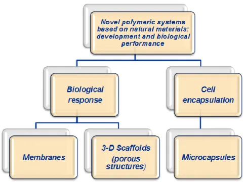
The present thesis, focused on the development and characterization of distinct natural origin polymeric systems that can find different applications in tissue engineering.
The first system studied consists of membranes and 3D-scaffolds based on chitosan, a biodegradable polymer of natural origin that shows adequate properties for several potential biomedical applications such as wound healing, skin substitutes, drug delivery, cell encapsulation and others. In this work, it was evaluated the general cytotoxicity and biocompatibility of several different chitosan membranes and 3D-chitosan scaffolds formulations, but the major focus was on the assessment of the effect of plasma surface treatments on the ability of chitosan membranes to promote cell adhesion and proliferation. For this purpose, the surface of the chitosan based membranes was modified by plasma treatment, using argon and nitrogen gas. Different in vitro cell culturing assays performed (MEM, MTT, MTS) showed that such treatments were useful to obtain enhanced cell attachment and proliferation on chitosan membranes. In fact, chitosan membranes treated by plasma, demonstrated a high potential for biomedical applications, such as substrates for skin tissue engineering. Additionally, plasma treatment procedure seems to have potential to be implemented in other chitosan-based materials, such as scaffolds for tissue engineering.
The second part of the work developed under this thesis was focused on the development of a novel cell encapsulation system based on two different natural origin polymers, namely alginate, a well know polymer widely studied for cell encapsulation, drug delivery, and carrageenan, a natural origin polymer that has not been significantly studied for biomedical applications despite its intrinsic interesting properties. Cell encapsulation systems allow to obtain constructs with a homogenous
cell distribution that can adapt to the shape of the tissue defect to be treated, as the microcapsules containing the cells can be easily injected into a tissue defect, using non-invasive surgical procedures.
Microcapsules were developed using kappa and iota-carrageenan, as well as a mixture of sodium alginate and iota-carrageenan. In all cases, chitosan was used to form the membrane of the capsules. All the developed capsules exhibited spherical shape, smooth surface and revealed good stability in PBS and in culture medium. The obtained results demonstrate that the incorporation of iota-carrageenan improves the stability of alginate based capsules and suggest that these capsules show adequate permeability for cell encapsulation without the need for additional citrate treatments (as it happens for alginate capsules). In fact, cell encapsulation experiments, using sodium alginate and iota-carrageenan (30/70%) capsules demonstrated that is possible to maintain cellular viability, demonstrating the potential of the developed system for tissue engineering applications.
R
ESUMO
A engenharia de tecidos tem sido estudada extensamente como uma alternativa para o tratamento de tecidos danificados por uma doença ou trauma. A maioria destes estudos utiliza materiais desenvolvidos para o efeito com formas/estruturas diferentes, como suportes para a proliferação e adesão de células (ou encapsulamento de células em sistemas poliméricos).
O trabalho apresentado, focou-se no desenvolvimento e caracterização de sistemas poliméricos de origem natural distintas com aplicações diferentes em engenharia de tecidos.
Na primeira parte do trabalho estudou-se à biocompatibilidade de membranas e estruturasporosas(“3D-scaffolds”) baseadas no quitosano, um polímero biodegradável de origem natural, que exibe propriedades adequadas para diversas potenciais aplicações biomédicas, relacionadas com a cicatrização de feridas, suportes para engenharia de tecidos, libertação controlada de fármacos e encapsulamento de células, entre outras. Foi avaliada a citotoxicidade e a biocompatibilidade de membranas com diferentes formulações e estruturas tridimensionais porosas baseadas no quitosano, sendo o objectivo principal a avaliação do efeito do tratamento de plasma na superfície das membranas de quitosano na promoção a adesão e proliferação de células. A superfície das membranas de quitosano foi modificada utilizando um tratamento por plasma, usando árgon e azoto. Foram realizados diferentes testes de culturas celulares in vitro (MEM, MTT, MTS), os quais demonstraram que os tratamentos por plasma, melhoram significativamnte á adesão e proliferação de células nas membranas de quitosano. De facto, as membranas de quitosano tratadas por plasma demonstraram um elevado potencial para aplicações em engenharia de tecidos.
A segunda parte do trabalho consistiu no desenvolvimento de um novo sistema para encapsulamento de células baseado em dois polímeros diferentes, ambos de origem natural, nomeadamente o alginato, um conhecido polímero extensamente estudado para encapsulamento de células e em sistemas para a libertação de fármacos, e o carragenano que, apesar exibir algumas propriedades intrínsecas
interessantes, não tem sido muito estudado para aplicações biomédicas. Os sistemas de encapsulamento de células permitem obter sistemas híbridos tridimensionais de células e materiais (constructs) com uma distribuição celular homogénea que podem adaptar-se à forma do defeito do tecido a ser tratado, uma vez que as microcápsulas podem ser facilmente injectadas no defeito a tratar, utilizando técnicas cirurgicas de invasão mínia. Estes sistemas híbridos podem ser usados para a regeneração de diversos defeitos tecidulares e podem também ser usados simultaneamente como veículos para a libertação de agentes bioactivos.
As microcápsulas foram desenvolvidas usando kappa-carragenano e o iota-carragenano assim como misturas de alginato de sódio e de iota-iota-carragenano. Em todos os casos foi utilizado o quitosano para formar a membrana envolvente das cápsulas. Todas as cápsulas desenvolvidas exibem uma forma esférica, uma superfície lisa e estabilidade adequada em PBS e no meio de cultura. Os resultados obtidos demonstraram que a incorporação do iota-carragenano melhora a estabilidade das cápsulas baseadas em alginato, o que indica que estas cápsulas apresentam permeabilidade adequada para o encapsulamento de células sem a necessidade de um tratamento adicional de citrato (como acontece para as cápsulas de alginato). Assim sendo, os ensaios de encapsulamento de células em cápsulas feitas de alginato de sódio e iota-carragenano (30/70%) provaram que é possível manter a viabilidade celular, demonstrando o potencial do sistema desenvolvido para aplicações em engenharia de tecidos.
C
ONTENTS
Pag.
1 INTRODUCTION ... 1
1.1 POLYMERS FOR BIOMEDICAL APPLICATIONS ... 1
1.1.1 Natural origin polymers for tissue engineering applications ... 4
1.1.2 Processing and format of natural polymers ... 12
1.2 BIOCOMPATIBILITY ... 21
1.2.1 In vitro cytotoxicity/biocompatibility assays ... 22
1.3 REFERENCES ... 29
2 MATERIALS AND METHODS ... 51
2.1 MATERIALS DEVELOPMENT AND PREPARATION ... 51
2.1.1 Chitosan membranes ... 51
2.1.2 Chitosan scaffolds ... 52
2.1.3 Development of carrageenan and alginate based microcapsules ... 53
2.2 CELL CULTURE STUDIES ... 57
2.2.1 Cells used ... 57
2.2.2 Cytotoxicity testing ... 58
2.2.3 Cell seeding/culturing onto scaffolds ... 63
2.2.4 Cell viability of encapsulated cells using fluorescent stains ... 66
2.3 REFERENCES ... 68
3 CELL ADHESION AND PROLIFERATION ONTO CHITOSAN BASED MEMBRANES TREATED BY PLASMA SURFACE MODIFICATION ... 73
3.2 MATERIALS AND METHODS ... 76
3.2.1 Test materials ... 76
3.2.2 Cytotoxicity assays ... 76
3.2.3 Direct Contact assay – assessment of cell adhesion and proliferation of L929 cells seeded onto chitosan membranes ... 80
3.2.4 Statistical analysis ... 81
3.3 RESULTS AND DISCUSSION ... 81
3.3.1 In vitro cytotoxicity assessment ... 81
3.4 CONCLUSIONS ... 90
3.5 REFERENCES ... 91
4 DEVELOPMENT OF A NOVEL CELL ENCAPSULATION SYSTEM BASED ON NATURAL ORIGIN POLYMERS FOR TISSUE ENGINEERING APPLICATIONS ... 97
4.1 INTRODUCTION ... 98
4.2 MATERIALS AND METHODS ... 102
4.2.1 Microcapsules development ... 102
4.2.2 Morphological characterization of the developed microcapsules ... 104
4.2.3 Evaluation of mechanical stability ... 105
4.2.4 Assessment in vitro cytotoxicity of the developed capsules ... 105
4.2.5 Cell encapsulation ... 107
4.2.6 Morphological characterization in encapsulated cells ... 108
4.2.7 Assessment of the viability of encapsulated cells ... 108
4.2.8 Statistical analysis ... 109
4.3 RESULTS AND DISCUSSION ... 109
4.3.1 Preparation of the microcapsules ... 109
4.3.3 In vitro cytotoxicity assessment ... 115
4.3.4 Encapsulated cell viability and proliferation ... 116
4.4 CONCLUSIONS ... 120
4.5 REFERENCES ... 121
5 PRELIMINARY STUDIES OF CELL ADHESION AND PROLIFERATION OF OSTEOBLASTIC-LIKE CELLS ON CHITOSAN POROUS HYBRIDS SCAFFOLDS ... 128
5.1 INTRODUCTION ... 128
5.2 MATERIALS AND METHODS ... 130
5.2.1 Test materials ... 130
5.2.2 Cell culture studies ... 131
5.2.3 Seeding and culturing osteoblastic-like cells into 3D-chitosan scaffolds 131 5.2.4 DNA Quantification in lysed cells ... 132
5.2.5 Alkaline phosphatase (ALP) assay ... 132
5.2.6 SEM analysis of the cell seeded scaffolds ... 133
5.3 RESULTS AND DISCUSSION ... 133
5.3.1 Cellular adhesion and proliferation-SEM analysis ... 133
5.3.2 DNA Analysis ... 135
5.3.3 ALP analysis ... 137
5.4 CONCLUSIONS ... 138
5.5 REFERENCES ... 138
L
IST OF TABLES
Table N⁰
Page
Table 1-1. Polymeric biomaterials: main properties and applications. Adapted from [4] ... 3
Table 1-2. Somestudiesdevelopedbythe3B’sResearch Group on chitosan based materials. . 7
Table 1-3. Main characteristics of the different forms of carrageenan. Adapted from [114]. ... 9
Table 1-4. Characteristics of some polyelectrolytes used for cell encapsulation. Adapted from [178]. ... 20
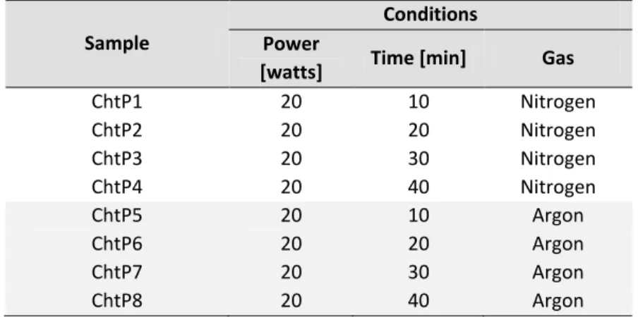
Table 2-1. Plasma conditions used to treat the surface of chitosan membranes ... 52
Table 2-2. Conditions used in the preparation of the several chitosan porous hybrid scaffolds 52 Table 2-3. Scoring for morphological changes, confluence and cell death, used in the cytotoxicity tests ... 60
Table 2-4. Cytotoxicity indexes used to classify the reactivity of tested samples - cytotoxic response ... 60
Table 2-5. Preparation of standard solutions - DNA assays ... 65
Table 2-6. Preparation of standard solutions - ALP assays ... 66
Table 3-1. Plasma conditions used on chitosan membranes ... 76
Table 3-2. Scoring for morphological changes, confluence and cell death, used in the cytotoxicity tests ... 78
Table 3-3. Cytotoxicity indexes used to classify the reactivity of tested samples - cytotoxic response ... 79
Table 3-4. Growth inhibition and cytotoxic response in short-term MEM extraction test after 72h testing ... 82
Table 5-1. Conditions of the several chitosan porous hybrid scaffolds ... 131
L
IST OF FIGURES
Figure N⁰
Page
Figure 1-1. Factors to consider when designing a new biomaterial. Biological, medical and
engineering properties must be integrated to achieve successful biomaterials for tissue
regeneration. Adapted from [5]. ... 2
Figure 1-2. Chitosan structure, adapted from [70]. ... 5 Figure 1-3. Examples of chitosan processing for uses in tissue engineering: Cells may be
encapsulated in gels or seeded in porous matrices including sponge-like or fibrous structures. Combinations of chitosan with other biocompatible materials, such as calcium phosphate or gelatine, are applied to modify biomechanical and cell-matrix-interaction properties. Adapted from [68]. ... 6
Figure 1-4. The main carrageenan chain consists of alternating 3-linked-β-D-galactopyranose and 4-linked- -D-galactopyranose units. Adapted from [115]. ... 8
Figure 1-5. Chemical structures of kappa, iota and lambda carrageenans. Adapted from [117]. 8 Figure 1-6. Alginate structure. Adapted from [143]. ... 11 Figure 1-7. Some forms based on natural polymers that can be used in different biomedical
applications, such as tissue regeneration and delivery systems... 13
Figure 1-8. Schematic representation of the research approach followed in this thesis. ... 14 Figure 1-9. Membrane prepared by solvent casting – schematic representation. ... 15
Figure 1-10. Schematic representation of a typical encapsulation process involving ionic
cross-linking of alginate. Adapted from [178]. ... 19
Figure 1-11. Schematic representation of the set up for a Direct Contact Assay, where the cell
suspension is seeded onto a membrane or scaffold materials. ... 23
Figure 1-12. Possible processing procedures for plasma-induced chemical micropatterning.
Adapted from [215]. ... 26
Figure 1-13. Approaches for enhancement of cell growth on surfaces. Adapted from [145]. ... 26 Figure 1-14. Morphology of some cell lines used in biocompatibility tests. A and B images
showed SaOS-2 cell line; C and D images showed L929 cell line. All photos were taken using inverted microscope using the objectives 10X (left images) and 20X (right images). ... 28
Figure 2-1. Chitosan membrane ... 51 Figure 2-2. Chitosan scaffolds ... 53
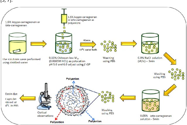
Figure 2-3. Graphical representation of the procedure used for the capsules development
using oppositely charged polysaccharides (carrageenan as polyanion and chitosan as
polycation)... 54
Figure 2-4. Variation of the pH relativetoamountofβ-GPwithrespecttochitosan(weightβ -GP/weight chitosan g/g), at room temperature ... 55
Figure 2-5. Graphical representation of capsules produced by ionotropic gelation method .... 56 Figure 2-6. Graphical representation of the cell encapsulation procedure carried out using the
ionotropic gelation method ... 57
Figure 2-7. Graphical representation of the procedure used to prepare extracts of the chitosan
membranes ... 59
Figure 2-8. Graphical representation of the procedure used for cell seeding onto chitosan
membranes ... 62
Figure 2-9. Graphical representation of the procedure used for cell seeding onto chitosan
scaffolds ... 64
Figure 2-10. Graphical representation of cell fluorometric determination of living cell using
Calcein-AM. Adapted from [41, 43]. ... 67
Figure 2-11. Graphical representation of the procedure used for Calcein – DAPI fluorescescent staining to assess the viability of encapsulated cells ... 68
Figure 3-1. Morphology of L929 cells incubated with extracts of: a) negative control (DMEM
culture medium), b) Positive control (latex extract), c) ChtP1 extract, d) ChtP5 extract, e) ChtP3 extract, f) ChtP7 extract, g) Cht extract (untreated membrane), over a 72 h period
(Photographs were taken with an inverted light microscope using a 20X magnification). ... 83
Figure 3-2. Percentage of cell viability, obtained from the MTT assay, of the fibroblast exposed
to extracts of chitosan membranes surface modified by argon and nitrogen plasma (at
different conditions). ... 84
Figure 3-3. SEM (left) and optical micrographs after blue methylene staining (right-10X) of L929
fibroblasts cultured on untreated chitosan membranes (control) and chitosan membranes modified by nitrogen plasma (using different conditions) after 7 days of culture: A1-A2: Cht (untreated chitosan membranes); B1-B2: ChtP1; C1-C2: ChtP2, D1-D2: ChtP3; E1-E2: ChtP4. .. 86
Figure 3-4. SEM (left) and optical micrographs after blue methylene staining (right-10X) of L929
fibroblasts cultured on untreated chitosan membranes (control) and chitosan membranes modified by argon plasma (using different conditions) after 7 days of culture: A1-A2: Cht (untreated chitosan membranes); B1-B2: ChtP5; C1-C2: ChtP6, D1-D2: ChtP7; E1-E2: ChtP8. .. 87
Figure 3-5. Cell viability and proliferation results obtained using the MTS test. Cell density used
was 8 x 10⁴ cells/cm2. For the control, the same amount of cells was seeded on 24 culture well plates. Cells were kept in culture for 3, 7 and 14 days. A) Optical density (OD) obtained in chitosan membranes treated by nitrogen plasma, B) OD obtained in chitosan membranes treated by argon plasma. ... 89
Figure 4-1. VariationofthepHrelativetoamountofβ-GP with respect to chitosan (weightβ -GP/weight chitosan g/g), at room temperature ... 103
Figure 4-2. Optical microscopy images taken at pH 5.0 of the obtained microcapsules based on
A1-A3) kappa-carrageenan and B1-B3) iota-carrageenan. ... 110
Figure 4-3. Optical microscopy images of solid capsules obtained using different polymer ratios
and at different pH of the chitosan solution: A1-A2) microcapsules obtained from a mixture of sodium alginate/iota-carrageenan (30/70) at pH5; B1-B2) microcapsules obtained from a mixture of sodium alginate/iota-carrageenan (30/70) at pH6; C1-C2) microcapsules obtained from a mixture of sodium alginate/iota-carrageenan (50/50) at pH5; D1-D2) microcapsules obtained from a mixture of sodium alginate/iota-carrageenan (50/50), at pH6. The
microcapsules (A, B, D) were dyed with eosin for a better observation of the chitosan
membrane. ... 112
Figure 4-4. Percentage of ruptured microcapsules (correspondent to different sodium
alginate/iota-carrageenan ratios) as a function of time. ... 113
Figure 4-5. Optical microscopy images of the capsules after the short-term stability study
carried out at different time periods. A) 30/70 solid capsules, B) 30/70 liquefied capsules, C) 50/50 solid capsules, D) 50/50 liquefied capsules. Photographs were taken with a
stereomicroscope. ... 114
Figure 4-6. Morphology of fibroblasts exposed to: a) 30/70 SA-IC capsules extracts; b) 50/50
SA-IC capsules extracts; c) latex extracts (positive control) and d) culture medium (negative control)incubated72hat37⁰C.Photographsweretakenwithaninverted optical microscope (20X). ... 115
Figure 4-7. Metabolic activity of the L929 fibroblasts cultured with extracts obtained from
30/70 and 50/50 SA-IC solid capsules for 24, 48 and 72 hours, determined using the MTT test. ... 116
Figure 4-8. Optical microscopy pictures showing encapsulated fibroblasts (density 5x10³ cells per ml) within sodium alginate/iota-carrageenan (30/70) solid capsules pH 5.0. A1, A2 and A3) cells within solid capsules in culture medium (5X and 10X respectively); B1) cells within solid
capsules in culture medium (20X); B2 and B3) Cells within solid capsules stained with blue methylene (20X and 40X respectively), after 72 h. ... 117
Figure 4-9. Cell viability and proliferation of fibroblast entrapped within SA (sodium
alginate)/IC (iota- carrageenan) capsules and in the polymeric solution SA/IC (ratio: 30/70), determined using the MTS test. ... 118
Figure 4-10. Images obtained in the fluorescent microscope of encapsulated fibroblasts stained
with Calcein-AM and DAPI dyes after: (A1-A3) 24 h, (B1-B3) 48 h, (C1-C3) 72 h and (D1-D3) 7 days. ... 120
Figure 5-1. SEM micrographs showing SaOS osteoblasts cultured on chitosan scaffold after 3, 7
and 14 days of cell culture. A1 to A3) CT5 scaffold, B1 to B6) CT6 scaffold, C1 to C3) CT7
scaffold. ... 134
Figure 5-2. Number of cells on chitosan based scaffolds with different formulations after 3, 7
and 14 days of cell culture obtained in the DNA Quantification assay. ... 136
Figure 5-3. ALP activity of osteoblasts cells after 3, 7 and 14 days of cell culture on chitosan
C
HAPTER
1
1
INTRODUCTION
1.1
POLYMERS FOR BIOMEDICAL APPLICATIONS
In the past years, the field of Tissue Engineering has opened a new and exciting range of applications for biodegradable polymers.
Tissue Engineering is an emerging interdisciplinary field and one of the major components of regenerative medicine which follows the principles of cell transplantation, materials science, and engineering towards the development of biological substitutes that can restore and maintain normal tissue function [1].
Biomaterials, in general, provide a space (2D or 3D) for the cells to form into new tissue with appropriate structure and function. Moreover, the biomaterials may allow for the delivery of cells and appropriate bioactive factors, to the desired site in the body [1, 2]. As the majority of mammalian cell types are anchorage dependent, and will die if no cell adhesion substrate is available, the biomaterials play an important role as support structures for tissue regeneration. For this type of applications, the ideal biomaterial should be biodegradable and bioresorbable to support the replacement of normal tissue without inflammation [1, 2]. Degradation products, if produced, should be removed from the body via metabolic pathways at an adequate rate in order to keep the concentration of these degradation products in the tissue at a tolerable level [1, 3].
Although most of the research on Tissue Engineering has focused on polymers that have already been studied (and in some cases approved) for other clinical applications, the specific and highly demanding requirements for this type of use has encouraged the development of new and improved systems, both from natural and synthetic origin, as well as combination of natural with synthetic polymers.
Polymers are a promising class of biomaterials that can be engineered to meet specific end-use requirements. They can be selected according to key 'device' characteristics such as mechanical resistance, degradability, permeability, solubility
and transparency, but most of the currently available polymers need to be improved by altering their surface and bulk properties in order to make then suitable for biomedical applications. The design of macromolecules must therefore be carefully tailored in order to provide the combination of chemical, interfacial, mechanical and biological functions necessary for the manufacture of new and improved biomaterials (Figure 1-1) [4].
Figure 1-1. Factors to consider when designing a new biomaterial. Biological, medical and engineering
properties must be integrated to achieve successful biomaterials for tissue regeneration. Adapted from [5].
Polymers have been used widely in medicine and biotechnology [4], in a wide range of applications such as surgical devices [6], implants [7], drug delivery systems , carriers of immobilized enzymes and cells [8], biosensors, bioadhesives, ocular devices [9] and materials for orthopaedic applications [10].
Natural origin polymers are abundant and usually biodegradable. Table 1-1, presents some of the most important natural and synthetic origin polymers and their main biomedical applications. The principal disadvantages of these polymers is related to the difficulty in the development of reproducible production methods, because their complex structure often renders modification and difficult purification [4]. In the case of synthetic polymers, these are available in a wide variety of compositions with readily adjusted properties. The main disadvantage of synthetic polymers is the
general lack of biocompatibility in the majority of cases, often associated with inflammatory reactions [4].
The work developed in this thesis has focused on the use of natural origin polymers such as chitosan, carrageenan and alginate and therefore, in the next sections, these materials will be further described.
Table 1-1. Polymeric biomaterials: main properties and applications. Adapted from [4] Polymer Main applications and comments Ref NATURAL POLYMERS
Collagen Absorbable sutures, wound dressing sponge, drug delivery microspheres.
[11], [12, 13]
Albumin Cell and drug microencapsulation. [14-18]
Poly(amino acids): poly(L- lysine), poly(L- glutamic).
Non toxic, non antigenic and biocompatible. Used as
oligomeric drug carriers. [19-21]
Carboxymethyl cellulose
Cell immobilization via a combination of ionotropic gelation and polyelectrolyte complex formation (e.g. with chitosan), drug-delivery systems and dialysis membranes.
[22-24]
Cellulose sulphate
Component of polyelectrolyte complex for immunoisolation of cells. Complex-forming ability is highly sensitive to degree of acetylation.
[25, 26]
Agarose Largely used as supporting materials in clinical analysis
and as an immobilization matrix. [27, 28]
Alginate
Excellent gel-formation properties; its relative biocompatible; microstructure and viscosity are dependent on the chemical composition (batch to batch variations). Used as immobilization matrices for cells and enzymes, controlled release of bioactive substances, injectable microcapsules for treating neurodegenerative and hormone-deficiency disease.
[6, 29, 30]
Carrageenan Excellent thermoreversible properties. Used for
microencapsulation of microorganisms. [31-33] Hyaluronic acid Excellent lubricant, potential therapeutic agent. [11, 34] Heparin and heparin-like
glycosaminoglycanes
Antithrombotic and anticoagulant properties. Extensively used in surgery. Some are candidates for ionotropic gelation and capsule formation.
[35]
Dextran and its derivates Excellent rheological properties. Plasma expander.
Widely used as a drug carrier. [36, 37]
Chitosan and its derivates
Biocompatible, non toxic, excellent gel and film forming ability, natural polycation. Widely used in controlled delivery systems (e.g. gels, membranes, microspheres).
Cont.
Polymer Main applications and comments Ref SYNTHETIC POLYMERS
Poly (lactic acid), poly (glycolic acid) and their copolymers
Used in sutures, drug delivery systems and in tissue engineering. Biodegradable. Often copolymerized to regulate degradation time.
[3, 47, 48] Poly (hydroxyl butyrate), poly
(caprolactone) and copolymers, poly (alkylene succinate), etc.
Biodegradable, used as a matrix for drug delivery systems, cell microencapsulation. Properties can be changed by chemical modification, copolymerization and blending.
[49]
Polyamides (nylons) Sutures, dressing, haemofiltration and blending. [50] Polyethylene (low density) Sutures, catheters, membranes, in surgery. [51, 52] Poly (vinyl alcohol) Gels and blended membranes are used in drug
delivery and cell immunoisolation. [53] Poly (ethylene oxide)
Highly biocompatible. Different polymer derivatives and copolymers have been utilized in a variety of biomedical applications.
[54]
Poly (hydroxyethyl methacrylate)
Hydrogels have been used as soft contact lenses, for drug delivery, as skin coatings and for immunoisolation membranes.
[9, 55]
Poly (methyl methacrylate) This and its copolymers are used as dental implants
and in bone replacement. [56, 57]
Poly (tetrafluroethylene) (Teflon®) Vascular grafts, clips and sutures, coating. [58] Polydimethylsiloxanes A silicone. Implants in plastic surgery, orthopaedics,
blood bags and pacemakers. [59]
Poly (ortho esters) Surface eroding polymers. Application in sustained
drug delivery, ophthalmology. [60-62] Polyanhydrides Biodegradable, useful in tissue engineering and for the
release of the bioactive molecules. [63, 64]
1.1.1 NATURAL ORIGIN POLYMERS FOR TISSUE ENGINEERING APPLICATIONS
Naturally derived materials have been used for tissue engineering because they offer several advantages related to biological recognition (being similar, often identical to macromolecular substances present in the ECM), by the biological environment, which is then prepared to recognize and to deal metabolically with these materials [4, 65].
1.1.1.1 Chitosan
Chitosanisalinearpolysaccharidecomposedofrandomlydistributedβ -(1-4)-linked D-glucosamine (deacetylated unit) and N-acetyl-D-glucosamine (acetylated unit)
Figure 1-2), commercially produced by deacetylation of chitin [44, 66]. Chitin is a natural polymer found in many arthropods as shells of marine crustaceans (crab, shrimp and shellfish), insects and cell walls of fungi. This polysaccharide is the second most important compound in nature after cellulose. It is not toxic and is insoluble in water, as well in many other solvents such as acetic acid [39, 44, 67-69]. Due to the existence of primary amine groups, the structure can be protonated at low pH, and chitosan is found to be soluble in moderated acidic solutions.
Figure 1-2. Chitosan structure, adapted from [70].
Chitosan can be formulated into a variety of forms including powders [71], gels [40], scaffolds [41, 72, 73], particles [41, 74], microspheres [75], microcapsules [29, 75, 76] and films [42, 77, 78] with an array of biological properties and applications (Figure 1-3) [39]. Furthermore, chitosan formulations allow a wide range of molecules to be attached. This property allows the delivery of growing factors that could help to optimize the physical and biological properties of this material as a matrix scaffold and vehicle system in tissue regeneration [39].
Chitosan was discovered in 1859, when Rouget boiled chitin in a concentrated potassium hydroxide solution, promoting the deacetylation of chitin [79]. Fundamental research on chitosan did not start until about a later century [29]. This natural polymer can selectively bind substances such as cholesterol, fats, metal ions, proteins and tumour cells. Others properties include antifungal effects, antibacterial [80], acceleration of wound healing [81, 82], stimulation of the immune system and acceleration of plant germination [29].
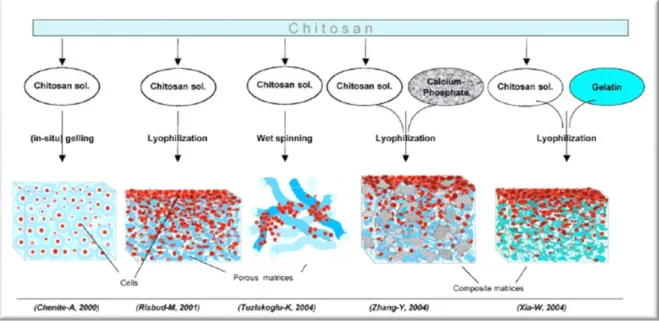
Figure 1-3. Examples of chitosan processing for uses in tissue engineering: Cells may be encapsulated in
gels or seeded in porous matrices including sponge-like or fibrous structures. Combinations of chitosan with other biocompatible materials, such as calcium phosphate or gelatine, are applied to modify biomechanical and cell-matrix-interaction properties. Adapted from [68].
Several research works have shown that membranes formed from this polymer could be explored for surgical dressing, guided tissue regeneration and controlled release applications [1, 67].
Chitosan has been the best researched version of the biopolymer because of its ready solubility in dilute acids, rendering the material more accessible for utilization and chemical reactions [41]. Chitosan in aqueous acid solutions reacts with anionic polysaccharides such as: carboxymethylcellulose [83], xanthan [84, 85], alginate [86, 87], carrageenan [31, 66], gellan [88], oxychitin and oxypullulan [89-91], chondroitin and hyaluronan [92, 93], poly (galacturonic) [94], poly ( -L-glutamic acid) [95] as well as synthetic polyanions such as poly (acrylic acid) [96], to give origin to the formation of polyelectrolyte complexes [97].
The3B’sResearchGroupoftheUniversityofMinhohasbeenstudyingchitosanfor a wide range of biomedical applications, summarized on Table 1-2, that includes some studies based on chitosan and others polymers for tissue engineering applications. These materials have been developed into different forms such as membranes [98-101], 3D-scaffolds [38, 73, 74, 102] and fibres [69] aiming at different tissue engineering applications. One of the aims of this work was to assess the biological
response of chitosan based membranes and scaffolds developed for biomedical applications.
Table 1-2. Some studies developed by the3B’sResearchGrouponchitosanbasedmaterials.
Name article Refs.
Multichannel mould processing of 3D structures from microporous coralline hydroxyapatite
granules and chitosan support materials for guided tissue regeneration/engineering. 2003. [105] Starch-chitosan hydrogels prepared by reductive alkylation crosslinking. 2004 [106] Production and Characterization of Chitosan Fibers and 3D Fiber Mesh Scaffolds for Tissue
Engineering Applications. 2004. [69]
Preparation and characterisation in simulated body conditions of glutaraldehyde crosslinked
chitosan membranes. 2004. [99]
Influenceofβ-Radiation Sterilization in Properties of New Chitosan/Soybean Protein Isolate
Membranes for Guided Bone Regeneration. 2004. [98]
Hydroxyapatite Reinforced Chitosan and Polyester Blends for Biomedical Applications. 2005. [38] Physical properties and biocompatibility of chitosan/soy blended membranes. 2005. [101] Functional nanostructured chitosan/siloxane hybrids. 2005. [107] Properties of Melt Processed Chitosan and Aliphatic Polyester Blends. 2005. [108] Use of Chemically Modified Chitosan and other natural-origin polymers in tissue engineering
and drug delivery In Biodegradable Systems in Tissue Engineering and Regenerative Medicine. 2005.
[109] Chitosan particles agglomerated scaffolds for cartilage and osteochondral tissue engineering
approaches with adipose tissue derived stem cells. 2005. [74] Study of the fosfosal controlled permeation through glutaraldehyde crosslinked chitosan
membrane. 2006. [100]
Physicochemical Characterization of Novel Chitosan-Soy Protein/TEOS Porous Hybrids for
Tissue Engineering Applications. 2006. [102]
Enzymatic degradation behaviour of starch conjugated phoshorylated chitosan. 2006. [110]
1.1.1.2 Carrageenan
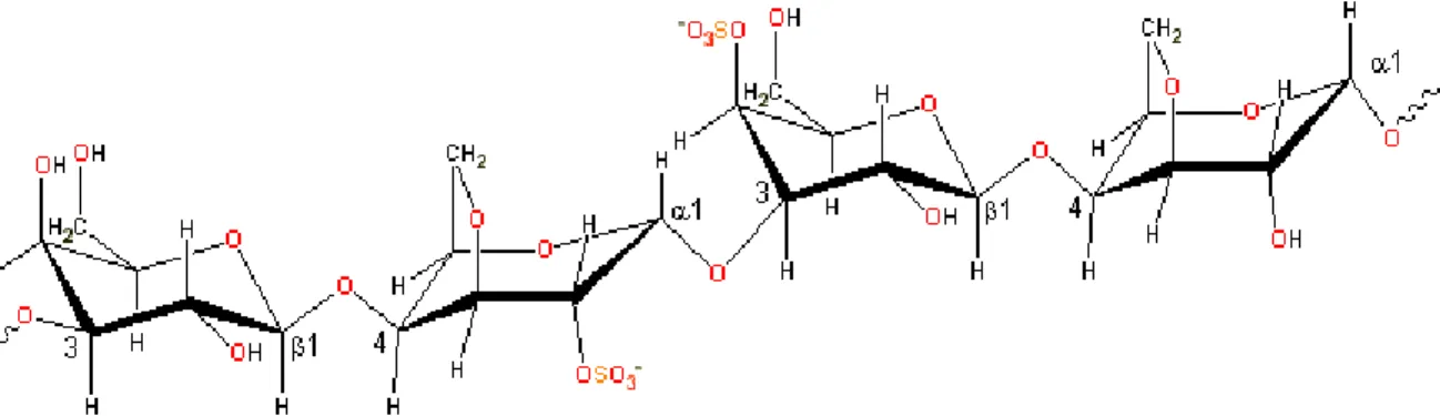
Carrageenanisalinearheteropolysaccharidewithestersulphategroups.It’smain chain consists of alternating copolymers of 1,4- and 1,3-β-D-galactopyranose and 3,6-anhydro-D-galactopyranose [103, 104] (Figure 1-4). Carrageenan can be found in them three major forms that are designated by means of the Greek letters κ (kappa), i (iota) and λ (lambda). The main structural difference among them is in the sulphate group degree of substitution (Figure 1-5).
Figure 1-4. The main carrageenan chain consists of alternating 3-linked-β-D-galactopyranose and 4-linked- -D-galactopyranose units. Adapted from [115].
All carrageenans are highly flexible molecules which, at higher concentrations, organize into double-helical structures. Gel formation in kappa- and iota-carrageenans involves helix formation on cooling from a hot solution together with gel-inducing and gel-strengthening; the helix–helix aggregation relies upon specific cation presence (Ca2+ and K+), which can screen electrostatic repulsive forces between the participating chains by packing within the aggregate structure [31, 111, 112].
Kappa-carrageenan forms firm and strong rigid gels, while iota-carrageenan
constitutes soft and elastic gels [31, 113]. Lambda-carrageenan does not gel as the high density of charged sulphate encourages an extensive conformation [114]. Table 1-3 presents some of the basic characteristics of different types of carrageenans.
Table 1-3. Main characteristics of the different forms of carrageenan. Adapted from [114].
Chemical composition
Hydrocolloid composed of -D-1.3andβ-D-1.4 galactose residues that are sulphated at up to 40% of the total weight. Strong negative charge over normal pH range. Associated with ammonium, calcium, magnesium, potassium or sodium salts.
Solubility
λ is readily soluble in cold or hot aqueous solution.
K is soluble in hot solution; treatment of aqueous solution with potassium ion
precipitates.
Gel formation
λ does not form gels.
λ and i form right-handed helices
Potassium chloride promotes gel formation of k. Calcium ion promotes gel formation of i.
Gel formation in k and i carrageenans involves helix formation on cooling from a hot solution together with gel-inducing and gel-strengthening K+ or Ca2+ cations respectively
Metabolism HydrolisisofglycosidiclinkagesatlowerpH,especiallypH≤3.0;also
desulphation by sulphatases.
Source Red algae; predominantly aqueous extraction from Chondrus, Gigartina, and
various Eucheuma species.
Viscosity
Near logarithmic increase in viscosity with increasing concentration. Viscosity of food-grade carrageenan defined as not less than 5 cps at 75ºC for a 1.5% solution; viscosity ranges from 5 to 800 cps for 1.5% solution at 75ºC.
Molecular weight
Discrepancies in definitions. Native carrageenan reported to have average molecular weight of 1.5x10⁶ – 2x10⁷, food grade carrageenan reported as 100.000-800.000 or 200.000-400.000. Degraded carrageenan (poligeenan) has average molecular weight of 20.000-30.000.
Properties λandkcombine easily with milk proteins to improve solubility and texture,
serve as thickening agent, emulsifier, stabilizer.
Major uses Milk products, processed meats, dietetic formulations, infant formula,
toothpaste, cosmetics, skin preparations, pesticides, laxatives.
Concentration in
food products 0.005-2.0% by weight.
Carrageenan has a structural role in the intercellular matrix material of numerous species of seaweeds of the class Rhodophyta, where its amorphous structure provides
the flexibility required to adapt to the varying stress of tidal and wave motion [103, 116]. The main substituent is the hemiester sulphate group. These strongly ionic groups mutually repel each other to maintain the molecule in a highly extended, flexible configuration [103].
Carrageenans are widely used in the food and other industries (cosmetic products, pesticides and pharmaceutical), as thickening and stabilizing agents. However, its predominant role has been in food preparations (a wide variety of food groups), because the carrageenans have the ability to substitute fat and to combine easily with milk proteins to increase solubility and improve texture [114].
Several investigations have associated carrageenan with the induction and promotion of intestinal neoplasm and ulceration in numerous animal experiments [118-121]. This behaviour is related with gastrointestinal metabolism of carrageenan, where food-grade carrageenan may be contaminated with low molecular weight carrageenan that may arise during food processing [114]. On the other hand, previous studies have demonstrated the benefits of carrageenans in health applications, and considered a useful microbicide that inhibits the human immunodeficiency virus (HIV-1) infection of epithelial at nanomolar concentrations. Moreover, carrageenans have also been considered as possible active agent against the cell-associated and cell-free virus in genital fluids [122]. The microbicide behaviour, was supported with recent studies where it has been demonstrated that carrageenans, as sulphated polysaccharides, are potent inhibitors of the infectivity of genital by human papilloma virus (HPV) and pseudovirus (PsV) in vitro [113, 123].
In the present work, carrageenan is proposed as an alternative polymer for the development of cell encapsulation systems. The use of carrageenans in the encapsulation field has rarely related in the literature. Previous studies have reported on capsules produced by carrageenan and oligochitosan polymer [31], but concerning cell encapsulation the reports are only related to the use of carrageenan (in special k-carrageenan) for the encapsulation of microbial cells [32, 33, 122].
1.1.1.3 Alginate
Alginate is a linear polymer of (1-4) - β-D-mannuronopyranosyl and (1-4)- -L-guluronopyranosyl units in a copolymer that contains homopolymeric sequences
(figure 1-6). They occur naturally as the major structural polysaccharides of brown marine algae (Phaeophyceae) [124, 125] and as extracellular mucilages secreted by certain species of bacteria (Azobacter Vinelandii) [30, 124].
Figure 1-6. Alginate structure. Adapted from [143].
Guluronic and manuronic acid contain negatively charged functional groups. As a result, these negatively charged regions within the alginate structure can parallel the electrostatic conditions created by sulphated proteoglycans in native tissue and might offer to some cells, such as chondrocytes, a more conductive environment [126] .
Alginate remains the most widely used biomaterial for immobilizing cells, because of the many advantages it offers [127-129]. First, alginate has the capacity to gel under physiological conditions that are compatible with cell survival [128, 130]. The gelling reaction does not require toxic solvents and does not generate harmful by products. Second, the rapidity of the gelling process is a further critical advantage [128].
This polymer develops a simple and rapid gelation with divalent metal ions such as Ca2+, through binding of consecutives blocks of G-molecules on individual or different molecules [124, 131-133]. Therefore, many researchers used alginate as the matrix to prepare microcapsules to be used as an injectable cell delivery vehicle [134] and cell immobilization matrix [135-137], involving different cell sources such as myoblasts [138, 139], epithelial cells [139] hybridoma cells [8, 140], fibroblasts [134, 137], pancreatic islets [6, 134] and microbial cells [141, 142]. Additionally, alginate is relatively biocompatible and approved by the Food and Drug Administration (FDA) for human use as wound dressing material [1].
As a natural polymer, alginate is limited by its tendency to be largely contaminated. Additionally, the industrial process used for extracting alginates from seaweed can introduce additional contaminants into the raw materials [128]. Consequently, the purity of the alginate has been found to be a pertinent factor in the biocompatibility of alginate [133]. For instance, when non purified alginate was used for islet encapsulation and transplantation into diabetic animals, a severe reaction against the microcapsule occurred immediately after implantation, and blood glucose was not normalized or was normalized only for a few days [128, 138]. On the other hand, alginate purification allowed the successful encapsulation and transplantation of islets, enabling the control of the levels of glucose in the blood of diabetic animals [128, 144].
Thepresenceofimmunologicresponsecanbepartlyforlinkingtoβ(1-4) glycosidic linkages, since other homopolymeric di-equatorial polyuronates, like D-glucuronic acid (C6-oxidized cellose), exhibit this feature. In vivo animal models have revealed the immunologic potential of polymannuronate in diverse areas for protection against lethal bacterial infections and irradiation, and for increasing non-specific immunity [124].
Due to its interesting and promising properties, alginate was also studied in this thesis for the development of a novel encapsulation system. However, our aim was to combine this polymer with other (namely with carrageenan) in order to enhance the properties of this system as a cell encapsulation device, envisioning the future development of highly functional systems.
1.1.2 PROCESSING AND FORMAT OF NATURAL POLYMERS
Polymeric materials have been applied in many different biomedical applications, and therefore may be found in a variety of forms such as membranes, films, fibres, gels, hydrogels, capsules, spheres, particles and 3D-structures (scaffolds) (Figure 1-7).
In fact, in most cases, the criteria for the design of the polymeric systems depend on the target application/site of implantation [4]. Careful design of the structure and of the interfacial properties of the matrix and its morphology and pore size are
particularly important for biomaterials used as cell-growth scaffolds, and as supports for immobilized biomolecules/cells [4, 145].
Figure 1-7. Some forms based on natural polymers that can be used in different biomedical applications,
such as tissue regeneration and delivery systems.
Others requirements and properties such as durability, elasticity, tensile strength, adhesion should be considered in polymers for using as sutures and long-term skin substitutes. Polymers for matrices or carrieres in controlled-delivery devices require the consideration of characteristics like molecular weight, adhesion and solubility, depending on the type of system, action and the target site in the body. Polymeric materials for drug delivery must satisfy additional requirements such as environmental responsiveness (pH or temperature dependent phase or volume transformation) [4, 126].
In this work, we have focused on three particular designs/formats, based on different polymers (Figure 1-8), that may found use in particular tissue engineering applications or in other cell based therapies. For this reason, in the next sections we will develop the description of membranes, 3D-porous scaffolds and capsules, their main applications, features, and processing methodologies.
Figure 1-8. Schematic representation of the research approach followed in this thesis.
1.1.2.1 Membranes
Polymeric membranes have been developed for a variety of industrial applications such as microfiltration, ultrafiltration, reverse osmosis and gas separation [146, 147]. In biomedical applications, polymer membranes are used specially for skin regeneration, maxillofacial and pharmaceutical applications [126]. Each application needs to meet specific requirements regarding the material and structure [146]. The final morphology of the membranes obtained depends on the material properties and the processing conditions. The majority of membranes are prepared by methods based on a controlled phase separation of polymer solutions into two phases: one with a high polymer concentration and another with low polymer concentration [146]. Membrane systems prepared by this method take advantage of their selectivity, high surface-area-per-unit-volume, and their potential for controlling the level of contact and/or mixing between two phases [147].
Nevertheless, membranes can also be obtained by an interfacial polymerisation technique. In this case, the polymer is transformed in a controlled manner from a liquid to a solid phase. The concept of phase inversion involves a variety of different techniques such as precipitation by controlled solvent evaporation, thermal precipitation, precipitation from vapour phase, immersion precipitation, etc. [148].
Another method for preparing membranes is solvent casting, where the polymer is dissolved into a suitable organic solvent (e.g. chitosan can be dissolved into aqueous acetic acid) and then the solution is cast into a petri dish or into an appropriate mould [101] (Figure 1-9).
Figure 1-9. Membrane prepared by solvent casting – schematic representation.
Chitosan, for example is a polymer easily processed into membranes. In fact, chitosan membranes may be prepared by various ways such as evaporation of chitosan solvents, cross-linking with bifunctional reagents, chelating with anionic counterions or complexion with polymers and proteins [29]. Chitosan membranes have been used in several biomedical applications, such as controlled release [76, 149, 150], wound healing and surgical dressing [42, 151, 152]. In this work, solvent casting was the methodology used to obtain the chitosan based membranes.
1.1.2.2 Scaffolds design and fabrication
Three-dimensional (3-D) porous constructs have been one of the most widely studied scaffolds format in tissue engineering, as they provide the necessary support for cells to proliferate and maintain their differentiated function, and their architecture may define the ultimate shape of the new tissue to be formed, such as bone and cartilage [153, 154].
Factors governing scaffold design are complex and include considerations of matrix architecture, pore size and morphology, mechanics versus porosity, surface properties and degradation rate [153, 155].
A number of fabrication technologies have been applied to process biodegradable and bioresorbable materials into 3-D polymeric scaffolds of high porosity and surface area. The conventional techniques for scaffold fabrication include fibre bonding [156, 157], solvent casting [157-161], particulate leaching [162], gas foaming [163-165], phase separation/emulsification [166-169], membrane lamination and melt molding [153, 170]. Additionally, others methods include extrusion, in situ polymerization [73, 171], 3D-printing and selective laser sintering [155, 172, 173]. Below some methods for scaffolds fabrication that were used in this thesis are described.
Solvent-casting and particle leaching. The solvent casting and particle leaching is
one of the most widely used methodologies to obtain scaffolds and membranes for biomedical applications. This method involves the use of a water soluble porogen, such as salt (NaCl) [159, 160, 174] to create pores. The solvent casting and particle leaching method consist in dispersing a sieved mineral (e.g. NaCl) or organic (e.g. saccharose) particles in a polymer solution [73]. This dispersion is then processed either by casting or by freeze-drying in order to produce porous bi and three-dimensional supports [175].
Freeze-Drying. The basic principle of the freeze-drying process relies on a
thermally induced phase separation, which occurs when the temperature of a homogenous polymer solution, previously poured into a mould, is decreased. Once the phase-separated system is stabilized, the solvent-rich phase is removed by vacuum sublimation leaving behind the polymeric foam [175]. The foam morphology is of course controlled by any phase transition that occurs during the cooling step, i.e., liquid-liquid or solid-liquid demixing [175].
In this work, chitosan based porous structures were produced by means of combining a sol-gel process and the freeze-drying technique. This technique has been used in previous studies by our group for the development of chitosan based porous structures, where the chitosan sol-gel was poured into a petri dish and allowed to dry
at room temperature for several days [102]. By its turn, the porous hybrids were obtained by transferring the sol-gel into a mould, freezing at -80⁰C overnight, followed by freeze-drying for several days to completely remove the solvent [102]. The properties, such as scaffold porosity, are controlled by several treatments including crosslinking, neutralization with alkali solutions and control of the pH of the initial polymeric solution [73].
1.1.2.3 Capsules
Cell encapsulation technology is an exciting field of biotechnology that has emerged and developed rapidly in the past decade [176]. One of its strategies is cell microencapsulation, which aims to overcome the present difficulties relating to whole organ graft rejection and, consequently, the requirements for the use of immunomodulatory protocols or immunosuppressive drugs [136]. Therefore, cell microencapsulation has also raised much interest in part due to the advancement and optimization of the biomaterials used to develop the capsules [177].
Capsules in the 0.3-1.5 mm range have been traditionally refereed to as microcapsules in the cell encapsulation field. Their relatively small size is considered advantageous from a mass transport perspective. Microcapsules are typically more durable than macrocapsules and difficult to mechanically disrupt. Numerous microencapsulation techniques, different in the nature of entrapment mechanism, have been developed. Besides traditional capsules with a well-defined shell-and-core structure, encapsulation in microbeads without a distinct membrane has been successful used in some specific applications [133].
Some known methodologies for capsules formation namely, complex coacervation, coextrusion and emulsion, polyelectrolyte complex and ionotropic gelation [129] are described below.
Coacervation: This methodology consists in using a system with some parameters
such as temperature, pH, or composition, that can be adjusted in a liquid phase of pre-membrane component separates from a polymeric solution and wraps the liquid core as a uniform layer. The liquid core droplet iscalled“coacervate”.Thepre-membrane component is solidified by means of heat, crosslinking, or solvent removal techniques. An hydrophilic pre-membrane component, such as gelatin and gelatin-gum acacia, is
required for the encapsulation of citrus oil, vegetable oil, and water-soluble vitamins. When the core material is water soluble or immiscible, the pre-membrane component is hydrophobic. Coacervation is an efficient but expensive process [141].
Emulsion/interfacial polymerization: In this method, interfacial polymerization
occurs between monomers dissolved in the respective immiscible phases. Aqueous drops containing the water-soluble monomer are dispersed in the organic phase by stirring. The capsule membrane is then formed by adding the other organic solvent-soluble monomer to the continuous organic phase [141].
Polyelectrolyte complex: This method is formed by mixing polysaccharides of
opposite charge, with a high potential for using in drug delivery systems and as well as in various biological and biotechnological applications. The characteristics of the polyelectrolytes, such as charge density, chain conformation, response to variation in pH, ionic strength and temperature, offer a wide set of variables that can be use to tailor the capsules to be used in different applications such as pharmaceutical formulations and cell encapsulation [66].
Ionotropic gelation: This is a very mild process formed with a variety of
counterions or polymers, such as alginate, carrageenan, carboxymethylcellulose, pyrophosphate, octylsulphate, etc. [29, 178], where, the counterion polymers are prepared by reacting the polymer with divalent ions such as calcium [29].
1.1.2.4 Cell encapsulation methods
Cell encapsulation technology has the potential to treat a wide range of diseases by the controlled and continuous delivery of biological products to the host [176]. The aim of encapsulation is the immunoisolation of cells (avoiding immunologic response). The technique consists on surrounding cells with a thin microporous semipermeable membrane. The principle of encapsulating cell is that the permeability of the membrane is engineered to allow the efficient passage of oxygen, important nutrients and cellular products [179].
Many types of natural and synthetic polymers are being explored for cell encapsulation application, but the development of an ideal material to form a capsule is challenging. The majority of the literature describes the use of sodium alginate (polyanion) and poly-L-lysine (PLL-polycations) [136].
The polymers used for cell encapsulation must have specific characteristics such as, mechanical resistance, degradability, permeability, solubility and transparency [4, 129, 136], but the currently available polymers need to be improved by altering their surface and bulk properties in order to meet those requirements. The design of macromolecules must therefore be carefully tailored in order to provide the combination of chemical, interfacial, mechanical and biological functions necessary to manufacture new and improved biomaterials [4, 129].
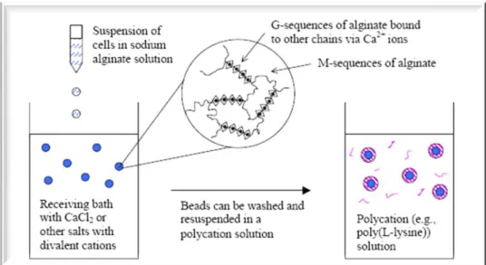
Polyelectrolyte gels and polyelectrolyte complexes have played an important role in the microencapsulation of cells, with alginate and alginate/poly(L-lyine) complexes representing the most widely studied materials for these application. In the case of ionic cross-linking (figure 1-10), cells are first suspended in a polymer solution (e.g. sodium alginate), which is subsequently dropped into a receiving bath containing a multivalent cation (e.g. Ca2+, Ba2+) to create beads. This gentle gelation process is cell-compatible. Additionally, a polycation is used to form the outer layer to the beads (e.g. chitosan, poly(L-lysine), which often provides additional mechanical integrity [178] to the capsules.
Figure 1-10. Schematic representation of a typical encapsulation process involving ionic cross-linking of
A wide variety of polyelectrolyte gels and complexes have been investigated for cell encapsulation. The Table 1-4 lists some physical characteristics of polyelectrolyte commonly used in cell encapsulation applications [178].
Table 1-4. Characteristics of some polyelectrolytes used for cell encapsulation. Adapted from [178]. Polyelectrolyte Reported Characteristics Ref. Alginate Strong polyanion. In vivo and in vitro applications with
long-term of cell viability.
[124, 138, 180, 181]
Carrageenan
Strong polyanion. κappa and ιota-carrageenan form
thermoreversible gels on cooling in the presence of appropriate counterions. Microbial cell viability.
[31]
Poly (styrene sulphonate)
Strong polyanion. A process for depositing alternating
layers and microencapsulation.
Cell viability and functionality was maintained.
[182, 183]
Carboxymethlcellulose
Polyanion. A sign of insufficient biocompatibility was
suppressed by altering the external surface material from Alg to CMC.
[184]
Cellulose sulfate
Polyanion. Preclinical studies of the biological properties,
being developed for use as a topical contraceptive antimicrobial agent.
[26]
Heparin
Polyanion. Heparin is a highly sulfated glycosaminoglycan
widely used as an injectable anticoagulant. It is also used to form an inner anticoagulant surface on various experimental and medical devices.
[185, 186]
Poly (methylene– co-guanidine)
Polyanion. Microcapsule has been developed based on
the polyelectrolyte complexation which was successfully tested in rodent animal models.
[187]
Poly (diallyldimethyl ammonium chloride)
Strong polycation. Microcapsules developed by
polyelectrolyte complexes, suitable for the encapsulation of these biological substances.
[188]
Chitosan Weak polycation, pKa of primary amine 6.3-6.8 Polymer
usedformembrane’scapsules. [31, 189]
Poly (L-lysine)
Weak polycation, pKa of primary amine ~ 10.5
A novel technique for creating supported lipid membranes on polyelectrolyte multilayers is described. Encapsulated CHO cells 90% viability.
[190, 191]
Poly (vinylamine) hydrochloride
Weak polycation. Mechanically stable microcapsules
have been produced for applications involving living cells and controlled delivery.
[192]
Poly (allylamine) hydrochloride
Weak polycation, pKa of amine group ~ 8.5
After encapsulation, cells preserve their metabolic activities and they are still able to divide.
1.2
BIOCOMPATIBILITY
One of the main aims of this thesis was to study the biocompatibility of chitosan based membranes and scaffolds as well as to assess the functionality i.e., the enhancement of cell adhesion on the surface of chitosan membranes due to several different plasma surface treatments.
Therefore, in this section, it will be described the importance of biocompatibility testing and the main types of cytotoxicity/biocompatibility assays that can be used to assess the biological behaviour of materials being developed for biomedical applications.
Biocompatibility of polymeric materials refers to the reaction of polymers with blood and tissue, depending on the site and purpose of use [126, 145]. Biocompatibility must be articulated within the context of an end-use application and has measurable dimensions only within this context [193].
Theterms“biocompatibilityassessment”and“safetyassessment”havebeen generally considered to be synonomous. The safety assessment of biomaterials, prostheses, artificial organs and other medical devices generally is considered to be the determination of the biological interactions of the medical device in an in vivo environment [194]. The goal of safety testing is to evaluate if a medical device or biomaterial support presents potential harm to the patient or user under conditions simulating use. A broad definition of biocompatibility applied to tissue engineering implants includes the determination of the biological interactions of the device, i.e. biomaterial component, tissue component, and combination of biomaterial and tissue components, in an in vivo environment [194, 195].
One of the major problems in the use of polymers as biomaterials is to make sure that they are biocompatible by themselves, and that the use of particular additives and/or processing technologies required to obtain different properties and or configurations will not interfere with the biocompatible behaviour [196, 197].
In terms of biocompatibility, the requirements that degradable materials must fulfil are much more demanding than those for non-degradable materials. In addition to the potential problem of toxic contaminants leaching out from the implant, such as residual monomers, stabilisers, emulsifiers and many other types of additive, it is also
necessary to considerer the potential toxicity of the degradation products and subsequent metabolites [198]. Natural origin polymers, besides being biodegradable, present some characteristics that may affect their biocompatibility behaviour.
In fact, these polymers usually contain domains that can send important signals to guide cells at various stages of their development [175, 199, 200]. However, this bioactivity can cause problems with antigenicity [175, 200], because when the material is implanted in a host, this might stimulate an immune response, potentially leading to immune rejection. Additionally, the degradation of natural polymers often relies on enzymatic processes that produce variation in the degradation rate. Nevertheless, natural origin polymers were the first to be used as scaffold materials for tissue regeneration [175, 200].
1.2.1 IN VITRO CYTOTOXICITY/BIOCOMPATIBILITY ASSAYS
Cytotoxicity testing represents the initial phase in testing the biocompatibility of potential biomaterials and medical devices. Its purpose is to act as a reliable, convenient, and reproducible screening method to detect, at an early stage in the testing process, cell death or other serious negative effects on cellular functions [201-205].
Theterm“cytotoxicity”meanstocausetoxiceffects(death,alterationsincellular membrane permeability, enzymatic inhibition, etc.) at the cellular level. It is clearly different from physical factors that affect cellular adhesion (surface charge of a material, hydrophobicity, hydrophilicy, etc.) [206]. A toxic material releases a chemical in sufficient quantities to affect cells directly or indirectly through inhibition of metabolic pathways [206].
Cytotoxicity in vitro assays are referenced in national and international standards [194, 206, 207]. Such methods can be applied to both direct and indirect testing assays. These assays differ mainly in the manner in which the test material is exposed to the cells. The choice of the method varies with the characteristics of the test material, the rationale for doing the test and the application of the data for evaluating biocompatibility [206, 208, 209]. Regarding indirect contact two methods can be considered [208, 209]. The first consists of the separation of the material from cells by
![Table 1-4. Characteristics of some polyelectrolytes used for cell encapsulation. Adapted from [178]](https://thumb-eu.123doks.com/thumbv2/123dok_br/17959551.854312/35.893.121.763.246.1153/table-characteristics-polyelectrolytes-used-cell-encapsulation-adapted.webp)
![Figure 1-13. Approaches for enhancement of cell growth on surfaces. Adapted from [145]](https://thumb-eu.123doks.com/thumbv2/123dok_br/17959551.854312/41.893.222.647.814.1104/figure-approaches-enhancement-cell-growth-surfaces-adapted.webp)