Vol.58, n.5: pp. 732-740, September-October 2015 http://dx.doi.org/10.1590/S1516-89132015050228
ISSN 1516-8913 Printed in Brazil
BRAZILIAN ARCHIVES OF BIOLOGY AND TECHNOLOGY
A N I N T E R N A T I O N A L J O U R N A L
Extract of the Bark of
Bathysa cuspidata
Attenuates the
Development
of
Chemically-Induced
Preneoplastic
Colorectal Lesions in Rats
Damiana Diniz Rosa
1, Sandra Aparecida dos Reis
1, Nathane Pais Siqueira
1, Reggiani Vilela
Gonçalves
2, Alessandra Barbosa Ferreira Machado
3, Natália Filardis Tafuri
1, João Paulo
Viana Leite
4, Sérgio Luís Pinto da Matta
5and Maria do Carmo Gouveia Peluzio
1*1Departamento de Nutrição e Saúde; Universidade Federal de Viçosa; Viçosa - MG - Brasil. 2Departamento de
Biologia Animal; Universidade Federal de Viçosa; Viçosa - MG - Brasil. 3Departamento de Microbiologia, Imunologia e Parasitologia; Universidade Federal de Juiz de Fora; Juiz de Fora - MG – Brasil. 4Department de Bioquímica e Biologia Molecular;Universidade Federal de Viçosa; Viçosa - MG - Brasil. 5Department de Biologia Geral; Universidade Federal de Viçosa; Viçosa - MG - Brasil
ABSTRACT
The aim of this study was to investigate the effect of the bark extract Bathysa cuspidata on chemically induced preneoplastic colorectal lesions in Wistar rats. Forty male rats were randomly divided into four groups (n = 10 each): saline (control group, oral administration of saline solution 0.9%); dimethylsulfoxide (DMSO, vehicle control), B200 (treated with 200 mg/kg bark extract of B. cuspidata), and B400 (treated with 400 mg/kg bark extract of B. cuspidata). Administration of treatments was carried out by the gavage. The animals received four subcutaneous injections of 1,2-dimethylhydrazine (DMH, 40 mg/kg) in the initial two weeks of the experiment to induce preneoplastic colorectal lesions. After 15 weeks, the animals were euthanized and the presence of aberrant crypt foci (ACF), body weight, biochemical analyses, and oxidative stress markers were measured. The extract of B. cuspidata decreased the levels of superoxide dismutase (SOD), but did not influence the levels of catalase (CAT), malondialdehyde (MDA), nitric oxide or protein carbonyl, compared with the saline group. The animals supplemented with a more concentrated B. cuspidata extract (B400) showed a significant reduction in the number of ACF in all the portions of the intestinal mucosa. The study demonstrated that the bark extract of B. cuspidata at 400 mg/kg reduced the preneoplastic colorectal lesions in an animal model of colon cancer and that the effect could be dose-dependent.
Key words:Bathysa cuspidata, animal model, oxidative stress, aberrant crypt foci
*Author for correspondence: [email protected]
INTRODUCTION
Colorectal cancer is a significant cause of mortality in both men and women and its etiology is multifactorial and complex (Jemal et al. 2011). According to the World Health Organization, colorectal cancer is the fourth most common type of cancer, with an estimated 940,000 new cases
number of such foci is a useful marker to determine the risk of developing colorectal cancer (Stevens et al. 2007). As colon cancer is a public health issue, new strategies are needed to reduce its high prevalence. It is estimated that 80% of the world population uses the products derived from the plants in their basic healthcare. Biodiversity is a great reservoir of bioactive secondary metabolites that are used for the production of therapeutic drugs for the treatment of different human pathologies, including cancer (Ranawat et al. 2010).
Bathysa cuspidata (A. St. Hil.) Hook f. belongs to
the Rubiaceae family. An infusion of its stem bark is used in popular medicine as an anti-inflammatory and healing agent, and for the treatment of various disorders, including stomach and liver problems (Botsaris 2007; Novaes et al. 2012). Studies have shown positive effects in the prevention and treatment of pulmonary and liver disorders by using ethanolic extracts of the leaves and bark of this species with no mutagenic effects. Use of the B. cuspidata extract to treat pulmonary and liver disorders is believed to be associated with an antioxidant potential effect, mainly in the animals exposed to chemical agents, such as carbon tetrachloride (CCl4) and paraquat (Goncalves et al. 2012; Novaes et al. 2012). Given the demonstrated benefits of the extract of
B. cuspidata and the interest in developing new
drugs from natural plant products, the purpose of this study was to evaluate the chemo-preventive effect of B. cuspidata extract on the rats with preneoplastic colorectal lesions, induced with the carcinogen 1,2-dimethylhydrazine (DMH).
MATERIAL AND METHODS
Preparation of the plant extract
Samples of B. cuspidata were collected in a biome of Brazilian Atlantic forest in the Minas Gerais state, Brazil and deposited in the herbarium of the Federal University of Viçosa under registration VIC 21559. The bark of B. cuspidata was air-dried at 38°C for two days and then triturated. The powdered bark (500 g) was exhaustively extracted by percolation with 95% ethanol. The extract was concentrated under vacuum at 45°C using a rotary evaporator and then lyophilized until complete removal of the solvent, yielding an ethanolic extract of 135 g.
Phytochemical characterization of the extract
To measure the total phenol and proanthocyanidin contents, the powdered stem bark (1.0 g) was extracted with 200 mL of water at 100°C under reflux for 30 minutes. The concentration of total
phenols was determined colorimetrically
(absorbance at 760 nm) using the Folin–Ciocalteu method (Verza et al. 2007). The proanthocyanidin content was determined according to the method of Price et al. (1980).The results were expressed in milligram catechin equivalents per gram of dry matter. The content of total flavonoids was determined using rutin as the reference compound. This method was based on the formation of a flavonoid–aluminum trichloride complex, with maximum absorption at 420 nm. The absorption of standard rutin solution in methanol was measured under the same conditions. All the determinations were performed in triplicate and the results were averaged (Boll et al. 2001). Chromatographic analysis was performed on a Shimadzu LC-20 AD UFLC system (Shimadzu Corp., Tokyo, Japan).
Ethical considerations
The experimental procedures were conducted in accordance with the Ethical Principles in Animal Experimentation, adopted by the Brazilian National Council for the Control of Animal Experimentation (CONCEA), with the approval of the Ethics Committee of the Department of Veterinary Medicine of the Federal University of Viçosa in Brazil (approval protocol 169/2009).
Animals and experimental design
The case–control study lasted for 15 weeks. Forty male rats (Rattusnorvegicus: var. albinus, Rodentia, Mammalia), 72 days old, with an average initial weight of 315 ± 22 g were used for the study. The rats were obtained from the Central Animal House at the Biological Sciences Center at the Federal University of Viçosa. Preneoplastic lesions were induced in all the 40 animals with DMH. The animals were randomly divided into four experimental groups, with treatments administered by gavage: saline (control group, oral administration of saline solution 0.9%); DMSO (vehicle control); B200 (treated with 200 mg/kg of bark extract), and B400 (treated with 400 mg/kg of bark extract). Each experimental group contained ten animals that received standard Nuvilab® food (composition: 19.0% protein, 56.0% carbohydrate, 3.5% fat, 4.5% cellulose, 5.0% vitamins and minerals, totaling 13.87 kJ/g) and filtered water ad
The ethanolic bark extract was re-suspended in 1.0 mL (w/v) of vehicle DMSO (w/v) before being administered by gavage. The animals received treatment every 48 h for a period of 15 weeks. The animals were maintained in individual cages at 22 ± 1ºC, relative air humidity of 60–70%, and controlled light/dark cycle of 12 h. The body weights were measured each week during the experimental period.
Colorectal carcinogenesis protocol
Preneoplastic colorectal lesions were induced by giving an intraperitoneal injection of 40 mg DMH/kg body weight, two times a week over two weeks for a total of four applications, as previously reported by Larangeira et al. (1998). The DMH was dissolved in a solution of 0.9%
NaCl, 1.5% EDTA
(ethylenediaminetetra-aceticacid) and 10 mM sodium citrate and the final pH was adjusted to pH 8.0.
Euthanasia, material collection and aberrant crypt foci counting
After 15 weeks of treatment, the animals were anesthetized with Halothane (Tanohalo®,Cristália, Brazil). The livers were removed and weighed and then stored at -80°C for later analysis. The hepatosomatic index (LSI) was calculated as LSI = (liver weight/body weight) × 100 (Fassini et al. 2011). The large intestine from the cecum to the anus was removed for the analysis of the ACF. It
was washed in saline solution, opened
longitudinally along the counter-mesenteric border, and fixed in 10% formaldehyde. After 48 h of fixation, the intestine was divided into three fragments (proximal, middle, and distal regions) of equal length. The fragments were stained with 0.1% methylene blue for approximately two minutes, washed in phosphate buffer, and examined using optical microscopy at ×40 magnification, according to the technique described by Bird (1987). The number of ACF on the mucosal surface of the colon was counted from the cecum to the rectum by two observers in a double-blind manner. The ACF were categorized as foci with fewer than three crypts, or foci with more than three crypts (Rosa et al. 2012).
Determination of biochemical parameters in the serum
The serum was obtained after centrifugation at 3000 x g and stored at -80 C for later analysis. Aspartate amino transferase (AST), alanine amino
transferase (ALT), and alkaline phosphatase (ALP) were analyzed in the serum using diagnostic test kits (Bioclin®, Diagnostica®, Belo Horizonte, Brazil) in an auto analyzer equipment (COBAS MIRA Plus, Roche Diagnostic Systems, Branchburg Inc., NJ, USA). Nitric oxide (NO) production was quantified by the standard Griess reaction according to the method of Ricart-Janéet al. (2002).
Oxidant status markers in the liver Antioxidant system biomarkers
Each liver sample (100 mg/mL buffer) was homogenized in 50 mM phosphate buffer (pH 7.0), with 1% Triton X-100 (pH 7.0). The homogenate was centrifuged at 11 290 x g at 4 C for 10 min, and the supernatant was used for biochemical analysis. The catalase (CAT) activity was evaluated according to the method described by
Aebi (1974) by measuring the rate of
decomposition of hydrogen peroxide; the results were expressed as units of catalase/milligram of protein. For the analysis of superoxide dismutase (SOD), the hepatic tissue homogenate (100 mg/mL buffer) was homogenized in 50 mM of phosphate buffer (pH 7.0). The SOD activity was determined by an adapted method of Dieterichet al. (2000). The degree of inhibition of pyrogallol
(1,2,3-trihidroxybenzen) auto-oxidation in the
supernatant was measured and absorbance was read at 570 nm. The results are expressed as units SOD/mg of protein
Peroxidation biomarkers
Determination of protein concentration
Protein concentration in the tissue homogenates was measured by the method of Lowry et al. (1951) using bovine serum albumin as a reference.
Statistical analysis
Results are presented as the mean values with standard errors. Statistical significance of the differences among the groups was assessed by one-way analysis of variance (ANOVA), followed
by Dunn’s, or Tukey’s post hoc multiple
comparison tests, using the GraphPad Prism (GraphPad Software Inc, San Diego, CA, USA). For statistical analysis, p < 0.05 was considered to be statistically significant.
RESULTS
Chemical profile of the extract
The ethanolic extract of B. cuspidata gave rise to four major peaks related to the constituents belonging to the class of polyphenols. The total phenol and proanthocyanidin content comprised of 45.0 mg/g of dry matter (expressed as pirogalol)
and 27.9 mg/g of dry matter (expressed as catechin), respectively. The total flavonoid content was 6.0 mg/g dry matter using the calibration curve of rutin.
Effect of treatment with B. cuspidata extract on
the physiological parameters in rats
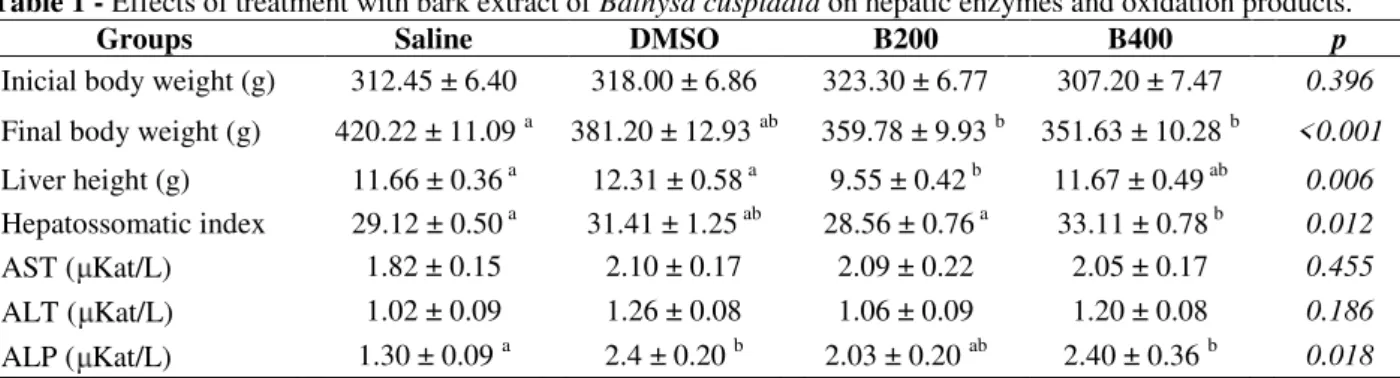
The results of the final bodyweight, liver height, and LSI are presented in Table 1. The animals in the four treatment groups had similar body weight at the beginning of the experiment (p = 0.396). However, after 15 weeks, the animals treated with the bark extract of B. cuspidata (B200 and B400) had lower body weight (p< 0.001) than the animals in the saline (control) group. Only the B200 group had lower liver weight relative to the saline group (p = 0.006). The B400 group had a higher LSI than all the other groups (p = 0.012). There were no significant differences in AST and ALT among the groups (Table 1). The serum levels of ALP were significantly higher in the DMSO and B400 groups, compared to the saline group (p = 0.018). The supplementation of animals with B. cuspidata extract did not affect the plasma levels of nitric oxide (NO) (p > 0.05, data not shown).
Table 1 - Effects of treatment with bark extract of Bathysa cuspidata on hepatic enzymes and oxidation products.
Groups Saline DMSO B200 B400 p
Inicial body weight (g) 312.45 ± 6.40 318.00 ± 6.86 323.30 ± 6.77 307.20 ± 7.47 0.396
Final body weight (g) 420.22 ± 11.09 a 381.20 ± 12.93 ab 359.78 ± 9.93 b 351.63 ± 10.28 b <0.001
Liver height (g) 11.66 ± 0.36 a 12.31 ± 0.58 a 9.55 ± 0.42 b 11.67 ± 0.49 ab 0.006
Hepatossomatic index 29.12 ± 0.50 a 31.41 ± 1.25 ab 28.56 ± 0.76 a 33.11 ± 0.78 b 0.012
AST ( Kat/L) 1.82 ± 0.15 2.10 ± 0.17 2.09 ± 0.22 2.05 ± 0.17 0.455
ALT ( Kat/L) 1.02 ± 0.09 1.26 ± 0.08 1.06 ± 0.09 1.20 ± 0.08 0.186
ALP ( Kat/L) 1.30 ± 0.09 a 2.4 ± 0.20 b 2.03 ± 0.20 ab 2.40 ± 0.36 b 0.018
DMSO, dimethylsulfoxide, AST: Aspartate aminotransferase, ALT: alanine aminotransferase, ALP: alkaline phosphatase; B200, animals group treated with the extract of B. cuspidata 200 mg/kg by gavage; B400, animals group treated with the extract of B.
cuspidata 400 mg/kg by gavage. Data are given as means ±SEM (n=10). Averages followed by the same letter in the column did
not differ by the Tukey test (p <0.05).
Oxidative stress markers and antioxidant enzymes
Enzymes involved in the endogenous antioxidant defense system were evaluated to measure the antioxidant activity of B. cuspidata extract (Fig. 1). No differences in CAT were found among the treatments (p = 0.08, Fig.1A). The DMSO, B200, and B400 groups had lower hepatic activity of SOD compared to the saline group (p = 0.0003, Fig. 1B). The products of lipid and protein oxidation were also evaluated. The DMSO group had higher levels of MDA compared to the saline
group (p = 0.0096, Fig. 1C). The consumption of
B. cuspidata extract did not affect the levels of
protein carbonyl (p> 0.05, Fig. 1D).
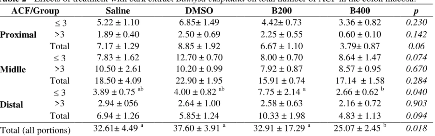
Aberrant crypt foci count in the colonic mucosa
group (p = 0.040). There was a reduction of ACF in the animals treated with B400 extract compared
to the saline, DMSO, and B200, across all portions of the intestinal mucosa of the animals (p = 0.018).
Figure 1 - Effects of the treatment with the bark extract of B. cuspidata on superoxide dismutase (SOD), catalase (CAT), Malondialdehyde (MDA) and protein carbonyl in liver tissue of Wistar rat. Saline (control group, saline solution 0.9%); DMSO (vehicle control); B200 (treated with 200 mg/kg bark extract) and B400 (treated with 400 mg/kg bark extract). The data are expressed as means ± SEM. a,b Different letters in columns indicate statistical difference between the groups (P< 0.05) and groups that have some common letter do not
differ statistically, anova followed by Tukey’s test.
Table 2 - Effects of treatment with bark extract Bathysa cuspidata on total number of ACF in the colon mucosa.
ACF/Group Saline DMSO B200 B400 p
Proximal
3 5.22 ± 1.10 6.85± 1.49 4.42± 0.73 3.36 ± 0.82 0.230
>3 1.89 ± 0.40 2.50 ± 0.69 2.25 ± 0.55 0.60 ± 0.10 0.142
Total 7.17 ± 1.29 8.85 ± 1.92 6.67 ± 1.10 3.79± 0.87 0.06
Midlle
3 7.83 ± 1.62 12.70 ± 0.70 8.00 ± 0.70 8.64 ± 1.47 0.074
>3 10.50 ± 2.61 10.20 ± 0.99 7.92 ± 0.87 8.57 ± 0.95 0.670
Total 18.50 ± 4.09 22.90 ± 1.95 15.91 ± 0.74 17.14 ± 1.58 0.284
Distal
3 3.89 ± 0.75 ab 4.00 ± 0.82 ab 7.75 ± 2.14 a 2.66 ± 0.62 b 0.040
>3 2.94 ± 056 2.64 ± 1.00 2.58 ± 0.63 2.16 ± 0.72 0.903
Total 6.94 ± 1.26 5.85± 1.24 10.33 ± 1.98 4.83 ± 1.13 0.094
Total (all portions) 32.61± 4.49 a 37.60 ± 3.91 a 32.91 ± 17.29 a 25.07 ± 2.45 b 0.018 DMSO, dimethylsulfoxide; B200, animals group treated with extract 200 mg/kg by gavage; B400, animals group treated with extract 400 mg/kg by gavage. Data are given as means ±SEM (n=10). Averages followed by the same letter in the column do not differ by the Tukey test (p <0.05).
DISCUSSION
Medicinal plants have a long history of use in the therapy throughout the world. In the 1960s, the
extracts must be safe and effective to be used in the formulation of therapeutic drugs (Cai et al. 2004; Atanassova et al. 2011). With that in mind, and considering the increase in the incidence of colorectal cancer worldwide, the effect of the extract of the bark B.cuspidata was investigated on the appearance of ACF in rats, after induction with DMH, as well as its effect on the physiological and biochemical parameters of the
animals. Chemically-induced models of
preneoplastic colorectal lesions and carcinogenesis in rodents have been shown to be adequate for the study of risk factors, development, and prevention of malignancy (Bird 1987). The dose of the bark extract of B. cuspidata used —200 or 400 mg⁄kg of body weight — may be classified as the normodose or standard dose. The extract of bark of
B. cuspidata showed significant hepatoprotective
effects, stimulated the antioxidant defense system and reduced the morphological and functional liver damage in Wistar rats exposed to carbon tetrachloride (CCl4) (Gonçalves et al. 2014). The antioxidant activity of some aqueous and ethanolic extracts of Chinese medicinal plants have been positively correlated to the content of polyphenols and flavonoids (Cai et al. 2004; Zhang et al. 2011). Other similar studies have
demonstrated the antioxidant activity of
polyphenols from Solanum melongena(Kimura et al. 1999) and Croton cajucara (Tieppo et al. 2006), which presented a high concentration of phenolic compounds that were able to suppress the oxidative stress in liver tissue. In the present study, the preliminary phytochemical analysis indicated the presence of flavonoids and polyphenolic compounds — important antioxidant compounds It has already been established that the toxicity of DMH manifests as systemic effects such as lethargy, anorexia, and progressive weight loss in the animals. In this study, none of these effects were observed in the DMH-treated animals; there was weight gain in all the groups. The LSI was higher in the groups treated with DMSO and B400, suggesting increased hepatic fibrosis in these groups. However, LSI is a low-specificity marker and further analysis, such as, a count of collagen fibers is necessary to confirm the fibrosis. Traditionally, measurement of liver-specific serum enzyme levels has served as a good indicator of liver damage (Sreelatha et al. 2009; Ranawat et al.
2010). AST enzyme is found in high
concentrations in a number of tissues, such as heart, liver, skeletal muscle, kidney, and pancreas.
ALT is primarily limited to the cytosol of hepatocytes and is considered a highly sensitive indicator of hepatocellular damage, and within certain limits, can provide a quantitative ratio of the degree of damage suffered by the liver (Al-Habori et al. 2002). In this study, the AST and
ALT levels, important markers of liver
dysfunction, were not influenced by the
consumption of the bark extract of B. cuspidata. In contrast, higher levels of ALP were found in the animals receiving DMSO, B200, and B400, compared to the saline group. The concentration of ALP could be used as a biomarker of diseases, and monitoring ALP levels would be useful to assess the liver when medication, or drugs damaging to the liver are taken; the same could also be true for monitoring the effectiveness of the treatments. For DMH to exert a carcinogenic effect, it must be metabolized in the liver (Rosenberg et al. 2009).Its metabolites are then taken to the colon through the blood and bile (Sengottuvelan et al. 2006). There, DMH can induce precancerous lesions as the metabolites generate reactive carbon ions that are capable of methylating DNA, RNA, and colonocyte proteins (Rosenberg et al. 2009). DMH also induces point mutations, micronuclei, and sister chromatid exchanges, and produces free radicals (Sengottuvelan et al. 2006). These lead to apoptosis of colonocytes and stimulate cell proliferation, which causes the appearance of precancerous lesions in the colon tissue (Newell and Heddle 2004).To determine the effects of B.
cuspidata on the hepatic antioxidant enzymes in
the rats after the induction of the ACF with DMH, the CAT and SOD activities were measured. The DMSO, B200, and B400 groups presented lower SOD activity than the saline group, indicating that the DMSO and extract protected the liver and prevented increased expression of SOD, probably by suffering less tissue destruction by the action of free radicals that were generated by the application of DMH. No differences were found in the levels of CAT. In addition, the extract B.
cuspidata (B200 and B400) did not influence the
The DMSO group showed higher levels of lipid oxidation products, verified by the quantification of MDA. Despite being a substance widely used in the biological assays, DMSO has generated controversy, leading to conflicting opinions. There is no consensus on the volume, dilution, and total amount of DMSO that should be used in the experimental animals (Melo et al. 2008). DMSO induces side effects. When administered in small doses, it may cause toxic effects to the liver and in larger doses, can cause hepatic steatosis (Mathew et al. 1980). However, the use of this substance in this study is justified by the good solubility of bark extract and the ability of DMSO to easily penetrate the organs, tissues, and membranes, including cellular and intracellular penetration after an intraperitoneal injection (Szmant 1975).
Treatment with the extract of the bark at 400 mg/kg for 15 weeks reduced the total number of preneoplastic lesions in the intestinal mucosa of rodents, indicating that the extract in high concentrations protected the colonic mucosa from developing ACF. This result could be explained by high levels of phenolic compounds found in the extract. Treatment with a lower dose of extract (200 mg/kg) did not exert a chemo-preventive effect. The high concentration of antioxidant compounds present in the B400 group could explain the reduction in preneoplastic lesions. Shwter et al (2014) evaluated the anticarcinogenic activity of the ethanolic extract of Gynura
procumbens on colorectal cancer induced by
azoxymethane in Sprague–Dawley rats. The doses of extract (250 and 500 mg/kg body weight) used decreased the incidence of ACF in the colons of the animals after 10 weeks of treatment. Phenolic compounds were the predominant antioxidant constituents in G. procumbens. A similar result was found by Almagrami et al (2014) in the rats with induced colorectal cancer and treated with the ethanolic extract of Acanthus ilicifolius (250 and 500 mg/kg body weight) for eight weeks.
Polyphenols and flavonoids may inhibit the carcinogenesis by affecting the molecular events in the initiation, promotion, and progression stages. The mechanisms used by these compounds to inhibit the carcinogenic process include antioxidant action, modulation of several protein functions, modulation of the secretion of protein kinases in tumor cell proliferation, induction of the expression of anticarcinogenic enzymes, inhibition of induction of cancer-promoting enzymes, activation of detoxifying enzymes, and increased
cell-to-cell communication, among others (Cai et al. 2004; Shwter et al. 2014).
This study demonstrated that the extract of the bark of B. cuspidata at 400 mg/kg for 15 weeks reduced preneoplastic colorectal lesions in an animal model of colon cancer. The results indicated a possible dose-dependent effect of B.
cuspidata extract. Further pharmacological
evaluations are essential to elucidate the detailed mechanism of action of this extract, which may have a high potential for the prevention and treatment of preneoplastic colorectal lesions.
ACKNOWLEDGMENTS
We would like to thank the Coordination for Enhancement of Higher Education Personnel
(PNPD, CAPES, Brazil, process no.2980/2010) for financial support of the project. The authors are grateful for financial support provided by the Research Support Foundation of Minas Gerais State (FAPEMIG, Brazil) and the National
Council of Technological and Scientific
Development (CNPq, Brazil).
REFERENCES
Aebi H. Catalase. In: Berg Meyer H, editor. Methods of Enzymatic Analysis. 2. Weinheim1974. p. 673-84. Al-Habori M, Al-Aghbari A, Al-Mamary M, Baker M.
Toxicological evaluation of Catha edulis leaves: a long term feeding experiment in animals. J Ethnopharmacol. 2002; 83(3): 209-217.
Almagrami AA, Alshawsh MA, Saif-Ali R, Shwter A, Salem SD, Abdulla MA. Evaluation of chemopreventive effects of Acanthus ilicifolius
against azoxymethane-induced aberrant crypt foci in the rat colon. Plos One. 2014; 9(5).
Atanassova M, Georgieva S, Ivancheva K. Total phenolic and total flavonoid contents, antioxidant capacity and biological contaminants in medicinal herbs. J Univ Chem Technol Metallurgy. 2011; 46(1): 81-88.
Bird RP. Observation and quantification of aberrant crypts in the murine colon treated with a colon carcinogen: Preliminary findings. Cancer Lett. 1987; 37(2): 147-151.
Bose A, Elyagoby A, Wong TW. Oral 5-fluorouracil colon-specific delivery through in vivo pellet coating for colon cancer and aberrant crypt foci treatment. Int J Pharmaceut. 2014;468(1–2):178-186.
Botsaris A. Plants used traditionally to treat malaria in Brazil: the archives of flora medicinal. J Ethnobiol Ethnomed. 2007; 3:18.
Buege JA, Aust SD. Microsomal lipid peroxidation. In: Gleischer S, Packer L, editors. Methods Enzymol. 52C. New York: Academic Press; 1978. p. 302-310. Cai Y, Luo Q, Sun M, Corke H. Antioxidant activity
and phenolic compounds of 112 traditional Chinese medicinal plants associated with anticancer. Life Sci J. 2004; 74: 2157 - 2184.
Dieterich S, Bieligk U, Beulich K, Hasenfuss G, Prestle J. Gene expression of antioxidative enzymes in the human heart: increased expression of catalase in the end-stage failing heart. Circulation. 2000; 101(1): 33-39.
Fassini P, Noda R, Ferreira E, Silva M, Neves V, Demonte A. Soybean glycinin improves HDL-C and suppresses the effects of rosuvastatin on hypercholesterolemic rats. Lipids Health Dis. 2011; 10(1): 165.
Goncalves RV, Novaes RD, Leite JP, Vilela EF, Cupertino MC, Nunes LG, et al. Hepatoprotective effect of Bathysa cuspidata in a murine model of severe toxic liver injury. Int J Exp Pathol. 2012; 93(5): 370-376.
Gonçalves RV, Matta SLPd, Novaes RD, Leite JPV, Peluzio MdCG, Vilela EF. Bark Extract of Bathysa cuspidata in the Treatment of Liver Injury Induced by Carbon Tetrachloride in Rats. Braz Arch Biol Technol. 2014; 57: 504-513.
Jemal A, Bray F, Center MM, Ferlay J, Ward E, Forman D. Global cancer statistics. CA Cancer J Clin. 2011; 61(2): 69-90.
Kimura Y, Araki Y, Takenaka A, Igarashi K. Protective effectsof dietary Nasunim on paraquat-induced oxidative stress in rats. Biosci Biothecnol Biochem. 1999; 63: 799-804.
Larangeira LLS, Taha MO, Ferme A, Lemos R, Plapler H. localização de lesões tumorais induzidas pela 1,2-dimetilhidrazina e seu grau de atipia no colon de ratos. Act Cir Bras. 1998;13.
Levine RL, Garland D, Oliver CN, Amici A, Climent I, Lenz AG, et al. Determination of carbonyl content in oxidatively modified proteins. Methods Enzymol. 1990; 186: 464-478.
Lowry O, Rosebrough N, Farr A, Randall R. Protein measurement with the folin phenol reagent. J Biol Chem. 1951; 193: 265-275.
Mathew T, Karunanithy R, Yee MH, Natarajan PN. Hepatotoxicity of dimethylformamide and dimethylsulfoxide at and above the levels used in some aflatoxin studies. Lab Investigat. 1980; 42: 257-262.
Melo JUS, Vasconcelos PRL, Santos JMV, Júnior MMC, Barreto MVB, Kimura OS. Efeitos do dimetilsulfóxido no estresse oxidativo e na regeneração hepática pós-hepatectomia em ratos. Rev Col Bras Cir. 2008; 35: 103-108.
Newell LE, Heddle JA. The potent colon carcinogen, 1,2-dimethylhydrazine induces mutations primarily in the colon. Mutat Res. 2004; 564: 1-7.
Novaes RD, Goncalves RV, Cupertino MC, Marques DC, Rosa DD, Peluzio Mdo C, et al. Bark extract of
Bathysa cuspidata attenuates extra-pulmonary acute lung injury induced by paraquat and reduces mortality in rats. Int J Exp Pathol. 2012; 93(3): 225-233.
Price ML, Hagerman AE, Butler LG. Tannin content of cowpeas, chickpeas, pigeon peas, and mung beans. J Agric Food Chem. 1980; 28(2): 459-461.
Ranawat L, Bhatt J, Patel J. Hepatoprotective activity of ethanolic extracts of bark of Zanthoxylum armatum DC in CCl4 induced hepatic damage in rats. J Ethnopharmacol. 2010; 127(3): 777-780.
Ricart-Jane D, Llobera M, Lopez-Tejero MD. Anticoagulants and other preanalytical factors interfere in plasma nitrate/nitrite quantification by the Griess method. Nitric Oxide. 2002; 6(2): 178-185. Rosa DD, Lourenco FC, da Fonseca AC, de Sales RL,
Ribeiro SM, Neves CA, et al. Fish oil improves the lipid profile and reduces inflammatory cytokines in Wistar rats with precancerous colon lesions. Nutrition and cancer. 2012; 64(4): 569-579.
Rosenberg DW, Giardina C, Takuji T. Mouse models for the study of colon carcinogenesis. Carcinogenesis. 2009; 30(2): 183-196.
Sengottuvelan M, Senthilkumar R, Nalini N. Modulatory influence of dietary resveratrol during different phases of 1,2-dimethylhydrazine induced mucosal lipid-peroxidation, antioxidant status and aberrant crypt foci development in rat colon carcinogenesis. Biochim Biopsy Acta. 2006; 1760: 1175-1183.
Shwter AN, Abdullah NA, Alshawsh MA, Alsalahi A, Hajrezaei M, Almaqrami AA, et al. Chemoprevention of colonic aberrant crypt foci by Gynura procumbens
in rats. J Ethnopharmacol. 2014; 151: 1194-11201. Sreelatha S, Padma PR, Umadevi M. Protective effects
of Coriandrum sativum extracts on carbon tetrachloride-induced hepatotoxicity in rats. Food Chem Toxicol. 2009; 47(4): 702-708.
Stevens RG, Swede H, Rosenberg DW. Epidemiology of colonic aberrant crypt foci: review and analysis of existing studies. Cancer Lett. 2007; 252(2): 171-183. Szmant HH. Physical properties of dimethyl sulfoxide
Tieppo M, Porawski M, Salvador M, Moreira AJ, Collado PS, Gonza´- lez-Gallego J, et al. Croton cajucara Benth. Leaf extract scavenges the stable free radical DPPH and protects against oxidative stress induced by paraquat. Biol Pharm Bull 2006; 29: 161-165.
Verza SG, Kreinecker MT, Reis V, Henriques AT, Ortega GG. Avaliação das variáveis analíticas do método de Folin-Ciocalteu para determinação do teor de taninos totais utilizando como modelo o extrato aquoso de folhas de psidium guajava L. Quim Nova. 2007; 30: 815-820.
World Health Organization. Global cancer rates could increase by 50% to 15 million by 2020.
http://wwwwhoint/mediacentre/news/releases/2003/pr 27/en/ (accessed 04/20/2015). 2013.
Zhang L, Ravipati AS, Koyyalamudi SR, Jeong SC, Reddy N, Smith PT, et al. Antioxidant and anti-inflammatory activities of selected medicinal plants containing phenolic and flavonoid compounds. J Agric Food Chem. 2011; 59: 12361-12367.