Exogenous ATP administration prevents ischemia/reperfusion-induced oxidative stress and tissue injury by... 1257
Exogenous ATP administration prevents ischemia/reperfusion-induced oxidative stress
and tissue injury by modulation of hypoxanthine metabolic pathway in rat ovary
Administração exógena de ATP impede isquemia/induzidas por reperfusão de estresse oxidativo de tecidos e lesões pela modulação da hipoxantina via metabólica em ovários de ratos
Serkan KumbasarI Nihal CetinII Omer Erkan YapcaIII Ebru SenerIV Unal IsaogluV Mehmet YilmazIII Suleyman SalmanVI Ayse Nur AksoyV Mehmet Ali GulVII Halis SuleymanII* ISSN 0103-8478
ABSTRACT
In this study, xanthine oxidase (XO), malondialdehyde (MDA), myeloperoxidase (MPO) and glutathione (GSH) levels in the ovarian tissues of rats during the development of ischemia and postischemia-induced reperfusion were investigated, and the effect of ATP on ischemia-reperfusion (I/R) damage was biochemically and histopathologically examined. The results of the biochemical analyses demonstrated that ATP signifi cantly reduced the level of XO and MDA and increased the amount of GSH in both ischemia and I/R-applied ovarian tissue at the doses administered. Furthermore, ATP signifi cantly suppressed the increase in MPO activity that occurred following the application of post ischemia reperfusion in the ovarian tissue. The biochemical results obtained in the present study coincide with the histological fi ndings.The severity of the pathological fi ndings, such as dilatation, congestion, haemorrhage, oedema and polymorphonuclear nuclear leukocytes (PMNLs), increased in parallel with the increase observed in the products of XO metabolism. In conclusion, exogenously applied ATP prevented I/R damage by reducing the formation of XO in ischemic ovarian tissue.
Key words:xanthine oxidase metabolism, ischemia/reperfusion injury, antioxidant, rat.
RESUMO
Neste estudo, a xantina oxidase (XO), o malondialdeído (MDA), mieloperoxidase ( MPO ) e glutationa (GSH) nos tecidos do ovário de ratos, durante o desenvolvimento de isquemia e reperfusão induzida por pós-isquemia foi investigada, e o efeito de ATP em isquemia e reperfusão (I/R). O
dano foi verifi cado por provas bioquímicas e por histopatologia. Os resultados das análises bioquímicas mostraram que o ATP reduziu signifi cativamente o nível de XO e MDA e aumentou a quantidade de GSH em ambas as isquemia e no tecido do ovário de I / R - aplicado nas doses administradas. Além disso, o ATP suprimiu signifi cativamente o aumento na atividade de MPO que ocorreu na sequência da aplicação de pós-isquemia reperfusão no tecido ovariano. Os resultados bioquímicos obtidos no presente estudo coincidem com os achados histológicos. A gravidade dos achados patológicos, como a dilatação, congestão, hemorragia, edema e polimorfonucleares leucócitos nucleares (PMNLs), aumentou em paralelo com o aumento observado nos produtos do metabolismo XO. Em conclusão, aplicando exogenamente ATP impedido de I/R, houve danos pela redução da formação de tecido de ovário de XO na isquemia.
Palavras-chave: metabolismo oxidativo da xantina, isquemia/ reperfusão, antioxidante, rato.
INTRODUCTION
Ischemia is defi ned as partial or complete oxygen (O2) deprivation in tissues due to reduced or completely interrupted blood fl ow. In ischemia, the synthesis of high-energy phosphates, such as ATP, is decreased in cells due to interruption of the O2fl ow into the tissue (JENNINGS & REIMER, 1991; REITER, 1995). The decrease in ATP leads to inhibition of the Na+, K+ ATPase pump in the cell membrane and
IDepartment of Obstetrics and Gynecology, Faculty of Medicine, Sakarya University, Sakarya, Turkey.
IIDepartment of Pharmacology, Faculty of Medicine, Recep Tayyip Erdogan University, 53000, Rize, Turkey, E-mail: [email protected]. *Autor para correspondência.
IIIDepartment of Obstetrics and Gynecology, Faculty of Medicine, Ataturk University, Erzurum, Turkey. IVDepartment of Pathology, Erzurum Region Education and Research Hospital, Erzurum, Turkey.
an increase in intracellular Na+ and Ca2+ concentrations (GREEN et al., 1989). Increased intracellular Ca2+ concentrations are reported to be cytotoxic (ORRENIUS et al., 1992). ATP, adenosine mono-phosphate (AMP), adenosine, inosine and hypoxanthine are produced from ATP, and ATP utilisation continues during the ischemia period. Furthermore, xanthine dehydrogenase (XD) is converted to xanthine oxidase (XO) in this period. In healthy tissues, hypoxanthine is metabolised by XD, whereas it is metabolised by XO in ischemic tissue (PARKS et al., 1988). Toxic O2 radicals are not formed in healthy tissues because nicotinamide dinucleotide is used in the metabolism of hypoxanthine by XD. Toxic O2 radicals are produced as intermediates in ischemic tissues because O2 is used in the metabolism of hypoxanthine by XO (GRACE, 1994; PARKS et al., 1988). Hypoxanthine cannot be metabolised by XO unless reperfusion occurs in ischemic tissue. As is known, the emptying of cellular energy stores may lead to cell death in long-lasting ischemia (PARKS et al., 1988; ZIMMERMAN & GRANGER, 1992). Therefore, the intervention in ischemic conditions has always been adequate perfusion of the tissue. During reperfusion, XO produced during ischemia converts hypoxanthine to xanthine using locally available O2, leading to excessive free O2 radical production and a decrease in antioxidant defence mechanisms (LINDSAY et al., 1990; LI & JACKSON, 2002). Previous studies showed that XO, the production of which increases during ischemia due to ATP defi ciency, is an essential component in the mechanism of reperfusion injury. This indicates that ATP administration may be benefi cial for the prevention of ischemia/reperfusion (I/R) injury.
Thus far, no studies have investigated the potentially protective effect of exogenous administration of ATP on I/R-induced ovarian injury in rats. Therefore, the aim of the present study was to investigate the effect of exogenous administration of ATP prior to experimentally induced ischemia and I/R of the rat ovary on biochemical variables and histopathological parameters.
MATERIAL AND METHODS
Animals
A total of 42 female adult Albino Wistar rats weighing between 220-235g were used for the experiment. The rats were obtained from Ataturk University Medical Experiments Research and Training Center (Erzurum, Turkey). The protocols and the procedures were approved by the local Animal Experimentation Ethics Committee (Date: 24. 04. 2012, meeting no: B.30.2.ATA.0.01.02/1845).
Chemical substances
Thiopental sodium was provided by IE Ulagay-Turkey and ATP was supplied by Zdorove Narodu Ukraine.
General procedure
The animals were allocated to 7 groups as Ovarian Ischemia (OI), Ovarian Ischemia/Reperfusion (OIR), 2mg kg-1 ATP + Ovarian Ischemia (AOI-2), 4mg kg-1 ATP + Ovarian Ischemia (AOI-4), 2mg kg-1 ATP+ Ovarian Ischemia/Reperfusion (AOIR-2), 4mg kg-1 ATP+ Ovarian Ischemia/Reperfusion (AOIR-4), and Control Group (Sham Operated- Healthy Control Group (CG). Surgical procedures were performed under sterile conditions in a suitable laboratory environment by administering 25mg kg-1 intraperitoneal (IP) thiopental sodium anesthesia.
Surgical and pharmacological procedures
ATP was injected into AOI-2, AOI-4, AOIR-2 and AOIR-4 groups via the intraperitoneal route 1h before anaesthesia. A 3h ischemia period was then simulated through vascular clamping to the lower part of the right ovaries of rats assigned to OI, OIR, AOI-2, AOI-4, AOIR-2 and AOIR-4 groups. At the end of this period, the ovaries of the rats in the OI, AOI-2, AOI-4 groups were removed. Reperfusion was provided for 2h by removing the vascular clamp in the OIR, AOIR-2 and AOIR-4 groups. The ovaries of the rats in the OIR, AOIR-2 and AOIR-4 groups were removed. Histopathological and biochemical analyses were performed subsequently in all the samples (KURT et al., 2013). A 2–2.5cm long vertical incision was made in the lower abdomen of each rat to reach the ovaries. Ischemia was not performed in the ovaries of the rats in CG. The ovarian tissue samples were collected from all the experimental groups, and the assay was conducted simultaneously.
Biochemical procedures
Biochemical analysis of ovarian tissue
The homogenates were prepared to measure the enzymatic activity in the ovarian tissues. The XO, malondialdehyde (MDA), myeloperoxidase (MPO) and total glutathione (tGSH) enzyme activities in the supernatants obtained from these homogenates were determined using established methods reported in the literature.
at 293nm. In this method, phosphate buffer with EDTA (pH 7.5) is used as the solvent that reaction takes place. A total of 50μl of the sample and 50μl of xanthine (4mM) were added to the phosphate buffer, and then the mixture was incubated for 30min at 37°. A total of 100% (w/v) of TCA was added to stop the reaction mixture. The mixture was centrifuged at 4500g for 20min. The increase in the absorbance of the supernatant was then measured at 293nm (ROWE et al., 1966).
Determination of malondialdehyde (MDA) activity The concentrations of ovarian lipid peroxidation were determined by estimating MDA activity using the thiobarbituric acid test (OHKAWA et al., 1979). The rat ovaries were promptly excised and rinsed with cold saline. The ovarian tissue, weighed and homogenised in 10ml of 100g l-1 KCl. The homogenate (0.5ml) was added to a solution containing 0.2ml of 80g l-1 sodium lauryl sulfate, 1.5ml of 200g l-1 acetic acid, 1.5ml of 8g l-1 2-thiobarbiturate and 0.3ml of distilled water. The mixture was incubated at 98°C for 1h. Upon cooling, 5ml of n-butanol:pyridine (15:l) were added. The mixture was vortexed for 1min and centrifuged for 30min at 4000rpm. The absorbance of the supernatant was measured at 532nm. The standard curve was obtained by using 1,1,3,3-tetramethoxypropane.
Determination of myeloperoxidase (MPO) activity The activity of MPO in the total homogenate was measured according to the method of Wei and Frenkel, with some modifications (WEI & FRENKEL, 1991). The sample was weighed and homogenised in a 2ml of 50mM phosphate buffer containing 0.5% hexadexyltrimethyl ammonium bromide (HDTMAB) and centrifuged at 3500rpm for 60min at 4°C. The MPO activity in the supernatant was determined using 1,3ml 4-aminoantipyrine-2% phenol (25mM) solution. A total of 25mM 4-aminoantipyrine–2% phenol solution and 0.0005% 1.5ml H2O2 were added and equilibrated for 3-4min. After establishing the basal rate, 0.2ml of sample suspension was added and quickly mixed. Increases in the absorbance at 510nm were recorded at 0.1min intervals for 4min. The absorbance was measured at 412nm using a spectrophotometer.
Determination of total glutathione (tGSH)
The amount of tGSH in the total homogenate was measured according to the method of Sedlak and Lindsay with some modifications (SEDLAK & LINDSAY, 1968).
The sample was weighed and homogenised in 2ml of 50mM Tris–HCl buffer containing 20mM of EDTA and 0.2mM of sucrose at pH 7.5. The homogenate was immediately precipitated with 0.1ml of 25% trichloroacetic acid, and the precipitate was removed after centrifugation at 4200rpm for 40min at 4°C. The GSH level of the supernatant was then determined. A total of 1500μl of measurement buffer (200mMTris–HCl buffer containing 0.2mM of EDTA at pH 7.5), 500μl of supernatant, 100μl of 5,5-dithiobis (2-nitrobenzoic acid) (DTNB) (10mM) and 7900μl of methanol were added to a tube, vortexed and incubated for 30min at 37°C. DTNB was used as a chromogen, and it formed a yellow-coloured complex with SH groups. The absorbance was measured at 412nm using a spectrophotometer. The standard curve was obtained using reduced glutathione.
Histopathological procedures
The ovaries that were removed from the rats were fi xed in 10% formalin solution, sections 4μm in thickness were obtained from the paraffin blocks obtained after the routine tissue follow-up process, and the sections were stained with hematoxylin&eosin (H&E) stain after deparaffi nization and rehydration. All the sections were encoded and examined under light microscopy (Olympus CX 51, Tokyo, Japan).
Statistical analysis
All data were subjected to a one-way analysis of variance (ANOVA) using Statistical Package for the Social Sciences (SPSS) 18.0 software. Multiple comparisons were analysed using the Duncan’s multiple range test. A P-value
0.05 was considered statistically signifi cant. The results are presented as the mean standard error of mean (SEM).
RESULTS
Biochemical results
and MPO levels during ischemia and postischemia-induced reperfusion in ovarian tissue and decreases in tGSH. No signifi cant change was observed in the MPO activity of the ischemic ovarian tissue.
Histopathological result
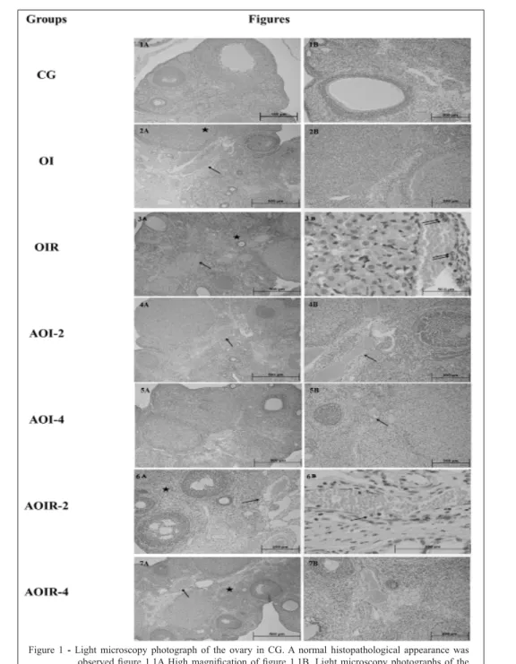
A normal histopathological appearance is observed in fi gure 1.1.A-B. A moderate dilation, congestion (arrow) and mild hemorrhage (star) were observed in the vessels of the ipsilateral ovarian stroma in the OI group (Figure 1.2A.) compared to a normal histopathological ovary in control group. High magnifi cation assessment provide a better insight in fi gure 1.2B. In fi gure 1.3A, vasodilation and congestion (arrow), edema, permanent and severe hemorrhage (star) were observed in the low power fi eld; in Figure. 1.3B, migration and adhesion of a large number of PMNLs (double arrow) to the endothelium were observed in the high power fi eld in the OIR group.
A mild congestion (arrow) and mild stromal hemorrhage were observed in fi gure 1.4A for the AOI-2 group. High magnifi cation assessment provide a better insight in fi gure 1.4B. In fi gure 1.5A., a near-normal histomorphological appearance was observed in the AOI-4 group. High magnifi cation assessment provide a better insight in fi gure 1.5B. In the AOIR-2 group, moderate congestion, dilation (arrow), moderate stromal hemorrhage and edema (star) were observed in the low power fi eld in fi gure. 1.6A, while migration and adhesion of PMNLs to the endothelium (arrow) were observed in fi gure. 1.6B. As seen in fi gure 2.7. mild hemorrhage (arrow) in the ovarian stroma, mild vascular congestion (star) and minimal edema were observed in the AOIR-4 group (Table 2). High magnifi cation assessment provide a better insight in fi gure 1.7B.
DISCUSSION AND CONCLUSION
This study investigated the effect of ATP on biochemical and histopathological damage caused by I/R. The results of the biochemical tests demonstrated that XO activity in the ischemia-induced OI group was 1.6 times higher than in the OIR group. In the literature, it has been reported that the presence of higher XO activity in ischemic tissue is due to the conversion of the xanthine dehydrogenase enzyme into XO by a calcium-mediated protease catalyst in the ischemia mediator (GRACE, 1994). In addition, the reduced occurrence of XO activity in the OIR group in comparison to the OI group could possibly be attributed to the XO activity being consumed in the production of XO oxidant. ATP used in doses of 2 and 4mg kg-1 in this study signifi cantly decreased XO activity during the formation of ischemia and postischemia-induced reperfusion in the ovarian tissue. The oxidation of sulfhydryl groups within XD molecules of cells leads to the formation of XO (HARRISON, 2002). Thus, the decrease in the formation of XO in the ovarian tissue of the rat groups that received ATP exogenously may be due to antioxidant activity associated with the generation of ATP. Similarly, the levels of oxidants, such as MDA and MPO, in the ovarian tissue of the OIR group signifi cantly increased in comparison to those in the OI group, and their tGSH levels decreased. Paller M.S. demonstrated that free radicals develop at a low rate during ischemia but that they are produced in greater numbers when re-oxygenation of the tissue by reperfusion takes place (PALLER, 1994). Another study also reported that while XO accumulates during the ischemia period, a large number of toxic radicals are produced by transforming hypoxanthine into xanthine using the O2 introduced by reperfusion, (CARDEN & GRANGER, 2000). In common with
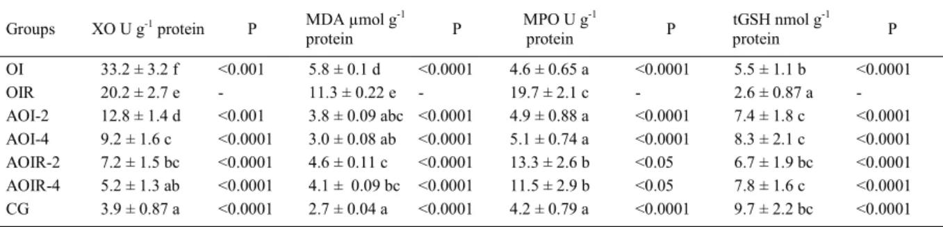
Table 1 –The effects of ATP on biochemical parameters.
Groups XO U g-1 protein P MDA μmol g-1
protein P
MPO U g-1
protein P
tGSH nmol g-1
protein P
OI 33.2 ± 3.2 f <0.001 5.8 ± 0.1 d <0.0001 4.6 ± 0.65 a <0.0001 5.5 ± 1.1 b <0.0001
OIR 20.2 ± 2.7 e - 11.3 ± 0.22 e - 19.7 ± 2.1 c - 2.6 ± 0.87 a
-AOI-2 12.8 ± 1.4 d <0.001 3.8 ± 0.09 abc <0.0001 4.9 ± 0.88 a <0.0001 7.4 ± 1.8 c <0.0001
AOI-4 9.2 ± 1.6 c <0.0001 3.0 ± 0.08 ab <0.0001 5.1 ± 0.74 a <0.0001 8.3 ± 2.1 c <0.0001
AOIR-2 7.2 ± 1.5 bc <0.0001 4.6 ± 0.11 c <0.0001 13.3 ± 2.6 b <0.05 6.7 ± 1.9 bc <0.0001
AOIR-4 5.2 ± 1.3 ab <0.0001 4.1 ± 0.09 bc <0.0001 11.5 ± 2.9 b <0.05 7.8 ± 1.6 c <0.0001
CG 3.9 ± 0.87 a <0.0001 2.7 ± 0.04 a <0.0001 4.2 ± 0.79 a <0.0001 9.7 ± 2.2 bc <0.0001
The effects of ATP on the XO, MDA, MPO and tGSH levels in rat ovarian tissue ischemia /reperfusion.OI: Ovarian Ischemia, OIR: Ovarian
Ischemia /Reperfusion, AOI-2: 2mg kg-1 + ATP Ovarian Ischemia, AOI-4: 4mg kg-1 + ATP Ovarian Ischemia, AOIR-2: 2mg kg-1 + ATP
Ovarian Ischemia/ Reperfusion, AOIR-4: 4mg kg-1 + ATP Ovarian Ischemia/ Reperfusion, CG: Healthy Control Group. Results are the
Figure 1 - Light microscopy photograph of the ovary in CG. A normal histopathological appearance was observed fi gure 1.1A High magnifi cation of fi gure 1.1B. Light microscopy photographs of the ipsilateral ovary in the OI group. A moderate dilation and congestion (arrow) and mild hemorrhage (star) were observed in the vessels of the ovarian stroma fi gure 1.2A. High magnifi cation of
fi gure 1.2B. Light microscopy photographs of the ipsilateral ovary in the OIR group. In fi gure 1.3A, vasodilation and congestion (arrow), edema, permanent and severe hemorrhage (star) were observed in the low power fi eld; in 3B, migration and adhesion of a large number of PMNLs (double arrow) to the endothelium were observed in the high power fi eld. Light microscopy photographs of ipsilateral ovaries in the AOI-2 group. A mild congestion (arrow) and mild stromal hemorrhage were observed in fi gure 1.4A. High magnifi cation of fi gure 1.4B. In Figure 1.5A light microscopy photographs of the ipsilateral ovaries in the AOI-4 group. High magnifi cation of
fi gure 1.5B. A near-normal histomorphological appearance was observed. In 1.6 light microscopy photographs of the ipsilateral ovaries in the AOIR-2 group. While moderate congestion and dilation (arrow), moderate stromal hemorrhage and edema (star) were observed in the low power
these studies, Ingec et al. reported that the amount of oxidant increased only in ischemia-induced ovarian tissue and that it was greatly intensifi ed by reperfusion (INGEC et al. 2011). As is well-known, AMP produced by aerobic tissues in the aerobic tissues provides the energy required to maintain cell viability by being broken down into adenosine, inosine and hypoxanthine. In ischemia, the consumption of ATP increases, and its production decreases (JENNINGS & REIMER, 1991). This event leads to the depletion of energy stores and the inhibition of the Na+, K+-ATPase pump found in the cell membrane. ATPase pump inhibition causes an excessive increase in intracellular Na+ and Ca2+ ion concentrations and leads to cell damage (GREEN et al., 1989). The prevention of ischemia and reperfusion damage by the administration of ATP in the experiment coincides with the information reported in the literature. The use of ATP in doses of 2 and 4mg kg-1 signi
fi cantly prevented the rise of MDA and the reduction of tGSH in ischemic ovarian tissue. At the same doses, ATP signifi cantly suppressed the levels of MDA and MPO, both of which had increased as a result of reperfusion. Free O2 radicals, which are known to mediate reperfusion, result in the release of toxic products, such as aldehydes and MDA, from lipids and stimulate oxidation in the cell membrane (GOULART et al., 2005). MDA, as the
fi nal product of lipid peroxidation, causes severe damage to the cell membrane and its intracellular structures (GOULART et al., 2005). The high level of MPO in reperfusion could possibly originate from the release of MPO by large numbers of polymorphonuclear nuclear leukocytes (PMNLs) in the blood entering the ischemic tissue during reperfusion (ZIMMERMAN & GRANGER, 1992). PNLs convert molecular O2 into superoxide and hydrogen peroxide. The hydrogen peroxide in the chloride ions is reduced to hypochlorous acid, a toxic and strong oxidant, via the MPO enzyme
found in the azurophilic granules of neutrophils (ZIMMERMAN & GRANGER, 1992). Studies have experimentally demonstrated that postischemia reperfusion leads to severe damage of other tissues, as well as the ovaries (BOREKCI et al., 2009; COSKUN et al., 2011; TOK et al., 2012). Moreover, previous studies reported that levels of ATP and Na+-K+-ATPase are decreased in ischemic tissue (GABRYEL et al., 2012; SINGH et al., 2012). The biochemical results obtained in the present study coincide with the histopathological fi ndings. In addition, the severity of the pathological fi ndings, such as dilatation, congestion, haemorrhage, oedema and PNLs, increased in parallel with the rise in XO metabolism products. In conclusion, exogenously administered ATP prevented the increase of XO, MDA and MPO, all of which are known to be oxidant parameters in ovarian tissue in ischemia and postischemia-induced reperfusion. These results suggest that the use of ATP at the clinic would be helpful as a preventive agent of both ovarian ischemia and post ischemia reperfusion damage. DECLARATION OF INTEREST STATEMENT
Authors have no confl icts of interest to declare.
ACKNOWLEDGEMENTS
Authors do not have fi nancial support.
REFERENCES
BOREKCI, B. et al. The protective effect of dehydroepiandrosterone on ovarian tissues after torsion-detorsion ınjury: a stereological and histopathological study. Eurasian Journal of Medicine, Erzurum, v.41, p.22-27, 2009.Available from: <http://www.eajm.org/ sayilar/180/buyuk/pdf_EAJM_154.pdf.>
CARDEN, D.L.; GRANGER, D.N. Phatophsiology of ischemia-reperfusion injury. Journal of Pathology,London, v.190, p.255-266, 2000. Available from: <http://onlinelibrary.wiley.com/
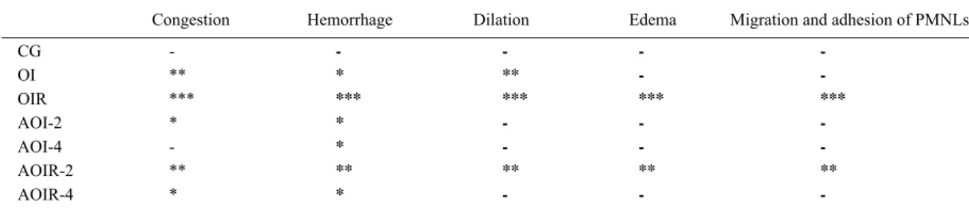
Table 2 – The histopathological results of ATP
Congestion Hemorrhage Dilation Edema Migration and adhesion of PMNLs
CG - - - -
-OI ** * ** -
-OIR *** *** *** *** ***
AOI-2 * * - -
-AOI-4 - * - -
-AOIR-2 ** ** ** ** **
AOIR-4 * * - -
doi/10.1002/(SICI)1096-9896(200002)190:3%3C255::AID-PATH526%3E3.0.CO;2-6/full>
COSKUN, A.K. et al. The effects of amlodipine on the biochemical and histopathological changes in the rabbit ileum subjected to ischemia-reperfusion. Eurasian Journal of Medicine, Erzurum, v.43, p.33-38, 2011. Available from: <http://www.eajm.org/ sayilar/178/buyuk/33-38.pdf>.
GABRYEL, B. et al. Neuronal autophagy in cerebral ischemia--a potential target for neuroprotective strategies? Pharmacological Reports, Poland, v.64, p.1-15, 2012. Available from: <http:// www.if-pan.krakow.pl/pjp/pdf/2012/1_1.pdf>.
GOULART, M. et al. Lipoperoxidation products and thiol antioxidants in chromium exposed workers. Mutagenesis, Oxford, v.20, p.311-315, 2005. Available from: <http://mutage. oxfordjournals.org/content/20/5/311.full.pdf+html>.
GRACE, P. A. Ischaemia-reperfusion injury. British Journal of Surgery, Glasgow, v.81, p.637-647, 1994. Available from: <http://onlinelibrary.wiley.com/doi/10.1002/bjs.1800810504/full DOI: 10.1002/bjs.1800810504>.
GREEN, C.J. et al. The importance of iron, calcium and free radicals in reperfusion injury: an overview of studies in ischaemic rabbit kidneys. Free radical research communications, Tokyo, v.7, p.255-264, 1989. Available from: <http://www.ncbi.nlm.nih. gov/pubmed/2684800>.
HARRISON, R. Structure and function of xanthine oxidoreductase: where are we now? Free radical biology and medicine, local, v.33, p.774-797, 2002. Available from: <http://dx.doi.org/10.1016/ S0891-5849(02)00956-5>.
INGEC, M. et al. Prevention of ischemia-reperfusion injury in rat ovarian tissue with the on-off method. Journal of Physiology and Pharmacology, Birmingham, v.62, p.575-582, 2011. Available from: <http://www.jpp.krakow.pl/journal/archive/10_11/pdf/575_ 10_11_article.pdf>.
ISAOGLU, U. et al. Biochemical and histopathological investigation of the protective effect of disulfi ram in ischemia-induced ovary damage. Gynecological Endocrinology,London, v.28, p.143-147, 2012. Available from: <http://informahealthcare. com/doi/abs/10.3109/09513590.2011.589922>.
JENNINGS, R.B.; REIMER, K.A. The cell biology of acute myocardial ischemia. Annual Review of Medicine, Palo Alto, CA v.42, p.225-246, 1991. Available from: <http://www.annualreviews. org/doi/pdf/10.1146/annurev.me.42.020191.001301> DOI: 10.1146/ annurev.me.42.020191.001301.
KURT, A. et al. An investigation about the inhibition of acut ischemia / reperfusion damage by dexmedetomidine in rat ovarian tissue. Gynecological Endocrinology, London, v.29, p.222-225, 2013. Available from: <http://informahealthcare.com/doi/abs/10.3 109/09513590.2012.665104>.
LI, C.; JACKSON, R.M. Reactive species mechanisms of cellular hypoxia-reoxygenation injury. American Journal of Physiology - Cell Physiology, Rockville Pike, v.282, p.227-241, 2002. Available from: <http://ajpcell.physiology.org/content/282/2/ C227.full-text.pdf+html>.
LINDSAY, T.F. et al. The effect of ischemia/reperfusion on adenine nucleotide metabolism and xanthine oxidase production in skeletal
muscle. Journal of Vascular Surgery, Chicago, v.12, p.8-15, 1990. Available from: <http://www.sciencedirect.com/science/article/ pii/074152149090360M>.
OHKAWA, H. et al. Assay for lipid peroxides in animal tissues by thiobarbituric acid reaction. Analytical Biochemistry, Oxford, v.95, p.351-358, 1979. Available from: <http://www.sciencedirect. com/science/article/pii/0003269779907383>.
ORRENIUS, S. et al. Calcium ions and oxidative cell injury. Annals of Neurology, Wiley-Blackwell, v.32, p.33-42, 1992. Available from: <http://onlinelibrary.wiley.com/doi/10.1002/ana.410320708/pdf>. PALLER, M.S. The cell biology of reperfusion injury in the kidney. Journal of Investigative Medicine,Philadelphia, v.42, p.632-639, 1994. Available from: <http://www.ncbi.nlm.nih.gov/ pubmed/8521026>.
PARKS, D.A. et al. Conversion of xanthine dehydrogenase to oxidase in ischemic rat intestine: a reevaluation. American Journal of Physiology,Rockville Pike, v.254, p.768-774, 1988. Available from: <http://ajpgi.physiology.org/content/254/5/G768>.
POLAT, B. et al. Antiulcerative effect of dexmedetomidine on indomethacin-induced gastric ulcer in rats. Pharmacological Reports, Poland, v.63, p.518-526, 2011. Available from: <http:// www.if-pan.krakow.pl/pjp/pdf/2011/2_518.pdf>.
REITER, R. J. Oxidative processes and antioxidative defense mechanisms in the aging brain. The FASEB Journal, Rockville Pike, v.9, p.526-533, 1995. Available from: <http://www.fasebj. org/content/9/7/526.abstract>.
ROWE, P.B. et al. The mechanism of dietary alterations in rat hepatic xanthine oxidase levels. Journal of Biological Chemistry, Rockville Pike, v.10, p.5571-5576, 1966. Available from: <http://www.jbc.org/content/241/23/5571.short>.
SEDLAK, J.; LINDSAY, R.H. Estimation of total, protein-bound, and nonprotein sulfhydryl groups in tissue with Ellman’s reagent. Analytical Biochemistry, Oxford, v.25, p.192-205, 1968. Available from: <http://www.sciencedirect.com/science/ article/pii/0003269768900924>.
SENER, M.T. et al. Biochemical and histologic study of lethal cisplatin nephrotoxicity prevention by mirtazapine.
Pharmacological Reports, Poland, v.64, p.594-602, 2012. Available from: <http://www.if-pan.krakow.pl/pjp/pdf/2012/3_594. pdf?origin=publication_detail>.
SINGH, A.P. et al. Animal models of acute renal failure.
Pharmacological Reports, Poland,v.64, p.31-44, 2012. Available from: <http://www.if-pan.krakow.pl/pjp/pdf/2012/1_31.pdf>. TOK, A. et al. Effect of mirtazapine on oxidative stress created in rat kidneys by ischemia-reperfusion. Renal Failure, London, v.34, p.103-110, 2012. Available from: <http://informahealthcare. com/doi/abs/10.3109/0886022X.2011.623499>.
WEI, H.; FRENKEL, K. In vivo formation of oxidized DNA bases in tumor promoter-treated mouse skin. Cancer Research, Philadelphia, v.5, p.4443-4449, 1991. Available from:<http:// cancerres.aacrjournals.org/content/51/16/4443.long>.
ZIMMERMAN, B.J.; GRANGER, D.N. Reperfusion injury.