DOI: 10.5935/2359-4802.20180081
ORIGINAL ARTICLE
Mailing Address: Silene Jacinto da Silva
Av. João Rita Dias, Q 105 Lt 06. Postal Code: 74.455-360, Jardim Leblon, Goiânia, GO - Brazil. E-mail: [email protected]
Influence of ACE Polymorphism on Echocardiographic Data of Patients with
Heart Failure
Silene Jacinto da Silva,1 Salvador Rassi,1 Alexandre da Costa Pereira2
Universidade Federal de Goiás (UFG),1 Goiânia, GO - Brazil
Faculdade de Medicina da Universidade de São Paulo (FMUSP),2 São Paulo, SP - Brazil
Manuscript received January 24, 2018; revised manuscript May 06, 2018; accepted June 26, 2018.
Abstract
Background: Angiotensin converting enzyme (ACE) polymorphism has been associated with different clinical and echocardiographic parameters in patients with heart failure (HF). However, no studies have been investigated such association with HF caused by Chagas disease.
Objectives: To perform a genetic study to evaluate the frequency of ACE polymorphism in patients with HF caused by Chagas disease attending a university hospital in the central-west region and its association with echocardiographic findings.
Methods: Descriptive study of ACE polymorphism (I/D) and echocardiographic data of 103 patients with HF caused by Chagas disease. Echocardiographic parameters were compared between the genotypes using the ANOVA test.
Results: Genotypic distribution of the ACE polymorphism was 16.5% DD, 57.3% DI and 26.2% II. There was no statistically significant difference in the distribution of genotypes between men and women. The echocardiographic findings were: left ventricular ejection fraction: 43.8 ± 14.8 (DD) vs. 42.3 ± 11.6 (ID) vs. 44.9 ± 13.0 (II), p = 0.664; left ventricular diastolic diameter: 59.2 ± 9.7 (DD) vs. 60.3 ± 7.6 (ID) vs. 59.7 ± 78.1 (II), p = 0.879; left ventricular systolic diameter: 48.6 ± 12.8 (DD) vs. 50.6 ± 9.7 (ID) vs. 49.3 ± 11.9 (II), p = 0.753; and left atrial volume: 44.9 ± 10.1 (DD) vs. 40.9 ± 9.6 (ID) vs. 38.2 ± 7.8 (II), p = 0.068. Significant correlation coefficients were found for gender, age, ethnicity, heart rate and dyslipidemia.
Conclusion: ACE polymorphism was not associated with echocardiographic findings in patients with HF caused by Chagas disease. (Int J Cardiovasc Sci. 2019;32(1)55-60)
Keywords: Heart Failure; Angiotensins; Polymorphism, Genetic; Chagas Disease; Echocardiography/methods.
Introduction
Chagas disease, described more than 100 years ago by Carlos Chagas, is considered one of the most neglected diseases in the world by the World Health Organization (WHO).1 It is an important cause of heart failure (HF) in
low socioeconomic regions, leading to high morbidity and
mortality.2 In the central-west region of Brazil, Chagas
disease has been considered the main cause of HF.3-5
Most health problems, including HF, have a multifactorial etiology, that involves environmental, lifestyle, and genetic factors.6 Multifactorial disorders
are characterized by genotypic contributions from many genes that interact with each other and with
environmental factors.6 Genetic composition, associated
with environmental factors, can predispose an individual to diseases and response to pharmacological interventions.7
Doppler echocardiography is a useful method for diagnostic confirmation, evaluation of etiology, pathophysiological and hemodynamic model, prognosis and indication of therapeutic alternatives for patients
with HF.8 Due to the importance and magnitude of
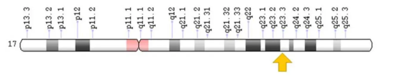
Figura 1 - Localization of the angiotensin converting enzyme (ACE) gene.
In this context, analysis of genetic polymorphisms is a non-invasive technique that may open new avenues and indicate potential preventive therapies for these patients.
Angiotensin converting enzyme (ACE) gene is located
on chromosome 17q23 and consists of 24 introns.9
Several approaches on ACE polymorphism in HF have been reported in the literature, including three clinical trials that did not find an association between ACE
polymorphism and HF.4,10,11 Other studies, however, have
suggested the DD phenotype as a risk factor for cardiac
hypertrophy and HF12 and associated this phenotype
with worse survival.13 Figure 1 shows the localization of
the gene that encodes ACE.14
In the last years, ACE polymorphism has drawn much attention due to conflicting results of the studies on HF. The present study aimed to identify ACE gene D/I (deletion/insertion) in patients with HF caused by Chagas disease and compare it with echocardiographic results.
Methods
This was an observational cohort study. Clinical data were collected from the medical records of patients seen at the Cardiology Unit of the Federal University of Goias, Brazil. Genetic analysis was performed at the Laboratory of Genetics and Molecular Cardiology (Incor/University of Sao Paulo, Brazil).
One hundred and three patients were evaluated from February 2014 to October 2015. All patients were on
optimized drug therapy according to current guidelines.8
All patients included in the study underwent complete genotyping process for I/D alleles. Alleles were determined after the patients were included in the study. All clinical data were extracted from medical records by the first author of this article, before patients’ visits, and ten patients were invited to participate in the study. Inclusion criteria were age older than 18 years, diagnosis of HF as established by the Framingham criteria, and doppler echocardiography using the Teichholz method (more recent and available tests were considered for analysis). Exclusion criteria were: unavailable or inadequate medical records, and HF caused by other than Chagas disease.
The study was analyzed and approved by the Ethics Committee of the General Hospital of the Federal University of Goias (approval number 908.870). All patients signed an informed consent form, and the study was conducted according to current ethical issues.
Echocardiographic variables
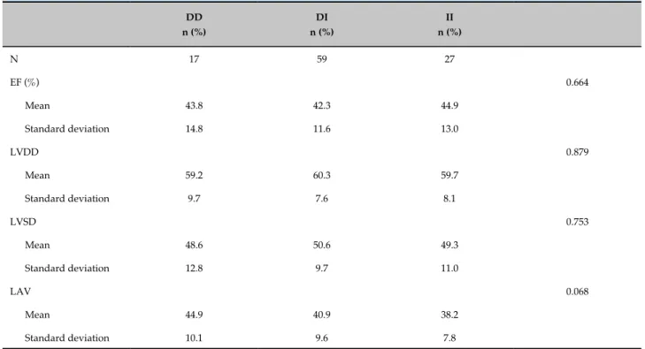
All patients had echocardiography results available in the medical records. The following echocardiographic parameters were evaluated (Table 1) – LAV: left atrial volume; EF: ejection fraction; LVDD: left ventricular diastolic diameter; LVSD: left ventricular systolic diameter (Teichholz method).
Echocardiographic measures of cavity diameter and muscle thickness were obtained following the American
Table 1 - Echocardiographic parameters according to angiotensin-converting enzyme (ACE) (Deletion/Insertion, D/I)
DD n (%)
DI n (%)
II n (%)
N 17 59 27
EF (%) 0.664
Mean 43.8 42.3 44.9
Standard deviation 14.8 11.6 13.0
LVDD 0.879
Mean 59.2 60.3 59.7
Standard deviation 9.7 7.6 8.1
LVSD 0.753
Mean 48.6 50.6 49.3
Standard deviation 12.8 9.7 11.0
LAV 0.068
Mean 44.9 40.9 38.2
Standard deviation 10.1 9.6 7.8
* ANOVA test. LAV: left atrial volume; DD: deletion/deletion; DI: deletion/insertion; LVDD: left ventricular diastolic diameter; LVSD: left ventricular systolic diameter; EF: ejection fraction; I/I: insertion/insertion.
Genetic analysis
Blood (8 mL) was collected in tubes containing EDTA, and submitted to DNA extraction. Subsequently, polymorphism genotyping was performed by polymerase chain reaction (PCR) and classified as DD, DI or II. One pair of primers was used to amplify the D and I alleles, resulting in 319-bp (base pairs) and 597 bp amplicons.
Hace 3,5’GCCCTGCAGGTGTCTGCAGCATGT3’
Hace 3,5’GGATGGCTCTCCCCGCCTTGTCTC3’
The protocol of PCR using a thermal cycle consisted of a cycle of denaturation at 94ºC for 30 seconds, annealing at 56ºC for 45 minutes and extension at 72ºC for 7 minutes. Amplification products of the D and I alleles were identified using an ultraviolet transilluminator. Since the D allele in heterozygous samples is preferentially amplified, each sample initially found to have the DD genotype was equally subjected to a second, independent amplification with a primer pair that recognizes an insertion-specific sequence with identical PCR conditions
except for an annealing temperature of 67ºC.16
Hace 5’,5’TGGGACCACAGCGCCCGCCACTAC3’
Hace 5’,5’ TCGCCAGCCCTCCCATGCCCATAA3’
Statistical analysis
The Shapiro-Wilk test was used for testing normality of data. Mean and standard deviation of EF, LVDD, LVSD and LAV were calculated for patients grouped according to ACE polymorphism (DD, DI and II). Differences in echocardiographic parameters between these groups were compared by analysis of variance (ANOVA, F test). P-values lower than 0.05 were considered statistically significant. Analysis was performed using the SPSS software version 18.0.
Echocardiographic parameters (EF, LVDD, LVSD and LAV) were treated as continuous variables with normal distribution, and the DD. DI and II genotypes treated as categorical variables.
Results
A total of 103 patients with HF caused by Chagas disease, mean age of 62.4 years (36 – 95 years), 63% men, were included in the study. All patients were on optimized drug therapy according to current guidelines.
significant difference was found in the distribution of genotypes between men and women, with a predominance of men with DI genotype (59.3%).
Functional class varied from II to IV; most patients were in NYHA functional class IV. Analysis of repeated measures for categorical data showed that there was no significant variation in functional class and D/I genotype (p = 0.472).
No difference was found in mean values of echocardiographic variables or genotypes. Patients with the DI genotype showed higher LVDD (60.3) as compared with the other genotypes.
Significant correlations were found for the variables sex, age, smoking, heart rate and dyslipidemia; male sex was associated with increased risk of HF (p = 0.023). Smoking was considered a risk factor for HF. Most patients did not consume alcohol, and thereby alcohol consumption was considered a protective factor (p = 0.008).
Dyslipidemia was associated with a five times higher risk for HF. DD, DI and II genotypes were not considered a risk factor or HF.
Discussion
Many researches involving ACE polymorphisms have been conducted to study the pathophysiology of HF and, so far, it is still controversial whether ACE polymorphism is associated with HF. Although ACE polymorphism has been associated with several pathophysiological events and with morbidity and mortality of cardiovascular diseases, in this study, we aimed to determine a relationship of this polymorphism with HF caused exclusively by Chagas disease.
Most of our patients were men (63%), similarly to
other studies.4,17,18 HF is an increasing epidemic that
affects mostly older men, and its increasing incidence has been associated with higher survival.4 No statistically
significant difference in genotype distribution was found between men and women, which is in agreement with a
previous study conducted in 2014.19
To evaluate an association of ACE polymorphism with HF severity, we investigated a possible association of D/I genotype with echocardiographic parameters, but no association was found. In contrast, a previous
national study20 reported an association of DD and ID
genotype with worse and better echocardiographic profile, respectively.20 We also did not find differences in
EF values between the two genotypes (p = 0.664).
HF is a common clinical condition with high morbidity and mortality, affecting 1.5% - 2.0% of the general population. Its prevalence increases with age and affects
approximately 10% of individuals older than 65 years.21
Our findings corroborate this finding, since we found a statistically significant association of age with HF in the study population.
We did not find any difference in the frequency of genotypes after adjustment for gender, which agrees with the results of a study conducted in a Chinese population.22
A national study performed in 200523 reported
augmented LVSD in patients with DD genotype, which was associated with higher morbidity and mortality in patients with different causes of HF. Our results differ from these findings, since we did not find any relationship between D/I genotypes and LVSD.
Similar occurrence of D/I genotypes was found in the study population, contradicting the hypothesis that D/I genotype is a risk factor for HF.4,24
We did not find any association of D/I genotypes
with HF severity, differently from previous studies25,26
reporting an association of the D allele with HF progression and higher mortality as compared with the I allele. Our study corroborates another Brazilian, clinical comparative study on 193 patients, that did not find any difference in the frequency of D/I genotype in patients with HF caused by Chagas disease without systolic dysfunction.27
Hospitalization for HF is an important public
health problem.28 Clinical treatment of this patients
involves multidisciplinary approach, since they have many comorbidities that may have an impact on the clinical course of the disease. There is evidence that the risk for HF in the general population depends on a genetic predisposition characterized by a very complex genetic architecture. Predisposition to individual conditions and genetic variations modulate
the pathophysiological responses.29
Possible limitations of this study include a small number of patients; however, there are available in the literature studies involving larger number of patients,10,23
but also others with smaller samples.27,30 Due to the lack
1. Murcia L, Carrilero B, Fau - Saura D, Iborra MA, Segovia M. Diagnosis and treatment of Chagas disease. Enferm Infec Mirobiol Clin. 2013;31(Suppl 1):26-34.
2. Carvalho G, Rassi S, Bastos JMDA, Câmara SSP. Coronariopatia assintomática em chagásicos com insuficiência cardíaca: prevalência e fatores de risco. Arq Bras Cardiol. 2011; 97(5):408-12.
3. Nogueira PR, Rassi S, Corrêa KS. Perfil epidemiológico, clínico e terapêutico da insuficiência cardíaca em hospital terciário. Arq Bras Cardiol. 2010;95(3)392-8.
4. Silva SJ, Rassi S, Silva C. Ausência de associação do polimorfismo dos alelos D/I do gene da enzima conversora de angiotensina (ECA) em pacientes com insuficiência cardíaca. Rev Bras Med. 2015,72(4):130-5.
5. Carvalho APPF, Rassi S, Fontana KE, Correa KS, Feitosa RHF. Influência da suplementação de creatina na capacidade funcional de pacientes com Insuficiência Cardíaca. Arq Bras Cardiol. 2012,99(1):623-9.
6. Skrzynia C, Berg JS, Willis MS, Jensen BC. Genetics and heart failure: a concise guide for the clinician. Curr Cardiol Rev. 2015;11(1):10-7.
7. Hamad E, Feldman AM. Pharmacogenetics in heart failure: how it will shape the future. Heart Fail Clin. 2010,6(1):1-10.
8. Bocchi EA, Braga FGM, Ferreira SMA, Rohde LEP, Oliveira WA, Almeida DR et al., Sociedade Brasileira de Cardiologia. III Diretriz brasileira de insuficiência cardíaca crônica. Arq Bras Cardiol. 2009;93(1 Suppl 1):1-71.
9. Cuoco MAR, Pereira AC, Mota GFA, Krieger JE, Mansur AJ. Polimorfismo genético, terapia farmacológica e função cardíaca sequencial em pacientes com insuficiência cardíaca. Arq Bras Cardiol. 2008;90(4):274-9.
10. Covolo L, Gelatti U, Metra M, Donato F, Nodari S, Pezzali N, et al. Angiotensin-converting-enzyme gene polymorphism and heart failure: a case-control study. Biomarkers. 2003;8(5):429-36.
11. de Groote P, Helbecque N, Lamblin N, Hermant X, Amouyel P, Bauters C, et al. Beta-adrenergic receptor blockade and the angiotensin-converting enzyme deletion polymorphism in patients with chronic heart failure. Eur J Heart Fail. 2004;6(1):17-21.
12. Schut AF, Bleumink GS, Stricker BH, Hofman A, Witteman JC, Pols HA, et al. Angiotensin converting enzyme insertion/deletion polymorphism and the risk of heart failure in hypertensive subjects. Eur Heart J. 2004;25(23):2143-8.
13. McNamara DM, Holubkov R, Postava L, Janosko K, MacGowan GA, Mathier M, et al. Pharmacogenetic interactions between angiotensin-converting enzyme inhibitor therapy and the angiotensin-angiotensin-converting enzyme deletion polymorphism in patients with congestive heart failure. J Am Coll Cardiol. 2004;44(10):2019-26.
14. Genetics Home Reference. Chromossome 17 [Cited in 2018 Jun 12]. Available from: https://ghr.nlm.nih.gov/chromosome/17
15. Porter TR, Mulvagh SL, Abdelmoneim SS, Becher H, Belcik JT, Bierig M et al. Clinical Applications of Ultrasonic Enhancing Agents in Echocardiography: 2018. American Society of Echocardiography Guidelines Update. J Am Soc Echocardiogr. 2018;31(3):241-74.
16. Lindpaintner K, Pfeffer MA, Kreutz R, Stampfer MJ, Grodstein F, LaMotte F et al. A prospective evaluation of an angiotensin-converting-enzyme gene polymorphism and the risk of ischemic heart disease. N Engl J Med. 1995;332(11):706-11.
17. Margoto G, Colombo RCR, Gallani MCBJ. Características clínicas e psicossociais do paciente com insuficiência cardíaca que interna por descompensação clínica. Rev Esc Enferm USP. 2009;43(1):44-53.
18. Barker WH, Mullooly JP, Getchell W. Changing incidence and survival for heart failure in a well-defined older population, 1970-1974 and 1990-1994. Circulation. 2006;113(6):799-805.
References
Conclusions
This study suggests that ACE (D/I) polymorphism is not related with echocardiographic parameters of patients with HF caused by Chagas disease.
Further larger, prospective studies are needed to determine which factors may be associated with HF caused by Chagas disease and investigate the best intervention with minimal adverse effects in this population.
Author contributions
Conception and design of the research: Silva SJ, Rassi S. Acquisition of data: Silva SJ. Analysis and interpretation of the data: Silva SJ, Rassi S, Pereira AC. Statistical analysis: Silva SJ. Obtaining financing: Pereira AC. Writing of the manuscript: Silva SJ. Critical revision of the manuscript for intellectual content: Rassi S, Pereira AC.
Potential Conflict of Interest
No potential conflict of interest relevant to this article was reported.
Sources of Funding
There were no external funding sources for this study.
Study Association
This article is part of the thesis of Doctoral submitted by Silene Jacinto da Silva, from Universidade Federal de
Goiás (Programa de Pós-graduação em Ciências da Saúde).
Ethics approval and consent to participate
19. Zhang YF, Cheng Q, Tang NL, Chu TT, Tomlinson B, Liu F, et al. Gender difference of serum angiotensin-converting enzyme (ACE) activity in DD genotype of ACE insertion/deletion polymorphism in elderly chinese. J Renin Angiotensin Aldosterone Syst. 2014;15(4):547-52.
20. Albuquerque FN, Brandao AA, Silva DA, Mourilhe-Rocha R, Duque GS, Gondar AF, et al. Angiotensin-converting enzyme genetic polymorphism: its impact on cardiac remodeling. Arq Bras Cardiol. 2014;102(1):70-9.
21. Carvalho Filho E, Curiati J. Como diagnosticar e tratar a Insuficiência Cardíaca no Idoso. Rev Bras Med. 2001;58(3):394-7.
22. Yang JK, Gong YY, Xie L, Lian SG, Yang J, Xu LY, et al. Lack of genetic association between the angiotensin-converting enzyme gene insertion/ deletion polymorphism and longevity in a Han Chinese population. J Renin Angiotensin Aldosterone Syst. 2009;10(2):115-8.
23. Cuoco MA, Pereira AC, de Freitas HF, Alves F, da Mota G , Fukushima JT, Krieger JE, et al. Angiotensin-converting enzyme gene deletion polymorphism modulation of onset of symptoms and survival rate of patients with heart failure. Int J Cardiol. 2005;99(1):97-103.
24. Pascuzzo-Lima C, Mendible JC, Bonfante-Cabarcas R. Polimorfismo I/D del gen de la enzima de conversión de angiotensina y progresión de la miocardiopatía chagásica. Rev Esp Cardiol. 2009; 62(03):320-2.
25. Huang W, Xie C, Zhou H, Yang T, Sun M. Association of the angiotensin-converting enzyme gene polymorphism with chronic heart failure in Chinese Han patients. Eur J Heart Fail. 2004,6(1):23-7.
26. Wu CK, Luo JL, Tsai CT, Huang YT, Cheng CL, Lee JK, et al. Demonstrating the pharmacogenetic effects of angiotensin-converting enzyme inhibitors on long-term prognosis of diastolic heart failure. Pharmacogenomics J. 2010;10(1):46-53.
27. Silva SJ, Rassi S, Pereira AC. Polimorfismo da enzima conversora da angiotensina (ECA D/I) em pacientes com insuficiência cardíaca de etiologia chagásica. Arq Bras Cardiol. 2017;109(4):307-12.
28. Mesquita ET, Jorge AJL, Rabelo LM, Souza CV Jr. Understanding hospitalization in patients with heart failure. Int J Cardiovasc Sci. 2017;30(1):81-90.
29. Lopes LR, Elliott PM. Genetics of heart failure. Biochim Biophys Acta. 2013;1832(12):2451-61.
30. Duque GS, Silva DA, Albuquerque FN, Schneider RS, Gimenez A, Pozzan R, et al . Influence of angiotensin-converting-enzyme gene polymorphism on echocardiographic data of patients with ischemic heart failure. Arq Bras Cardiol. 2016;107(5):446-54.