ACTA RADIOLÓGICA PORTUGUESA January-April 2018 Vol 30 nº1 15-22
Endovascular Treatment of Splanchnic Pseudoaneurysms Following
Blunt Solid Organ Injuries in Children
Tratamento Endovascular de Pseudo-Aneurismas após Trauma Abdominal Fechado
em Crianças
João André Oliveira1, Nuno Vasco Costa2, Tiago Bilhim2, Filipe Veloso Gomes2, Élia Coimbra2 1 Serviço de Radiologia, Hospital Geral de Santo
António, Centro Hospitalar do Porto, Portugal
2 Serviço de Radiologia, Hospital Curry Cabral,
Centro Hospitalar de Lisboa Central, Portugal
Address
João André Oliveira Rua das Artes Gráficas, 67-71 4100-092 Porto, Portugal
e.mail: joao_a_oliveira@hotmail.com
Resumo
A abordagem conservadora é atualmente o tratamento de primeira linha de lesões traumáticas de órgãos sólidos intra-abdominais de grau I-IV (escala da Associação Americana de Cirurgia de Trauma) em idade pediátrica. Embora pouco frequentes, os pseudoaneurismas esplâncnicos pós-traumáticos podem potencialmente originar hemorragia intraperitoneal ou retroperitoneal catastrófica. Na população adulta, a rotura tardia de pseudoaneurismas é uma das causas do insucesso da abordagem conservadora. A angioembolização seletiva destes pseudoaneurismas contribui para a diminuição da taxa de insucesso desta abordagem. Na população pediátrica, a relevância clínica e prognóstica dos pseudoaneurismas esplâncnicos não se encontra ainda clarificada, e atualmente, não há guidelines de alto nível de evidência para a sua abordagem terapêutica. Os autores deste artigo apresentam 3 casos de pseudoaneurismas abdominais pós-traumáticos em crianças que foram identificados em exames de imagem após tratamento conservador, tratados com sucesso por embolização seletiva, e apresentam uma revisão da literatura no que concerne a este tema. Apesar de estudos prospetivos randomizados serem ainda necessários para melhor definir a incidência, a história natural e a abordagem terapêutica dos pseudoaneurismas abdominais no trauma pediátrico, os autores defendem que a angioembolização seletiva representa uma abordagem segura e efetiva no tratamento desta entidade clínica e que deve ser integrada no protocolo da abordagem multidisciplinar do trauma pediátrico.
Palavras-chave
Trauma abdominal; Crianças; Pseudo-aneurisma; Radiologia de intervenção; Embolização.
Abstract
Non operative management is currently the standard treatment of blunt abdominal solid organ injuries grades I-IV (American Association for the Surgery of Trauma’s organ injury scale) in children. Even though post-traumatic splanchnic pseudoaneurysms are an infrequent complication, they may potentially lead to life-threatening intra-peritoneal or retrointra-peritoneal bleeding. In adults, the relationship between failure of conservative management in abdominal trauma patients and delayed rupture of a pseudoaneurysm identified in follow-up imaging is well established, as is the capability of selective angioembolization to decrease non operative management failure rate. In the pediatric population, the clinical and prognostic significance of splanchnic pseudoaneurysms remains controversial and, currently, there are no high-level evidence-based guidelines on its management. The authors of this paper present 3 cases of post-traumatic abdominal pseudoaneurysms in children which were identified in imaging exams after conservative management, successfully treated by selective embolization, and a review of the literature regarding this subject is also presented. Although prospective randomized-controlled trials are needed to better define the incidence, natural history and optimal management of abdominal PAs in pediatric blunt abdominal trauma, we believe that selective angioembolization provides a safe and effective therapy for its treatment and should be considered as part of the multidisciplinary trauma management protocol in children.
Keywords
Blunt abdominal trauma; Children; Pseudoaneurysm; Interventional radiology; Angioembolização.
Review Article / Artigo Revisão
Introduction
Abdominal post-traumatic pseudoaneurysms (PAs) are a relatively rare complication following blunt trauma, with disruption of the arterial wall and formation of a blood-perfused sac contained by the media/adventitia layers or in some cases by the soft-tissues encircling the injured vessel. Due to continuous high arterial pressure feeding the PA, there is a potential life-threatening risk of intra-abdominal or retroperitoneal bleeding.
In adults, the relationship between failure of conservative management in abdominal trauma patients and delayed rupture of a PA identified in follow-up imaging – namely in the liver, kidney and spleen territories - is well established, and most trauma centers perform prophylactic angiographic embolization of abdominal PA in hemodynamically stable patients.
In the pediatric population, clinical and prognostic significance of traumatic abdominal PAs is not well defined. For instance, small splenic artery PAs are thought to frequently resolve spontaneously with thrombosis of the
aneurismatic sac.1-7 On the other hand, hepatic artery PAs
have an up to 80% risk of rupture with potential catastrophic bleeding, and the incidence of spontaneous thrombosis is low.8,9 Thus, controversy remains regarding the management
of PAs identified on follow-up imaging, and strategies are often influenced by the clinicians’ preferences and expertise in each center.
The authors present 3 cases of post-traumatic abdominal PAs in children successfully treated with angioembolization at our institution reviewing the literature related with this matter.
Case 1
A 6-year-old boy came to our institution due to upper abdominal trauma after hitting a pool handrail. In the initial assessment, he was found to be conscious, tachycardic (119 BPM) but hemodynamically stable, with pain referred to the left upper quadrant. His complete blood count (CBC) showed a decreased hemoglobin level(10,4g/dL). A contrast-enhanced CT-scan was performed, revealing a grade III AAST spleen laceration involving the inferior pole, associated with a subcapsular hematoma and several small intraparenchymal hematomas, with no active extravasation of contrast (fig. 1). No other injuries, namely bone fractures in the thoracic cage or involving abdominal solid organs, were observed. In light of the standard treatment of isolated splenic injuries in a stable pediatric patient, he remained in the hospital for clinical surveillance. Although clinically asymptomatic during hospital stay, Doppler ultrasound (US) performed 11 days after trauma demonstrated an increase of the subcapsular hematoma, with internal vascularization and feeding vessels in its vicinity suggesting PA formation (fig. 2). This finding was confirmed with computer tomography angiography (CTA) (fig. 3), which demonstrated an hypervascular structure within the cleft of the splenic laceration. The patient underwent angiography via right femoral artery approach. Selective angiography of the splenic artery with Simmons 5Fr catheter (Tempo, Cordis® , Miami, FL, USA) demonstrated the presence of a PA in the lower pole (fig. 4a). There was no evidence of other PAs or contrast extravasation within the splenic vascular bed. After superselective catheterization with 2.7Fr microcatheter (Progreat, Terumo®, Tokyo, Japan), transcatheter embolization was first performed with N-butyl-cyanoacrilate (NBCA, Glubran Tiss, GEM, Viareggio, Italy) prepared with lipiodol (Guerbert, Aulnay-sous-Bois, France)
Figure 1 – Contrast-enhanced coronal (a) and axial (b) CT. Splenic parenchyma laceration evolving the lower pole with subacute hematoma (arrow).
a
b
Figure 2 – Doppler-US. Subcapsular splenic hematoma (star) and areas of arterial flow in its vicinity (arrow, left) with yin-yang sign (arrow, right), suggesting pseudoaneurysm.
1/3 ratio. It was not possible to achieve a distal position of the microcatheter tip inside the PA so NBCA was chosen to allow a distal penetration of the embolic from a proximal injection. Afterwards, Onyx 18® (Covidien Inc., Plymouth, MN, USA) was used for a more proximal embolization (fig. 4b). Post-embolization angiography showed exclusion of the PA (fig. 4c). Follow-up US-Doppler examination performed on post-procedural day 5 documented the thrombosed PA, with no vascular abnormalities associated. (fig. 5). The patient underwent an uneventful recovery and was discharged 6 days after angioembolization.
Case 2
A 12 year-old female with no significant past medical history presented herself to the referring hospital after sustaining an 8 meter fall, with blunt upper abdominal and facial trauma. The child was hemodynamically stable, and her vital signs were within normal limits on arrival to the emergency room, with a Glasgow Coma Scale (GCS) of 13. She presented several facial excoriations, a peri-orbital hematoma, lower lip laceration and upper superior abdominal tenderness upon physical exam. Her blood and urine tests showed elevated liver enzymes (AST – 807 u/L; ALT – 684 u/L) and microscopic hematuria. Her hemoglobin level was 7,6 g/ dL requiring red blood cell transfusion. Abdominal CT scan showed moderate volume hemoperitoneum, grade III AAST liver laceration in the right lobe with no evidence of active extravasation of contrast and grade I AAST contusion of the right kidney. There were also several maxillofacial fractures demonstrated in head CT, treated during hospital stay. Non operative management was decided for the abdominal injuries. 11 days after trauma follow-up abdominal CT was performed, showing a 15mm arterial PA in the right hepatic lobe and early opacification of a portal branch in its vicinity, suggesting an arterio-portal fistula. The patient was referred to our institution for angiographic embolization. After right common femoral artery retrograde puncture, a 5Fr sheath-introducer catheter was placed. Careful observation of the CTA prior to the procedure revealed replaced origin of the hepatic artery directly from the abdominal aorta. Selective catheterization and subsequent angiography of the right hepatic artery were performed using a Simmons 5 Fr catheter (Tempo, Cordis®, Miami, FL, USA) confirmed the PA and the arterio-portal fistula arising from a segmental branch (fig. 6a). Superselective angiography of the left hepatic artery, also branching from the aorta, excluded vascular abnormalities (fig. 6b). A 2.7 Fr microcatheter (Progreat, Terumo®, Tokyo, Japan) was advanced coaxially through the Simmons catheter with superselectivation of the segmental branch feeding the PA and arterio-portal fistula. Due to the high-flow fistulous connection of the PA to the portal vein, embolization was performed with Onyx 18® (fig. 6c), and repeated contrast injection into the right hepatic artery demonstrated no filling of the pseudoaneurysm and exclusion of the arterio-portal fistula (fig. 6d). No major nor minor complications were registered after procedure. The child was transferred back to her referring hospital and was maintained under conservative
Figure 3 – Contrast-enhanced sagittal CT, arterial phase. Contrast extravasation within a contained sac confirming a large pseudoaneurysm (arrow) in the inferior splenic pole.
Figure 4 – a) Celiac arteriogram revealing the pseudoaneurysm (arrow) in the inferior splenic pole. b) Embolization with Onyx 18® (arrowhead). c) Celiac aortogram post-embolization demonstrating vascular exclusion of the pseudoaneurysm.
a
b
c
Figure 5 – Doppler US. Thrombosed pseudoaneurysm with no internal vascularization.
treatment. Her liver enzymes returned to normal values (AST – 18 u/L; ALT – 39 u/L) and Doppler-US performed 2 days after endovascular intervention showed resolution of the vascular lesions. After an uneventful recovery, she was discharged 6 days after angioembolization. Follow-up CT performed 5 months later demonstrated vascular exclusion of the segmental branch with no evidence of PA or arterio-portal fistula, with a patent arterio-portal vein.
Case 3
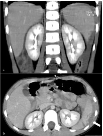
A 17-year old male adolescent was admitted to a regional hospital after sustaining a blunt lumbar trauma in a bicycle crash. Abdominal computed tomography was performed, revealing a grade III AAST injury in the right kidney, with parenchymal laceration and a large peri-renal hematoma (8,3x8cm) with no parenchymal contrast uptake in the upper third of the kidney. There was no evidence of collecting system lesion or active extravasation of iodine contrast. No other traumatic lesions were found in the abdominopelvic cavity. He was transferred to our institution for further management. On admission, he was conscious, hemodynamically stable, referring pain in the lumbar region. The patient’s physical examination revealed only excoriations in the right lumbar region. His hemoglobin level was 12,8g/dl. The patient underwent non-operative management and proceeded to an uneventful clinical recovery. Ultrasound performed at day 7 after trauma revealed slight decrease in the size of the hematoma and no active bleeding (fig. 7). He was discharged home the day after. At day 15 after trauma, the patient was readmitted to his hospital with acute and intense right lumbar pain. His CBC showed a decrease in hemoglobin level (11,8g/dL). He underwent CTA imaging (fig.8), revealing a pericentimetric PA in the upper third of the
Figure 6 – a) Selective angiography from the right hepatic artery. Large pseudoaneurysm (arrow) arising from a segmental branch, associated with early opacification of a portal venous branch suggesting arteriovenous fistula (arrowhead). b) Selective angiography from the left hepatic artery. c) Embolization with Onyx 18® (arrow). d) Post-embolization angiogram from a common arterial trunk (right hepatic and phrenic arteries) with no opacification of the pseudoaneurysm or arteriovenous fistula.
a b
c d
Figure 7 a) and b) - Doppler US. Peri-renal hematoma (arrows) with no vascular flow in the upper third of right kidney.
a
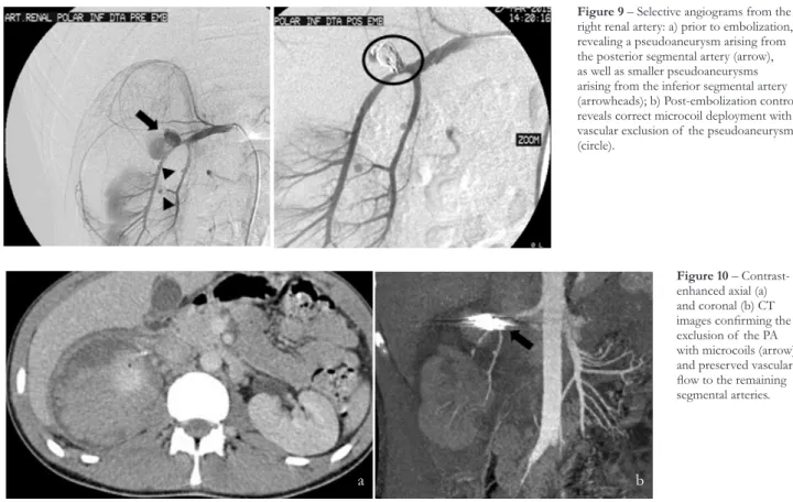
right kidney with active extravasation of contrast. There was also a slight increase in the size of the peri-renal hematoma. He was readmitted to our department for angiography. Using a right femoral approach, a selective angiography of the right renal artery was performed, that demonstrated the PA in the posterior segmental artery with active extravasation of contrast, as well as 2 other millimetric PAs (fig. 9a). Due to the anatomic location, aneurysm size and poor stability of the microcatheter tip inside the aneurysm sac, detachable coils were selected for embolization. Two 0.018 inch detachable microcoils of 15mm in diameter were deployed through a 2.7Fr coaxial microcatheter (Progreat®, Terumo, Tokyo, Japan) after superselective catheterization of the larger PA. It was decided to manage conservately the other PAs due to their small size and location. Control angiography revealed successful embolization of the pseudoaneurysm preserving vascular flow to the anteroinferior and inferior segmental arteries (fig.9b). CTA was performed 72 hours later, confirming the exclusion of the PA and preserved
Figure 8 – Contrast-enhanced axial and coronal images. Contrast-filled sac with relation with a segmental branch of the right renal artery suggesting pseudoaneurysm (arrow)
Figure 9 – Selective angiograms from the right renal artery: a) prior to embolization, revealing a pseudoaneurysm arising from the posterior segmental artery (arrow), as well as smaller pseudoaneurysms arising from the inferior segmental artery (arrowheads); b) Post-embolization control reveals correct microcoil deployment with vascular exclusion of the pseudoaneurysm (circle).
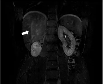
vascular flow to the remaining segmental arteries, as well as thrombosis of the smaller PAs (fig.10). The patient was discharged 12 days after embolization. During follow-up, he developed arterial hypertension, controled with an ACE inhibitor. MR imaging performed 14 months after treatment showed a small remaining right kidney with normal contrast uptake (fig.11). Further evaluation with dimercaptosuccinic acid (DMSA) renal scan showed decreased renal function in the right kidney (right kidney - 13%; left kidney – 87%). Although this could have been partially due to segmental angioembolization, we believe that the initial vascular injury to the upper hemikidney evidenced in post-trauma imaging studies played a major role in this clinical outcome. Selective angiography before embolization (fig. 9a) showed no parenchimal perfusion to the upper 2/3 of the right kidney, proving that vascular oclusion with devascularization of 2/3 of the affected kidney was already present before embolization.
Figure 10 – Contrast-enhanced axial (a) and coronal (b) CT images confirming the exclusion of the PA with microcoils (arrow) and preserved vascular flow to the remaining segmental arteries.
Discussion
Abdominal trauma remains one of the leading causes of mortality and morbidity in the pediatric population. During the past decades, there has been a major shift from surgical to selective non operative management (NOM) of traumatic blunt abdominal injuries, and in hemodynamically stable children with blunt solid organ injury grade I to IV (American Association for the Surgery of Trauma organ injury scale, table 1), NOM of blunt abdominal solid organ injuries is currently the standard of care. Operative intervention, however, is still performed in patients with hemodynamic instability and/or active hemorrhage, often leading to removal of the injured organ with significant clinical consequences, such as a lifelong risk of overwhelming post-splenectomy infection, and potential progression to chronic renal disease after unilateral nephrectomy due to long-term vicarious glomerular hyperfiltration in the contralateral kidney.
AAST blunt solid organ injury
grade Liver Spleen Kidney
I Subcapsular haematoma: <10% surface area Laceration: <1 cm depth Subcapsular haematoma: <10% of surface area Laceration <1 cm depth Contusion
Subcapsular haematoma: non expanding II Subcapsular haematoma: 10-50% surface area Intraparenchymal haematoma: <10 cm diameter Laceration: 1-3 cm parenchymal depth, <10 cm length Subcapsular haematoma: 10-50% of surface area Intraparenchymal haematoma: <5 cm in diameter
Laceration 1-3 cm in depth not involving trabecular vessels
Perirenal haematoma: non-expanding, confined to retroperitoneum
Laceration: <1 cm depth, not involving the collecting system
III Subcapsular haematoma: >50% surface area of ruptured subcapsu-lar or parenchymal haematoma Intraparenchymal haematoma: >10 cm or expanding
Laceration: capsular tear >3 cm parenchymal depth Subcapsular haematoma: >50% of surface area or expanding Intraparenchymal haematoma >5 cm or expanding Laceration: >3 cm in depth or involving trabecular vessels
Laceration: >1 cm without extension into the renal pelvis or collecting system
IV Laceration: parenchymal disruption involving 25-75% hepatic lobe or involves 1-3 Coinaud segments
Ruptured subcapsular or parenchymal haematoma Laceration: involving segmen-tal or hilar vessels with major devascularisation (>25% of spleen)
Laceration: extending to renal pelvis or urinary extravasation
Vascular: injury to main renal artery or vein with contained haemorrhage Segmental infarctions without associated lacerations
V Laceration: parenchymal disruption involving >75% of hepatic lobe or involves >3 Coinaud segments (within one lobe)
Vascular: juxtahepatic venous injuries (retrohepatic vena cava / central major hepatic veins)
Shattered spleen Hilar vascular injury with splenic devascularisation
Avulsion of renal hilum: devascularisa-tion of a kidney due to hilar injury Ureteropelvic avulsions
Complete laceration or thrombus of the main renal artery or vein
VI Vascular - hepatic avulsion
Table 1 - The American Association for the Surgery of Trauma grading system for blunt injury of the liver, spleen and kidney
Figure 11 – T1-weighted contrast enhanced coronal MR image. Small remnant right kidney with normal contrast perfusion on venous phase.
In adults sustaining blunt abdominal trauma injuries, angiographic selective embolization of solid organ vascular territories has increasingly been used as an adjunct to NOM allowing for control of hemorrhage and organ salvage with high success and safety rates reported by various authors, resulting in a decrease the NOM failure and allowing for an expansion of NOM indications to more severe trauma patients, such as blunt solid organ high grade injuries (blunt solid organ injury grade V), older age and associated injuries.4-6,10
Splanchnic PAs in blunt abdominal trauma setting develop following a disruption in arterial wall continuity. Due to sustained arterial pressure, blood dissects into the tissues around the damaged vessel and forms a perfused sac contained only by the outer vascular layers or simply by soft-tissue structures around the vessel.1 Its most common sites
include the spleen, liver and kidney vascular beds. Although the majority of them are asymptomatic, they can present with abdominal pain, obstructive jaundice, hemobilia or hematuria.11 PAs have been recognized as a significant risk
factor for NOM failure due to delayed hemorrhage, a low incidence (5–25% of splenic, 0–3.9% of hepatic and 0–9% of renal injuries, respectively) but life-threatening complication of NOM,12 manifesting as intra-abdominal or retroperitoneal
bleeding and requiring emergency treatment. Regarding blunt splenic trauma, delayed rupture is seen in 5% to 6% of cases and is often attributed to the rupture of a posttraumatic splenic PA.13,14 A study by Schurr et al demonstrated splenic
PA in 67% of adults who failed NOM.15,16 This relationship
between failed NOM and posttraumatic PAs has led to the practice in several trauma centers of semi-elective angiographic embolization of splanchnic PAs if detected in imaging studies.
The significance of post-traumatic splanchnic PAs in the pediatric population is less well defined, as it has until recently not been emphasized as a significant complication following abdominal trauma. There is also a lack of understanding regarding the natural history of traumatic abdominal PAs which are often asymptomatic and may not develop until days or even months after the initial injury, rendering its detection very difficult. The overall incidence of splenic and hepatic PAs reported in previous series is 2-27%2 and 1,7-25%,17,18
respectively. This discrepancy in numbers may be related to different strategies between pediatric trauma centers for follow-up imaging in children in NOM. However, there is a general consensus regarding the relationship between the frequency of PAs and the severity of blunt solid organ injury. For instance, in one series reporting 5,4% of splenic PAs and 1,7% of hepatic PAs in 101 children sustaining abdominal trauma, the incidence was significantly higher in grade IV injuries (17 and 27%, respectively).11
Currently, there are no high-grade recommendations for the screening and management of abdominal PAs in children, and the role of embolization as an adjunctive procedure is still unclear, partially due to higher success rates for NOM of spleen, liver, and renal injuries. In a recent guideline consensus published in 2017 by the World Society of Emergency Surgery regarding splenic trauma,18 a level 2C evidence
recommendation was attributed for angioembolization of post-traumatic splenic pseudo-aneurysms prior to patient discharge. PAs in these abdominal organs are thought to frequently self-tamponade and undergo spontaneous thrombosis due to children’s more elastic parenchyma and a thicker capsule, especially with small size PAs. It is however
mentioned that some authors recommend follow-up imaging to be performed prior to discharge, and in case of persistence of splenic PAs, angioembolization should be performed. The fear of delayed splenic rupture and other late complications of PAs such as arteriovenous fistula formation were cited as the reason for embolization in hemodynamically stable children. This is also the case in adolescents of more than 13-15 years old, that should be managed according to adults’ protocols (level 1C recommendation). In children of less than 13 years old that are more vulnerable to overwhelming post-splenectomy infection, angioembolization should be considered as well.18 Regarding other sites of PA formation,
namely liver and kidney, only case reports have been described in the literature.19-23 Most trauma centers opt to manage these
cases with prophylactic angiographic embolization due to the risk of catastrophic bleeding, especially in larger PAs that don’t resolve spontaneously or that increase in size in imaging follow-up, as in the first case presented in this paper, as the technique and success of angiographic embolization are quite satisfactory.
Overall, there is a lower risk of complications in the endovascular treatment of visceral PAs compared to surgical management, and mortality and major complications are rarely reported.12,19 Potential complications following
selective angioembolization in children are similar to those in adults, namely arterial puncture site hematoma, contrast nephropathy catheter or guidewire-related arterial injury, target organ ischemia, non-target organ embolization, and intra-procedural rupture of the PA. Post-embolization syndrome is frequently reported, consisting of abdominal pain, nausea, ileus and fever, but it is usually self-limited and tends to resolve spontaneously in 6 to 9 days. Splenic infarction areas are frequent, and several quantitative studies24-26 have
demonstrated preservation of the reticulo-endothelial function of the spleen after selective angioembolization as the trabecular distribution of the intraparenchymal splenic vessels allows for targeted embolization while preserving blood flow to noninjured areas of the organ. Regarding the liver vasculature, ischemia is a rare complication due to the dual blood supply from the hepatic artery and the portal vein.19 Delayed failure of embolization, although very rare,
has been reported due to recanalization of the embolized vessel and reconstitution of arterial flow to the PA.1
We presented 3 cases of abdominal PAs that didn’t resolve with spontaneous thrombosis or demonstrated progression in size or signs of active bleeding after conservative management. In this clinical scenario, as the risk of life-threatening intra-peritoneal hemorrhage is significantly higher, we believe that the benefits of prophylactic embolization clearly outweigh its potential complications, as early embolization under controlled circumstances is obviously preferable to radiologic or surgical intervention to treat PA rupture with catastrophic bleeding.
Conclusions
NOM is now the mainstay of treatment for clinically stable children with solid organ injury grade I-IV (AAST) after blunt abdominal trauma, with a success rate over 90% of cases. Delayed catastrophic bleeding requiring emergency treatment is, however, a potential life-threatening complication of this management approach, usually attributed to rupture of a splanchnic post-traumatic pseudoaneurysm. The capability of selective angioembolization to decrease NOM failure rate
Received /Recebido 03/12/2017 Acceptance / Aceite 04/02/2018 Ethical disclosures / Divulgações Éticas
Conflicts of interest: The authors have no conflicts of interest to declare. Conflitos de interesse: Os autores declaram não possuir conflitos de interesse. Financing Support: This work has not received any contribution, grant or
scholarship.
Suporte financeiro: O presente trabalho não foi suportado por nenhum subsídio
ou bolsa.
Confidentiality of data: The authors declare that they have followed the
protocols of their work center on the publication of data from patients.
Confidencialidade dos dados: Os autores declaram ter seguido os protocolos do
seu centro de trabalho acerca da publicação dos dados de doentes.
Protection of human and animal subjects: The authors declare that the procedures
followed were in accordance with the regulations of the relevant clinical research ethics committee and with those of the Code of Ethics of the World Medical Association (Declaration of Helsinki).
Protecção de pessoas e animais: Os autores declaram que os procedimentos
seguidos estavam de acordo com os regulamentos estabelecidos pelos responsáveis da Comissão de Investigação Clínica e Ética e de acordo com a Declaração de Helsínquia da Associação Médica Mundial
References
1. Saad NEA, Saad WE, Davies MG, et al. Pseudoaneurysms and the role of minimally invasive techniques in their management. Radiographics. 2005;25:173-90.
2. Martin K, Vanhouwelingen L, Bütter A. The significance of pseudoaneurysms in the nonoperative management of pediatric blunt splenic trauma. J Pediatr Surg. 2011;46:933-7.
3. Iacobellis ABF, Villamaina URE. Non operative management of blunt splenic trauma: a prospective evaluation of a standardized treatment protocol. Eur J Trauma Emerg Surg. 2016;42:1-6.
4. Haan JM, Bochicchio G V, Kramer N. Nonoperative management of blunt splenic injury: a 5-year experience. J Trauma. 2005;58:492-8. 5. Cheynel N, Loffroy R, Guiu B, et al. Transcatheter arterial embolization of splenic artery aneurysms and pseudoaneurysms: short- and long-term results. Ann Vasc Surg. 2008;22:618-26.
6. Finley DS, Hinojosa MW, Paya M, et al. Hepatic artery pseudoaneurysm: a report of seven cases and a review of the literature. Surg Today. 2005;35:543-7.
7. Lynn KN, Werder GM, Callaghan RM, et al. Pediatric blunt splenic trauma: a comprehensive review. Pediatr Radiol. 2009;39:904-16.
8. Soudack M, Epelman M, Gaitini D. Spontaneous thrombosis of hepatic posttraumatic pseudoaneurysms: sonographic and computed tomographic features. J Ultrasound Med. 2003;22:99-103.
9. Khoo I, Lim F, Miao P, et al. Prophylactic embolization of hepatic artery pseudoaneurysm after blunt abdominal trauma in a child. J Pediatr Surg. 2010;45:837-9.
10. Bhullar IS, Frykberg ER, Siragusa D, et al. Selective angiographic embolization of blunt splenic traumatic injuries in adults decreases failure rate of nonoperative management. J Trauma Acute Care Surg. 2012;72:1127-34.
11. Durkin N, Deganello A, Sellars ME, et al. Post-traumatic liver and splenic pseudoaneurysms in children: diagnosis, management, and follow-up screening using contrast enhanced ultrasound (CEUS). J Pediatr Surg. 2016;51:289-92.
12. Schuster T, Leissner G. Selective angioembolization in blunt solid organ injury in children and adolescents: review of recent literature and own experiences. Eur J Pediatr Surg. 2013;23:454-63.
13. Haan JM, Scalea TM. Blunt splenic injuries in the adolescent trauma population: the role of angiography and embolization. JEM. 2011;41:21-8. 14. Skattum J, Gaarder C, Aksel P. Splenic artery embolisation in children and adolescents - an 8 year experience. Injury. 2014;45:160-3.
15. Schurr M, Fabian T, Gavant M, et al. Management of blunt splenic trauma: computed tomographic contrast blush predicts failure of nonoperative management. J Trauma. 1995;39:507-13.
16. Kittaka H, Yagi Y, Zushi R, Hazui H, Akimoto H. The investigation of posttraumatic pseudoaneurysms in patients treated with nonoperative management for blunt abdominal solid organ injuries. PLoS One. 2015;10:1-12.
17. Safavi A, Beaudry P, Jamieson D, Murphy JJ. Traumatic pseudoaneurysms of the liver and spleen in children: is routine screening warranted? J Pediatr Surg. 2011;46:938-41.
18. Coccolini F, Montori G, Catena F, Kluger Y, Biffl W, Moore EE, et al. Splenic trauma: WSES classification and guidelines for adult and pediatric patients. World J Emerg Surg. 2017;12:1-26.
19. Kiankhooy A, Sartorelli KH, Vane DW, Bhave AD. Angiographic embolization is safe and effective therapy for blunt abdominal solid organ injury in children. J Trauma. 2010;68:526-31.
20. Yamaçake KGR, Lucon M, Lucon AM, et al. Renal artery pseudoaneurysm after blunt renal trauma: report on three cases and review of the literature. Sao Paulo Med J. 2013;131:356-362.
21. Garg A, Gokhale A, Garg P, et al. Endovascular treatment of a delayed renal artery pseudoaneurysm following blunt abdominal trauma. Urol J. 2007;4:184-6.
22. Hardcastle TC, Reitz D, Hollander Dd, Rodseth R, Muckart DJ. Posttraumatic intrahepatic pseudoaneurysm in a child managed by coil angioembolization: a case report and literature review. J Pediatr Surg. 2010; 45:1-3.
23. Ong C, et al. Primary hepatic artery embolization in pediatric blunt trauma. J Pediatr Surg. 2012;47:2316-20.
24. Skattum J, Jeanette R, Loekke V, Larsen T, Grete A, Aaberge IS, et al. Preserved function after angioembolisation of splenic injury in children and adolescents: a case control study. Injury. 2014;45:156-9.
25. Sclafani S, Shaftan G, Scalea T. Nonoperative salvage of computed tomography-diagnosed splenic injuries: utilization of angiography for triage and embolization for hemostasis. J Trauma. 1995;39:818-27.
26. Schimmer JAG, Steeg AFW Van Der, Zuidema WP. Splenic function after angioembolization for splenic trauma in children and adults: A systematic review. Injury. 2016;47:525-30.
27.Tinkoff G, Esposito TJ, Reed J, Kilgo P, Fildes J, Pasquale M, Meredith JW. American association for the surgery of trauma organ injury scale I: spleen, liver, and kidney, validation based on the national trauma data bank. Journal of the American College of Surgeons. 2008;207:646-55.
in adults with blunt abdominal solid organ injuries has been demonstrated. Although currently no high-level evidence-based guidelines exist regarding the management of abdominal PAs in children, we believe that selective angioembolization should be strongly considered as an adjunctive treatment to NOM in persistent or evolving posttraumatic abdominal PAs, as demonstrated in the cases presented in this article.
Current available data in the literature have also reported very satisfactory results regarding this matter, but are based only on cohort studies and case reports. Prospective randomized-controlled trials are lacking in order to define its incidence, natural history and management, as well as which patients will benefit from earlier angioembolization.