Non operative management of gunshot wounds on the right
Non operative management of gunshot wounds on the right
Non operative management of gunshot wounds on the right
Non operative management of gunshot wounds on the right
Non operative management of gunshot wounds on the right
thoracoabdomen
thoracoabdomen
thoracoabdomen
thoracoabdomen
thoracoabdomen
Tratamento não operatório do ferimento por arma de fogo na região
Tratamento não operatório do ferimento por arma de fogo na região
Tratamento não operatório do ferimento por arma de fogo na região
Tratamento não operatório do ferimento por arma de fogo na região
Tratamento não operatório do ferimento por arma de fogo na região
toracoabdominal direita
toracoabdominal direita
toracoabdominal direita
toracoabdominal direita
toracoabdominal direita
SIZENANDO VIEIRA STARLING, TCBC-MG1; BRUNODE LIMA RODRIGUES, ACBC-MG1; MARCELO PORTES ROCHA MARTINS1;
MARCELLE SOUZA ALVESDA SILVA1; DOMINGOS ANDRÉ FERNANDES DRUMOND, TCBC-MG2
A B S T R A C T A B S T R A C T A B S T R A C T A B S T R A C T A B S T R A C T
Objective: Objective: Objective: Objective:
Objective: To analyze the results obtained by the introduction of the non-operative management (NOM) of right thoracoabdominal gunshot wounds (GSW) protocol. Methods: Methods: Methods: Methods: Methods: A prospective study with data stored in an Excel spreadsheet, in the period from January 2005 to December 2011, with the inclusion criteria: GSW located only in the right thoracoabdominal region, hemodynamic stability, absence of signs of peritonitis and a CT scan carried out. Results: Results: Results: Results: Results: In the study 115 patients met the inclusion criteria. Most patients (95.6%) were men. The mean age was 25.8 years. Injury indices: RTS 7.7, ISS 14.8, and TRISS 97%. Most patients had thoracoabdominal injuries (62.6%) and 43 patients (37.4%) abdominal injuries. Liver injury occurred in 109 patients (94.8%) and kidney in 28 patients (24.4%). Hemothorax and concomitant intra-abdominal injuries were observed in 72 patients (62.6%). Associated injuries were present in 19 patients (16.5%) and complications in 12 patients (10.5%). NOM failure occurred with 4 patients (3.5%). In this study 2 patients (1.7%) died both due to TBI. The mean hospital stay was 9.4 days. Sixty-seven patients (58.3%) were under control after two months of injury. CT scans of the abdomen showed healing in 58 patients (86.5%). Conclusion: Conclusion: Conclusion: Conclusion: Conclusion: The choice of NOM for penetrating trauma from firearms in the right thoracoabdominal region should be viewed with caution and used only in selected cases using evidence-based protocols and in locations with all the necessary infrastructure.
Key words: Key words: Key words: Key words:
Key words: Abdominal injuries. Thoracic injuries. Liver. Kidney. Diaphragm.
Study carried out at Hospital João XXIII. Hospital Foundation of the State of Minas Gerais (FHEMIG). Belo Horizonte. MG. Brazil.
1 Principal surgeon at Hospital João XXIIII. Hospital Foundation of the State of Minas Gerais (FHEMIG). Belo Horizonte. MG. Brazil; 2 Head of Service
of General Trauma Surgery. Hospital João XXIIII. Hospital Foundation of the State of Minas Gerais (FHEMIG). Belo Horizonte. MG. Brazil.
INTRODUCTION
INTRODUCTION
INTRODUCTION
INTRODUCTION
INTRODUCTION
M
edical care for trauma victims is under constant improvement and has as its main purpose the correct treatment of the patient, with the goal of reducing mortality and occurrences of permanent sequelae.It can be said, without committing an injustice, that the advent of the use of computed tomography (CT) has changed substantially the approach to, and the treatment of, these patients, independent of the kind - blunt or penetrating, and the site of trauma - thoracic, cranial, abdominal or skeletal muscle. The preoperative diagnosis, provided by CT, allows a planned and safer approach, favoring the use of new therapeutic options for certain in-juries. The non-operative management (NOM) of solids abdominal organs due to blunt trauma is an excellent example of this change. The creation and use of well-designed and defined protocols shows that this approach is safe and reliable.
However, even with the progress of diagnostic imaging, there are still doubts on the approach and handling
of patients with penetrating abdominal or thoracoabdominal trauma.
The approach to patients suffering abdominal stab wounds must be different from that for victims of gunshot wounds (GSW). In abdominal trauma from stab wounds the selective treatment has been used, that is, surgery is performed on patients with signs of intra-abdominal injury, namely: evisceration, presence of hemodynamic instability, peritonitis or gastrointestinal bleeding. In GSW the possibility of intra-abdominal injuries is high and the necessity of surgical treatment is the rule. However, the selective approach, choosing not to operate on patients with GSW abdominal or right thoracoabdominal, has been proposed by some authors1-3. To perform this type of treatment, the
The objectives of this study are to verify that the non-operative management of patients with gunshot wounds of upper right thoracoabdominal injuries.
METHODS
METHODS
METHODS
METHODS
METHODS
Prospective study of victims of GSW in the right thoracoabdominal region attended at Hospital João XXIII (FHEMIG) in Belo Horizonte between January 2005 and December 2011. Patients who met the inclusion criteria determined by the protocol of the general and trauma surgery department of the said hospital (Algorithm 1) participated in the study. The study was approved by the Ethics Committee.
The data analyzed were age, gender, trauma indices, hemodynamic condition and abdominal examination on admission, CT result, existing injuries, serum levels of hemoglobin, change in symptoms, complications and their treatment, length of hospital stay, occurrence of death and patient follow up.
The criteria for inclusion in this study were patients with only upper right thoracoabdominal injuries caused by gunshot (entering between the ribs) and that, on admission, had hemodynamic stability, defined as systolic blood pressure greater than 90mmHg and heart rate lower than 110 bpm, and no signs of peritonitis. The right thoracoabdominal area is defined as that upper-bounded by the fourth upper right intercostal space, the sixth lateral right intercostal space and the seventh posterior right inter-costal space, and lower-bounded by a line marking the right costal border and medially by the midline of the abdomen. It should be noted that injuries below the costal margin and/or affecting the left side of the abdomen are not considered in the protocol.
The presence of these criteria allows the patient to be studied very carefully by imaging examinations. The performance of CT is part of the inclusion criteria and the examination is essential for the patient to satisfy the protocol criteria.
Patients with right thoracoabdominal injuries may fall into three groups: abdominal injuries only, thoracoabdominal injuries themselves and only thoracic injuries. Patients included in this latter group were excluded from the study. Patients classified in the other groups that show signs of peritonitis and hemodynamic instability on clinical examination or signs of injury to the gastrointestinal tract suggested by imaging studies and those on whom it was not possible to carry out a reliable physical examination were referred for surgical treatment and, therefore, were also excluded from the study.
Then the selected patient is referred to Trauma Support Room (TSR) where he is adequately monitored and subjected to a rigorous clinical examination and monitored at short time intervals, according to each specific protocol. If, during the period of hospitalization, signs of peritonitis appear or, if during monitoring, a persistent fall in hematocrit and hemoglobin levels occurs, then the treatment is discontinued and the patient is referred for surgical treatment or another available less invasive method. Failure of the NOM procedure, defined as the need for surgical intervention for injury treatment or its complications.
Discharge is granted when the patient is well fed, has a regular bowel habit and has no abdominal pain or fever. Patients with liver damage greater than or equal to grade IV, or renal injury greater than or equal to grade III, remain hospitalized until the seventh day when another CT scan is performed to analyze the injury evolution. All patients are instructed to return to outpatients control after fifteen, thirty and sixty days. In this last assessment a chest X-ray and a CT scan of the abdomen are requested to see if the injuries have already completely healed. Long-term monitoring is performed at 6, 12, 18 and 24 months after the date of the injury.
The variables were described using measures of central tendency. The student’s t-test was used to test
Figura 1 Figura 1 Figura 1 Figura 1
differences in the means. Pearson’s chi-squared test was used to test differences between proportions.
RESULTS
RESULTS
RESULTS
RESULTS
RESULTS
During the study period 115 patients met the inclusion criteria. Regarding gender distribution, 110 patients (95.6%) were male. The mean age was 25.8 years, ranging from 15 to 78. All patients were stable on admission and no signs of peritonitis. The mean overall trauma indices observed were RTS 7.7, ISS 14.8 and TRISS 97%. The average dosage of hemoglobin on admission was 10.7g/dL and at discharge was 8.9g/dL. Transfusion was performed on 17 patients (14.7%) with an average of 1.8 units of red blood cells per patient.
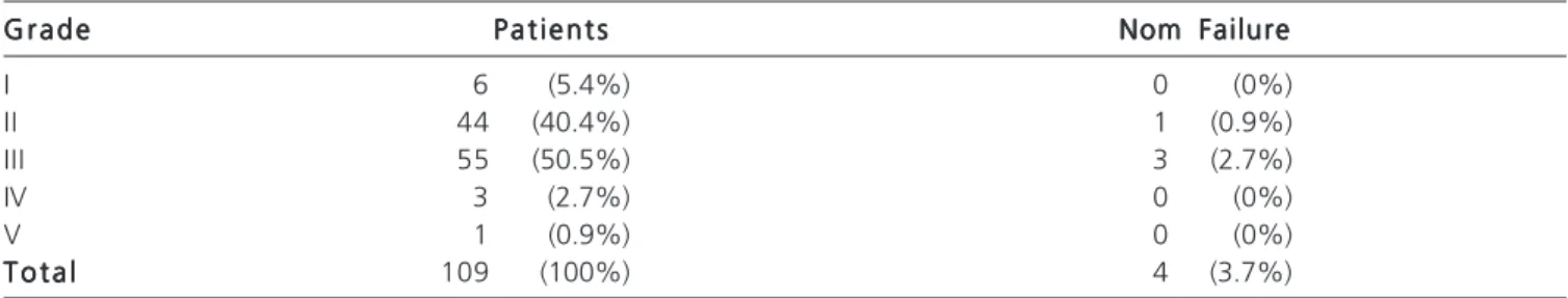
Computed tomography of the abdomen was performed on all patients and the lesions found are described in table 1. The vast majority of patients had thoracoabdominal injuries (62.6%) while in 43 patients (37.4%) the wound was entirely abdominal. Liver injuries were found in 109 patients (94.8%) and their classifications are shown in table 2. The commonest injuries were grades II and III. Renal injuries occurred in 28 patients (24.4%) and their classifications are shown in table 3. Hemothorax and concomitant intra-abdominal injury was found in 72 patients (62.6%), so in that particular group it can be inferred that a phrenic injury was present.
Injuries associated with other regions, excepting the chest and abdomen, were present in 19 patients (16.5%) the most common being fracture of the thoracolumbar spine. During the period 20 patients (17.4%) repeated imaging
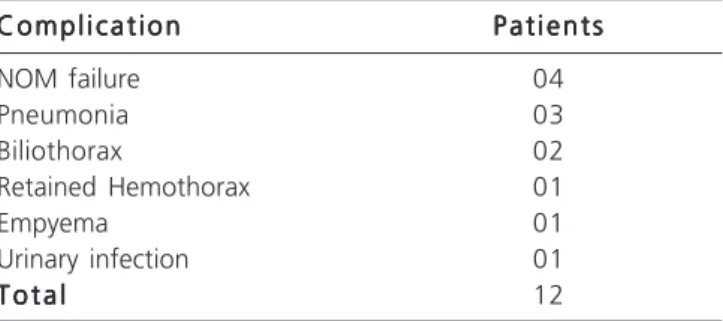
tests (ultrasound or CT). The main reasons for repeating the test were a change of symptoms and a progressive decrease in hemoglobin. Complications were present in 12 patients (10.5%) and are listed in table 4. Failure of the NOM procedure occurred in 4 patients (3.5%) and their characteristics are described in table 5. Interestingly, liver damage was present in all patients that failed NOM and the recommendation for surgery was directly related to it. In this study two patients (1.7%) died both due to associated TBI, one on the fifth day after injury and the other on the eleventh. The mean hospital stay was 9.4 days; for patients who didn’t developed complications it was 6 days, and for those who did, 12.5 days.
Regarding follow-up after hospital discharge, 67 patients (58.3%) had recovered two months after injury. All of them underwent CT of the abdomen which showed healing and an unaltered thorax in 58 patients (86.5%), a liver cyst in 3 patients (4.5%), a renal cyst in one patient (1.5%) and injuries with a reduced size and in the healing phase in five patients (7.5%). After two years of follow-up, only 45 patients showed up, even after active searches for the patients. It is interesting to note that the families of ten patients from this study said that they had been victims of new gunshot attacks and had subsequently died. For the 45 patients who showed up, their chest X-rays showed no direct or indirect alterations in the phrenic injury. The nine patients, who showed changes in their CT after two months, showed up to all reviews. In the last follow-up, after 24 months, only two patients remained with a liver cyst, although their sizes were diminishing.
DISCUSSION
DISCUSSION
DISCUSSION
DISCUSSION
DISCUSSION
Penetrating trauma injuries in the upper right thoracoabdominal area affect mainly the following organs: liver, right lung, right diaphragm, right kidney, duodenum, liver angle of the colon and the inferior vena cava. The colonic duodenal and inferior vena cava injuries require immediate and routine surgical treatment. Thoracic drainage is the treatment used routinely for lung injuries. The discussion about the best treatments for liver, renal and phrenic injuries still generates controversy. The option to perform NOM in carefully selected cases of GSW has already been discussed by various authors1-3,5-10 and there are several
Table 2 -Table 2 -Table 2 Table 2
-Table 2 - Classification of liver injuries by grade and their relationships to NOM failure.
G r a d e G r a d eG r a d e G r a d e
G r a d e P a t i e n t sP a t i e n t sP a t i e n t sP a t i e n t sP a t i e n t s Nom FailureNom FailureNom FailureNom FailureNom Failure
I 6 (5.4%) 0 (0%)
II 44 (40.4%) 1 (0.9%)
III 55 (50.5%) 3 (2.7%)
IV 3 (2.7%) 0 (0%)
V 1 (0.9%) 0 (0%)
T o t a l T o t a lT o t a l T o t a l
T o t a l 109 (100%) 4 (3.7%)
Table 1 Table 1 Table 1 Table 1
Table 1 - Principal injuries found.
I n j u r i e s I n j u r i e sI n j u r i e s I n j u r i e s
I n j u r i e s P a t i e n t sP a t i e n t sP a t i e n t sP a t i e n t sP a t i e n t s PercentagePercentagePercentagePercentagePercentage
Liver + Diaphragm 56 48.7%
Liver 29 25.2%
Liver + Diaphragm +Kidney 14 12.2%
Liver + Kidney 11 9.6%
Kidney 3 2.6%
Diaphragm + Kidney 2 1.7%
T o t a l T o t a lT o t a l T o t a l
justifications for adopting this type of conduct.
Until the advent of the use of imaging the vast majority of surgeons opted for mandatory laparotomy for penetrating abdominal and right thoracoabdominal injuri-es, principally those caused by firearms. In these cases it was not important whether the clinical examination was uncertain or not, nor the eventual projectile trajectory2. Even
in cases of doubt about penetration of the abdominal cavity, laparotomy was justified. However, these studies are not clear in relation to surgical findings during the perioperative period, in other words, what the real frequency of therapeutic and non-therapeutic laparotomy was. The arguments for this type of conduct can be summarized as follows: abdominal and right thoracoabdominal GSW are associated with a high frequency of intra-abdominal injury which requires surgical treatment11,12, a non-therapeutic
laparotomy is the safe procedure13, a delay in operating on
asymptomatic patients having abdominal injuries is associated with an increase in morbidity and, finally, the initial clinical examination is not a reliable method to exclude
abdominal11 injury, that is, the abdominal injuries cannot
be safely diagnosed without laparotomy.
In fact, many studies show that 20-30% of patients with abdominal gunshot wounds underwent unnecessary laparotomies1,2,10,14.15. Regarding the initial
physical examination, and principally subsequent examinations, their credibility and reliability, when performed by an experienced physician and by the same team, show criteria for a recommendation regarding the level 2 of evidence16. That credibility and reliability are even more
intense in patients with GSW than those with stab wounds, since in most cases, a larger number of intra-abdominal viscera are injured in patients with gunshot wounds, as well as the injuries being more serious and more severe when measured by trauma indices. Therefore, in these patients, signs of peritonitis are early and more exuberant1,2,7. Another
important and widely discussed argument is the incidence of morbidity and mortality, both locally (surgical site infection, incisional hernia, intestinal obstruction from adhesions, etc.) and systemically, (pneumonia, atelectasis, urinary tract infection, etc.) in non-therapeutic laparotomies. Several studies reported indices of complications ranging from 2.5 to 41.3%14,15,17,18.
A major problem in relation to NOM is the presence of injuries to hollow viscera not detected in the initial evaluation. To avoid this happening beyond the follow-up clinical examination it’s convenient to use sfollow-upplementary diagnostic methods such as arterial blood gas analysis, do-ses of serum lactate and repeated imaging exams as necessary. It should be emphasized that missed injuries treated within a few hours after injury, especially those of hollow viscera, are not related to a significant increase in
Table 3 Table 3 Table 3 Table 3
-Table 3 - Classification of kidney injuries by grade and their relationships to NOM failure.
G r a d e G r a d e G r a d e G r a d e
G r a d e P a t i e n t sP a t i e n t sP a t i e n t sP a t i e n t sP a t i e n t s Nom FailureNom FailureNom FailureNom FailureNom Failure
I 2 (7.2%) 0 (0%)
II 14 (50%) 1 (3.6%)
III 12 (42.8%) 1 (3.6%)
IV 0 (0%) 0 (0%)
V 0 (0%) 0 (0%)
T o t a l T o t a l T o t a l T o t a l
T o t a l 28 (100%) 2 (7.12%)
Table 4 Table 4 Table 4 Table 4
-Table 4 - Complications.
C o m p l i c a t i o n C o m p l i c a t i o n C o m p l i c a t i o n C o m p l i c a t i o n
C o m p l i c a t i o n P a t i e n t sP a t i e n t sP a t i e n t sP a t i e n t sP a t i e n t s
NOM failure 04
Pneumonia 03
Biliothorax 02
Retained Hemothorax 01
Empyema 01
Urinary infection 01
T o t a l T o t a l T o t a l T o t a l
T o t a l 12
Table 5 Table 5 Table 5 Table 5
Table 5 - Outcomes of patients failing NOM.
P t P t P t P t
P t I n j u r i e sI n j u r i e sI n j u r i e sI n j u r i e sI n j u r i e s Failure TimeFailure TimeFailure TimeFailure TimeFailure Time P o s t - o p e r a t i v eP o s t - o p e r a t i v eP o s t - o p e r a t i v eP o s t - o p e r a t i v eP o s t - o p e r a t i v e c o m p l i c a t i o n sc o m p l i c a t i o n sc o m p l i c a t i o n sc o m p l i c a t i o n sc o m p l i c a t i o n s H o s p i t a l i z a t i o nH o s p i t a l i z a t i o nH o s p i t a l i z a t i o nH o s p i t a l i z a t i o nH o s p i t a l i z a t i o n to surgery
to surgeryto surgery to surgery
to surgery (days)(days)(days)(days)(days)
1 Liver / Kidney 48hours Non-therapeutic No 9
2 Liver / Kidney / Htx 32hours Coleperitoneum No 7
3 Liver / Htx 48hours Coleperitoneum No 9
4 Liver / Htx 8hours Hemoperitoneum Yes / PNM 32
morbidity and mortality2, 6,7,19. Therefore these patients can
be safely observed until suggestive signs arise, on physical examination, which indicate the need for surgical treatment. A 24-hour observation period is considered sufficient to rule out associated abdominal injuries19, 20. If during this period
of observation doubt prevails then surgical treatment is necessary. In trauma, laparotomy continues to be the most accurate propaedeutic. Additionally, the incidence of missed injury in those patients selected for NOM is extremely small. The largest sample shows the frequency of missed injuries as 0.6% and the index of complications in patients operated on for late-diagnosed injuries was 6%8. In this study we did
not observe any missed abdominal injury and the only patient who was operated on with this suspicion had a non-therapeutic laparotomy.
With the advancement of trauma care already mentioned, patients with penetrating trauma, and who are admitted to emergency hemodynamically stable and without evident signs of peritoneal irritation, deserve a chance to be evaluated meticulously in search of a more detailed diagnosis concerning possible injuries present and therefore to avoid emergency surgery. Currently there are methods available that allow this precision. It is only after studies such as this, that the safest and most effective treatment can be chosen. The unequivocal progress and advancement in CT image quality, even in three-dimensions, can provide us a detailed study of the trajectory of a projectile from a firearm, determining whether or not there is penetration of the abdominal cavity. In addition, CT shows the presence of visceral injury (liver or kidney), its classification21, the
presence, location and estimated quantification of intracavity free fluid, allows diagnosis of possible complications and finally documents the evolution of the healing of injury. It has adequate specificity and sensitivity for these findings22,23.
Studies have shown CT to be safe, cost effective and fast in the evaluation of patients with right thoracoabdominal GSW24,25. Important also is that the
presence of intravenous contrast leak (blush) is indicative of active bleeding. This finding, in itself, is an important predictor of NOM failure26. In this circumstance, trying to
perform NOM necessitates the performance of angiography and embolization of the damaged vessel. Analyzing these characteristics it can be concluded that CT makes a selective approach to these patients safer, having evidence of recommendation level 216. It is an excellent tool that adds
important information to, but not replaces, the clinical examination of the patient as the main criterion for choosing NOM in thoracoabdominal right trauma by GSW. Therefore, there are occasions to institute NOM, but it is advisable and necessary that this group of patients be identified and safely selected in order to avoid an increase in morbidity and mortality12,27.
Undoubtedly the liver is the organ most damaged by this type of trauma. Liver injury has certain important features that permit the performance of NOM. This approach
was initially adopted in victims of blunt trauma. The success of NOM in cases of blunt liver injury is great reaching rates as high as 98.5% in multi-centric studies28. In the majority
of cases, bleeding from liver damage ceases spontaneously because it is of venous origin and of low pressure. This fact is often noted by the surgeon during surgery on a liver injury in a hemodynamically stable patient29,30. However, if during
surgery this injury were unpacked, voluminous bleeding often occurs, with a difficult hemostasis and requiring complex surgical maneuvers to stop the bleeding, but with high morbidity31. In some circumstances the bleeding is so
heavy and the injury so severe that a satisfactory hemostatic result cannot be achieved with conventional techniques, necessitating the use of maneuvers proposed by Damage Control Surgery. All this happens in a patient who was hemodynamically stable with no active bleeding!
Another important characteristic which must be considered is that to obtain a complete and secure approach to liver surgery, one should take into consideration the size and location of the organ. Due to this detail, during the act of surgery it is necessary to obtain a incision with ample access, using special adjustable retractors and release all hepatic ligaments, especially when there are posterior and superior injuries.
The liver parenchyma has great capacity for healing and regeneration, either from traumatic or surgical injury, keeping its tissue architecture preserved. Work in experimental models show that, 3-6 weeks after the occurrence of injury, the force required to break the scar that has formed is equal to that required to damage the normal parenchyma, regardless of whether the liver injury was sutured or not32. This healing ability is one of the most
important factors in the indication and success of non-operative treatment of liver injuries, even on those considered extensive.
Based on these characteristics, coupled with accurate diagnosis that CT allows, several surgeons have been successful in the non-surgical approach of penetrating hepatic injury3, 5,9,10,33. The analysis of results of existing
studies in literature are surprising and encouraging, showing success rates ranging from 67% to 100% 3,6,9,10,33.
Undoubtedly, the vast majority of patients with right thoracoabdominal GSW will require a laparotomy for treatment of their injuries. However there are a number of patients ranging from 6.5% to 40% who do not initially require surgery3, 6,9,10,33,34. The correct selection of these
The other viscus often injured in right thoracoabdominal GSW is the right kidney. The main goal of kidney injury treatment is to preserve the kidney. NOM also can be used with success in renal injuries. The use of excretory urography for diagnosis and guidance regarding the procedure is one of the factors that allows the use of NOM earlier on in the treatment of blunt renal trauma. In most cases, the bleeding arising from renal injury ceases spontaneously because it is self-limited and is contained in the retroperitoneum being buffered by the Gerota’s fascia. An excretory system lesion causing a urine leak, when present, is another major concern for the surgeon. With the advancement of endoscopic manipulation of the urinary tract, especially with the use of a Stent (double J) within the renal pelvis, injuries to the calyx and renal pelvis can be addressed in this way without the need for conventional surgery. An exception being when there is total rupture in the pyeloureteral junction.
In penetrating trauma, mainly by gunshot, the surgical treatment of renal lesions is the most suitable due to the high frequency of intra-abdominal associated injury 36-38. The major disadvantages of this approach are the high
rates of nephrectomy and unnecessary exploration of the kidney. Some authors advocate, even in the perioperative period, not to open the Gerota’s fascia and not to explore the injury in selected cases: in low-grade renal injuries and without extravasation of the CT and of lateral hematoma, or small and not expansive ones39. This is because in patients
with grade I and II renal injuries, when surgically explored, the bleeding has already ceased and any other hemostatic procedure is not necessary. This type of procedure is considered by these authors as non-operative treatment 39-41. The use of CT provides important information such as
injury classification, it studies the renal vasculature, checks for ischemic segment and principally it visualizes the contrast leak in the excretory system. Therefore it is important to perform this late stage in these patients.
Based on these assertions it was proposed, in well-selected cases, to use NOM for kidney injury itself, that is, not to operate on the patient. For penetrating GSW the selection of patients for NOM should be made carefully and strictly follow the proposed inclusion criteria. It is recommended to pay a lot of attention to the location of the perforations caused by the projectile, especially the entry point.
The anatomical location of the right kidney, especially considering its proximity to the liver, favors that this approach be adopted for right thoracoabdominal GSW, especially when the entry point is posterior. Nevertheless, it should be noted that the hepatic angle of the colon may, in some situations, stand between the right kidney and the liver. Therefore, we must be especially careful when the entry point is posterior and there is kidney damage and concomitant liver.
The reconstruction of the trajectory of the projectile by means of axial, coronal and sagittal views
during a CT scan with multi-detectors is essential to rule out injury to the large intestine. If in doubt, surgery is the safest option. The authors who propose this approach manage to achieve it with 10% to 40% of renal GSW, obtaining a success rate ranging from 91% to 100%37,39-42.
NOM reduces the index of nephrectomy because it provides a greater chance of renal preservation, a decrease in the rate of complications such as acute renal failure, and also in the possibility of unnecessary surgery40,41. NOM for GSW
was initially proposed for simpler injuries, but today it is also adopted for complex injuries43. Injuries with contrast
leak of the excretory system have a higher possibility of complication and NOM failure43.
In relation to the injury of the right diaphragm, without doubt, one can say that currently there is no consensus about what is the best approach to adopt when the diagnosis is made before surgery. There is much discussion about the possibility of hernia on the right. The natural evolution of an isolated injury to the right diaphragm is not yet known, in other words, if the injury heals spontaneously or not.
Experimental studies with pigs and mice suggest that there is strong evidence of spontaneous healing in around more than 90% of injuries44-47. In reviewing these
studies it is important to take into consideration the location and direction of the lesion (if it is in part of the tendon or diaphragm muscle) as well as its extension.
Another important factor to be remembered is the protection afforded by the liver that, theoretically, would block the hole of the injury thus preventing the migration of abdominal viscera into the thorax. However, there are no studies that effectively document spontaneous healing in all right diaphragmatic injuries. Indirect evidence exists. In patients who undergo NOM for right thoracoabdominal penetrating injuries in themselves, the diaphragmatic injury is not addressed, so theoretically, the frequency of complications such as biliothorax should occur with some frequency. However publications on this type of approach to this complication is rare or absent3,6,9,10,33,34.
Rezende Neto et al claim that the presence of bile in the liquid drained from the thorax does not exclude the possibility of performing NOM in cases of right thoracoabdominal GSW, provided that the patient is properly selected and monitored. However, persistent elevated bilirubin levels can be considered as a predictive factor in NOM failure.
R E S U M O R E S U M O R E S U M O R E S U M O R E S U M O
Objetivo: Objetivo: Objetivo: Objetivo:
Objetivo: Analisar os resultados obtidos com a introdução do protocolo de tratamento não operatório (TNO) dos ferimentos por arma de fogo (PAF) na transição toracoabdominal direita. Métodos: . Métodos: . Métodos: . Métodos: . Métodos: Estudo prospectivo com dados levantados no período de janeiro de 2005 a dezembro de 2011, tendo como critérios de inclusão: PAF localizado na região toracoabdominal direita, estabili-dade hemodinâmica, ausência de sinais de irritação peritonial e realização de tomografia computadorizada. Resultados: Resultados: Resultados: Resultados: Resultados: No estudo 115 pacientes preencheram os critérios de inclusão. A maioria dos pacientes (95,6%) era do sexo masculino. A média das idades foi 25,8 anos. A média dos índices de trauma: RTS 7,7; ISS 14,8; e TRISS 97%. A maioria dos pacientes era portadora de ferimentos toracoabdominais (62,6%) e 43 pacientes (37,4%), ferimentos abdominais. A lesão hepática ocorreu em 109 pacientes (94,8%) e a renal em 28 pacientes (24,4%). Hemotórax e lesão concomitante abdominal foram verificados em 72 pacientes (62,6%). As lesões associadas foram encontradas em 19 (16,5%) pacientes e as complicações, em 12 (10,5%). A falha do TNO aconteceu em quatro pacientes (3,5%). Nesta série, dois pacientes (1,7%) morreram, ambos devido a trauma cranioencefálico. A permanência hospitalar média foi 9,4 dias. Sessenta e sete pacientes (58,3%) compareceram no controle com dois meses de trauma. A tomografia de abdome mostrou lesão cicatrizada em 58 pacientes (86,5%). Conclusão: Conclusão: Conclusão: Conclusão: Conclusão: A opção por TNO do PAF na região toracoabdominal direita deve ser vista com cautela e empregada em casos selecionados através de protocolos bem fundamentados e em locais com toda infraestrutura necessária.
Descritores: Descritores: Descritores: Descritores:
Descritores: Traumatismos abdominais. Traumatismos torácicos. Fígado. Rim. Diafragma.
There is growing evidence that non-operative treatment of abdominal injuries of abdominal solids organs by NOM is feasible and safe. Around a third of all abdomi-nal trauma or thoracoabdomiabdomi-nal GSW can be approached non-operatively2,7,8,49. To perform NOM for right
thoracoabdominal GSW it is necessary to check the exact location(s) of perforation(s), conduct a thorough clinical evaluation with special attention to the hemodynamic condition and examination of the abdomen and have a detailed imaging study of the trajectory of the projectile. Another advantage of this approach is to allow less invasive techniques (endovascular, endoscopic and percutaneous) to be used in the treatment of injuries to the solids organs and their complicações10, 35. Como et al16 made the
following recommendations based on a level of evidence: a routine laparotomy is contraindicated in hemodynamically stable patients with abdominal injury GSW if the same were tangential and the patient had no signs of peritonitis (level 2); patients with isolated penetrating injuries in the right thoracoabdominal region can be treated without a laparotomy in the presence of stable vital signs, a reliable physical examination and with no or minimal abdominal pain (levels 2 and 3). The authors conclude the study by saying that NOM for penetrating injury trauma of solids organs (liver and kidney) require further studies. The data presented here corroborate, once again, the safety in performing NOM in selected cases of right thoracoabdominal GSW.
The approach to abdominal trauma is changing; the performance of NOM is possible both in blunt trauma and penetrating trauma. Undoubtedly one of the greatest dilemmas for the trauma surgeon, at the present time, is to decide whether routine surgical treatment is really the best option for patients with GSW to the upper right thoracoabdominal region or whether, under pre-established and well defined conditions, non-operative treatment can be performed safely.
In opting for NOM, the great challenge is to decrease the rate of unnecessary laparotomies without an increase in morbidity and mortality from intra-abdominal injury not diagnosed during the initial examination. Even authors who advocate mandatory surgical treatment recognize that, despite the risks, NOM could be performed provided that there is a protocol in place, so that it can be done safely and in the correct environment50.
Despite the good results achieved in this study, the option of performing NOM for penetrating GSW in the upper right thoracoabdominal region should be viewed with caution and used only in well selected cases using evidence-based procedures and in locations with all the necessary infrastructure. In the absence of professionals experienced in this type of approach, and qualified in selecting and properly monitoring patients, explorative surgery is still the safest method of treatment. However the option of performing NOM when the necessary conditions are present is not only scientifically correct but ethically justifiable.
REFERENCES
REFERENCES
REFERENCES
REFERENCES
REFERENCES
1. Muckart DJ, Abdool-Carrim AT, King B. Selective conservative management of abdominal gunshot wounds: a prospective study. Br J Surg. 1990;77(6):652-5.
2. Demetriades D, Charalambides D, Lakhoo M, Pantanowitz D. Gunshot wound of the abdomen: role of selective conservative management. Br J Surg. 1991;78(2):220-2.
3. Renz BM, Feliciano DV. Gunshot wounds to the right thoracoabdomen: a prospective study of nonoperative management. J Trauma. 1994;37(5):737-44.
5. Demetriades D, Rabinowitz B, Sofianos C. Non-operative management of penetrating liver injuries: a prospective study. Br J Surg. 1986;73(9):736-7.
6. Chmielewski GW, Nicholas JM, Dulchavsky SA, Diebel LN. Nonoperative management of gunshot wounds of the abdomen. Am Surg. 1995;61(8):665-8.
7. Demetriades D, Velmahos GC, Cornwel E 3rd, Berne TV, Cober S, Bhasin PS, et al. Selective nonoperative management of gunshot wounds of anterior abdomen. Arch Surg. 1997;132(2):178-83. 8. Velmahos GC, Demetriades D, Toutouzas KG, Sarkisyan G, Chan
LS, Ishak R, et al. Selective nonoperative management in 1,856 patients with abdominal gunshot wounds: should routine laparotomy still be the standard of care? Ann Surg. 2001;234(3):395-402; discussion 402-3.
9. Omoshoro-Jones JA, Nicol AJ, Navsaria PH, Zellweger R, Kriege JE, Kahn DH. Selective non-operative management of liver gunshot injuries. Br J Surg. 2005;92(7):890-5.
10. Navsaria PH, Nicol AJ, Krige JE, Edu S. Selective nonoperative management of liver gunshot injuries. Ann Surg. 2009;249(4):653-6. 11. Moore EE, Marx JA. Penetrating abdominal wounds. Rationale
for exploratory laparotomy. JAMA. 1985;253(18):2705-8. 12. Saadia R, Degiannis E. Non-operative treatment of abdominal
gunshot injuries. Br J Surg. 2000;87(4):393-7.
13. Shah R, Max MH, Flint LM Jr. Negative laparotomy: mortality and morbidity among 100 patients Am Surg. 1978;44(3):150-4. 14. Nance FC, Wennar MH, Johnson LW, Ingram JC Jr, Cohn I Jr.
Surgical judgment in the management of penetrating wounds of the abdomen: experience with 2212 patients. Ann Surg. 1994;179(5):639-46.
15. Renz BM, Feliciano DV. Unnecessary laparotomies for trauma: a prospective study of morbidity. J Trauma. 1995;38(3):350-6. 16. Como JJ, Bokhari F, Chiu WC, Duane TM, Holevar MR, Tandoh
MA, et al. Practice management guidelines for selective nonoperative management of penetrating abdominal trauma. J Trauma. 2010;68(3):721-33.
17. Ross SE, Dragon GM, O’Malley KF, Rehm CG. Morbidity of negative coeliotomy in trauma. Injury. 1995;26(6):393-4.
18. Morrison JE, Wisner DH, Bodai BI. Complications after negative laparotomy for trauma: long-term follow-up in a health maintenance organization. J Trauma. 1996;41(3):509-13. 19. Schmelzer TM, Mostafa G, Gunter OL Jr, Norton HJ, Sing RF.
Evaluation of selective treatment of penetrating abdominal trau-ma. J Surg Educ. 2008;65(5):340-5.
20. Inaba K, Barmparas G, Foster A, Talving P, David JS, Green D, et al. Selective nonoperative management of torso gunshot wounds: when is it safe to discharge? J Trauma. 2010;68(6):1301-4. 21. Moore EE, Cogbill TH, Jurkovich GJ, Shackford SR, Malangoni
MA, Champion HR. Organ injury scaling: spleen and liver (1994 revision). J Trauma. 1995;38(3):323-4.
22. Múnera F, Morales C, Soto JA, Garcia HI, Suarez T, Garcia V, et al. Gunshot wounds of abdomen: evaluation of stable patients with triple-contrast helical CT. Radiology. 2004;231(2):399-405. 23. Velmahos GC, Constantinou C, Tillou A, Brown CV, Salim A,
Demetriades D. Abdominal computed tomography scan for patients with gunshot wounds to the abdomen selected for nonoperative management. J Trauma. 2005;59(5):1155-60; discussion 1160-1. 24. Grossman MD, May AK, Schwab CW, Reilly PM, McMahon DJ, Rotondo M, et al. Determining anatomic injury with computed tomography in selected torso gunshot wounds. J Trauma. 1998;45(3):446-56.
25. Ginzburg E, Carrillo EH, Kopelman T, McKenney MG, Kirton OC, Shatz DV, et al. The role of computed tomography in selective management of gunshot wounds to the abdomen and flank. J Trauma. 1998;45(6):1005-9.
26. Renz BM, Bott J, Feliciano DV. Failure of nonoperative treatment of a gunshot wound to the liver predicted by computed tomography. J Trauma. 1996:40(2):191-3.
27. Degiannis E, Psaras G, Smith MD. Abdominal gunshot wounds— current status of selective non-operative management. S Afr J Surg. 2004;42(1):4-5.
28. Pachter HL, Knudson MM, Esrig B, Ross S, Hoyt D, Cogbill T, et al. Status of nonoperative management of blunt hepatic injuries in 1995: a multicenter experience with 404 patients. J Trauma. 1996;40(1):31-8.
29. Defore WW Jr, Mattox KL, Jordan GL Jr, Beal AC Jr. Management of 1,590 consecutive cases of liver trauma. Arch Surg. 1976;111(4):493-7.
30. Marr JD, Kriege JE, Terblanche J. Analysis of 153 gunshot wounds of the liver. Br J Surg. 2000;87(8):1030-4.
31. Pal KM, Khan A. Nonoperative management of penetrating liver trauma. Injury. 2000;31(3):199-201.
32. Dulchavsky SA, Lucas CE, Ledgerwood AM, Grabow D, An T. Efficacy of liver wound healing by secondary intent. J Trauma. 1990;30(1):44-8.
33. Demetriades D, Gomez H, Chahwan S, Charalambides K, Velmahos G, Murray J, et al. Gunshot injuries to the liver: the role of selective nonoperative management. J Am Coll Surg. 1999;188(4):343-8.
34. Demetriades D, Hadjizacharia P, Constantinou C, Brown C, Inaba K, Rhee P, et al. Selective nonoperative management of penetrating abdomnal solid organ injuries. Ann Surg. 2006;244(4):620-8.
35. DuBose J, Inaba K, Teixeira PG, Pepe A, Dunham MB, McKenney M. Selective non-operative management of solid organ injury following abdominal gunshot wounds. Injury. 2007;38(9):1084-90.
36. McAninch JW, Carroll PR, Armenakas NA, Lee P. Renal gunshot wounds: methods of salvage and reconstruction. J Trauma. 1993;35(2):279-83; discussion 283-4.
37. Wessells H, McAninch JW, Meyer A, Bruce J. Criteria for nonoperative treatment of significant penetrating renal lacerations. J Urol. 1997;157(1):24-7.
38. Kansas BT, Eddy MJ, Mydlo JH, Uzzo RG. Incidence and management of penetrating renal trauma in patients with multiorgan injury: extended experience at an inner city trauma center. J Urol. 2004;172(4 Pt 1):1355-60.
39. Voelzke BB, McAninch JW. Renal gunshot wounds: clinical management and outcome. J Trauma. 2009;66(3):593-600; discussion 600-1.
40. Velmahos GC, Demetriades D, Cornwell EE 3rd, Belzberg H, Murray J, Asensio J, et al. Selective management of renal gunshot wounds. Br J Surg. 1998;85(8):1121-4.
41. Bjurlin MA, Jeng EI, Goble SM, Doherty JC, Merlotti GJ. Comparison of nonoperative management with renorraphy and nephrectomy in penetrating renal injuries. J Trauma. 2011;71(3):554-8. 42. Navsaria PH, Nicol AJ. Selective nonoperative management of
kidney gunshot injuries. World J Surg. 2009;33(3):553-7. 43. Cheng DL, Lazan D, Stone N. Conservative treatment of type III
renal trauma. J Trauma. 1994;36(4);491-4.
44. Zierold D, Perlstein J, Weidman ER, Wiedeman JE. Penetrating trauma to the diaphragm: natural history and ultrasonographic characteristics of untreated injury in a pig model. Arch Surg. 2001;136(1):32-7.
45. Gamblin TC, Wall CE Jr, Morgam JH 3rd, Erickson DJ, Dalton ML, Ashley DW. The natural history of untreated penetrating diaphragm injury: an animal model. J Trauma. 2004;57(5):989-92.
46. Shatney CH, Sensaki K, Morgan L. The natural history of stab wounds of the diaphragm: implications for a new management scheme for patients with penetrating thoracoabdominal trauma. Am Surg. 2003;69(6):508-13.
47. Perlingeiro JA, Saad R Jr, Lancelotti CL, Rasslam S, Candelária PC, Soldá SC. Natural course of penetrating diaphragmatic injury: an expperimental study in rats. Int Surg. 2007;92(1):1-9.
48. De Rezende Neto JB, Guimarães TN, Madureira JL Jr, Drumond DA, Leal JC, Rocha A Jr, et al. Non-operative management of right side thoracoabdominal penetrating injuries—the value of testing chest tube effluent for bile. Injury. 2009:40(5):506-10.
50. Moore EE. When is nonoperative management of a gunshot wound to the liver apropriate? J Am Coll Surg. 1999;188(4):427-8.
Received on 15/01/2012
Accepted for publication 18/03/2012 Conflict of interest: none
Source of funding: none
How to cite this article: How to cite this article:How to cite this article: How to cite this article:How to cite this article:
Starling SV, Rodrigues BL, Martins MPR, Silva MAS, Drumond DAF. Non operative management of gunshot wounds of right thoracoabdomen . Rev Col Bras Cir. [periódico na Internet] 2012; 39(4). Disponível em URL: http://www.scielo.br/rcbc