ISABEL
MARIA
DA
SILVA
FONSECA
PREDICTIVE FACTORS OF GRAFT DYSFUNCTION AND
LONG-TERM KIDNEY ALLOGRAFT FAILURE
FATORES PREDITIVOS DE DISFUNÇÃO E
PERDA DO ENXERTO RENAL A LONGO PRAZO
Tese de Candidatura ao grau de Doutor em Ciências Biomédicas submetida ao Instituto de Ciências Biomédicas Abel Salazar da Universidade do Porto.
Orientadora – Doutora Denisa Mendonça
Categoria – Professora Associada (Aposentada)
Afiliação – Instituto de Ciências Biomédicas Abel Salazar da Universidade do Porto.
Coorientadora – Doutora Luísa Lobato Categoria – Professora Auxiliar Convidada
Afiliação – Instituto de Ciências Biomédicas Abel Salazar da Universidade do Porto.
Nothing in this world can take the place of persistence. Talent will not: nothing is more common than unsuccessful men with talent. Genius will not; unrewarded genius is almost a proverb. Education will not: the world is full of educated derelicts. Persistence and determination alone are omnipotent.
Financial support for this thesis was kindly provided by:
Centro Hospital do Porto
Unit for Multidisciplinary Research in Biomedicine, Porto
Abbott Laboratories (for providing the kits for measuring urinary NGAL in almost 200 samples)
Astellas Pharma (for supporting the acquisition of reagents for measuring oxidative markers)
Conflict of interest:
Preface
The work presented in this PhD thesis was conducted at the Department of Nephrology and Kidney Transplantation in close collaboration with the Department of Clinical Chemistry of Centro Hospitalar do Porto, Porto, Portugal.Because of the difficulties in obtaining funds for the prospective studies, the need for a period of interruption as a result the illness of a close relative, and my concurrent full-time employment, the work included in this PhD thesis was extended and performed from July 2008 to October 2014.
A PhD is a long, complex, meticulous, and, in most cases, laborious and painful process. The prospective cohort-based studies included in this thesis were purposely designed and completed for this PhD project and some of the biomarker analyses were performed for the first time in the Department of Clinical Chemistry. The financial support obtained for this part of the study was exhausted by the purchase of reagents. Thus, I had an active role in accomplishing of this task. Whenever a patient was called for transplant and agreed to participate in the study I was notified and then the study process was triggered. Every day for seven months, including weekends, I prepared and stored approximately 3000 blood samples (whole blood, serum, plasma and erythrocytes). And after that, I actively participated in the laboratory analyses, mainly in the oxidative stress measurements.
This thesis cannot express the long days (and nights!) spent in the lab but it represents a culmination of work, writing and learning. I have been responsible for the design and organization of the study herein, as well as almost all aspects of the data collection and processing. I have learned how to elaborate and conduct research in a complex field, how to collaborate with other researchers as a team and how to conduct research as an individual. This PhD was a challenging as well as a rewarding journey during which I have gained important knowledge and valuable skills. I have been the main author for all publications. With the exception of the competing risks analysis, which was performed by Laetitia Teixeira, one of my co-authors, all statistical analyses were performed by me (some for the first time) and guided by my main supervisor Denisa Mendonça.
This thesis is the report of this long process. It cannot account the long hours spent on computer with statistics and scientific writing. It cannot express the hope for good results and the sadness and tiredness with each manuscript rejection. But I hope that it expresses hard work, determination and persistence. Do not give up! I believe that this was the hardest lesson on this PhD journey.
Isabel Fonseca, Porto, November 2014
Acknowledgements
I would like to express my acknowledgement to CENTRO HOSPITAL DO PORTO who provided aresearch scholarship support for my doctoral study and also to my other funding sources: ABBOTT Laboratories, ASTELLAS Pharma and MULTIDISCIPLINARY UNIT FOR BIOMEDICAL
INVESTIGATION, Porto, Portugal. Without these financial supports, this study would not have
been possible. Thank you so much!
My acknowledgements are also extended to the PATIENTS who consented to taking part in the
studies included in this thesis.
Personnal Thanks
There is a long list of people who have supported, helped and encouraged me throughout my work with this thesis. I wish to emphasize my gratitude to some of you in particular.My PhD journey has been a process of experiencing, learning and maturing, both in terms of scientific knowledge and personal growth. This thesis would not have been possible without the confidence and motivation of my teacher and main supervisor PROF. DENISA MENDONÇA. She is the main individual responsible for ensuring that I initiated, continued, and completed my research towards my PhD. She did not allow me to give up. I am grateful for her guidance, mentorship, encouragement and friendship. Although I do not say it often, THANK YOU, for
being there every day, for giving me the opportunity to learn and work with you, and for exploring the field of scientific research with me. More than anyone else, I owe this thesis to you. THANK YOU SO MUCH FOR EVERYTHING!
I would also like to acknowledge my co-supervisors PROF. LUÍSA LOBATO and DR. ANTÓNIO
CASTRO HENRIQUES for their trust, valuable advice and encouraging guidance. Thank you DR.
CASTRO for your unconditional support throughout almost 20 years! Thank you LUÍSA for your
friendship and for helping me accomplish this achievement especially in these last months. And thank you both for your efforts in achieving funds for this PhD.
I am deeply indebted to LA SALETE MARTINS, MANUELA ALMEIDA, LEONÍDIO DIAS, JOSEFINA
SANTOS, SOFIA PEDROSO, and ANTÓNIO CASTRO HENRIQUES for their commitment in patient
recruitment and for their invaluable help with many aspects of the work presented in this thesis. Most of all, I would like to gratefully and sincerely thank for your invaluable friendship. I am also most grateful to DR.ANTÓNIO CABRITA and to all MY NEPHROLOGY CO-WORKERS who,
in one way or another, have supported me and without whom I would not have been able to conduct this PhD journey. Special thanks goes to DR.GUILHERME ROCHA andDR.JOÃO PEDRO
PIMENTEL for our daily exchange of ideas and thoughts, and for the fun moments together. I would like to express my appreciation to the NURSES of the Nephrology Department for
I also wish to thank DR. JOSÉ CARLOS OLIVEIRA for his advice on NGAL, adipokines and cystatin C and for allowing me to perform the oxidative stress analyses at his Department. Thank you for allowing me to use the Department’s facilities and for trusting me with the Department keys, including on weekends.
I am grateful to HENRIQUE REGUENGO and MARIA LUÍS CARDOSO, my lab instructors, for their friendship, expertise, and technical laboratory support. Thank you both for the incredible patience during my time as a lab assistant and for the long nights in the lab.
I would like to thank to the laboratory staff of the Department of Clinical Chemistry, particularly MADALENA CRUZ, for their precious help, technical assistance and positive attitude over the
months that I spent there.
I am also very grateful for the help of PROF. MARTINS DA SILVA for his ongoing support throughout the PhD process and of PROF. PEDRO OLIVEIRA for all valuable comments and advice to improve my thesis.
I appreciate the opportunity and experience of working with LAETITIA TEIXEIRA on an interesting
competing risk problem in kidney transplantation. Thank you for your friendship, helpful comments, and encouragements during the last phase of my work.
Thank you JORGE MALHEIRO for your insightful discussions about research, valuable inputs, and help with sampling and methodological issues. Thank you for general breadth of knowledge of all things and for meticulously editing my thesis.
Many friends have encouraged me during the years I have been working with this PhD thesis, and I would like to thank all of them, specially MARGARIDA LIMA, PEDRO AMORIM, LAURINDA
LAPA, JOSÉ MANUEL PEREIRA, TERESA MENDONÇA, MARIA MENEZES, PAULA GAMA, JOSÉ
QUEIRÓS,ANABELA RODRIGUES,IDALINA BEIRÃO, AND LAURINDA TEIXEIRA.
Special thanks goes to FREDERICO for being my every day computer consultant and for being a
source of relaxation through the ups and downs. Thank you for your never-ending support and for giving me happiness. Thank you so much for always being there for me!
Last but not the least, I would like to thank to my best friends, my sisters TITA E SUSANA and
my brother Rui,for their incentive and emotional support thoughout this effort. I could not have done it without you! You have helped me more than you know! Thank you, more than words can say.
To MY PARENTS, thank you for your unconditional love, support and encouragement. You two have always inspired me to work harder and pursue my dreams, and for that I will be forever grateful.
Abstract
Kidney transplantation is considered the treatment of choice for many patients with end-stage chronic kidney disease; however, despite advancements in short-term allograft survival, long-term survival has not paralleled this improvement. Due to the inevitable ischemic damage and associated reperfusion injury, delayed graft function (DGF) is a common complication after kidney transplantation, which may negatively affect graft survival. Because serum creatinine (SCr) and other traditional markers of kidney injury are insensitive and delayed in the detection of the early stages of kidney damage and DGF, there has been a keen interest in the identification of novel biomarkers for the early detection of allograft dysfunction that could expedite treatment and improve long-term patient and graft survival. Biomarkers are characteristics that can be objectively measured in a biological sample. In clinical settings, biomarkers enable the diagnosis of a dysfunction or disease and, in some cases, they are used to monitor a treatment or to make a prognosis regarding the future outcome of a patient. The analysis of predictive factors of graft dysfunction and long-term kidney allograft failure focusing on novel biomarkers was the major motivation for this work. Thus, the general aim of this thesis was to investigate the potential of different biomarkers to reliably diagnose and predict early graft dysfunction and their effect on long-term kidney allograft failure as well as to gain insight into the underlying mechanisms of graft dysfunction.
Patients and Methods: The study involved three cohorts of patients: two retrospective
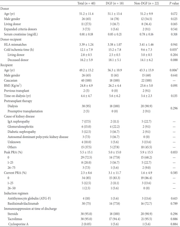
cohorts that included kidney transplant recipients selected from a database that contained transplant and follow-up information on kidney transplants performed between 1983 and 2008 (for the first retrospective cohort) or 2012 (for the second retrospective cohort); and one prospective cohort that included 40 patients undergoing kidney transplantation between December 2009 and June 2010. The first retrospective cohort was used to validate the one-year SCr as a surrogate endpoint of long-term graft survival, and the second retrospective cohort was considered to analyze the impact of DGF (defined by the need for dialysis during the first week after kidney transplantation) on graft and patient survival using a competing risks approach. The studies based on the prospective cohort had a longitudinal observational design, which was initiated at the time of transplantation; this cohort was used to examine nine potential candidate biomarkers for the early diagnosis of DGF (one biomarker in urine and eight biomarkers in blood): cystatin C (CysC), neutrophil gelatinase-associated lipocalin (N.GAL), leptin and adiponectin, malondialdehyde (M.D.A), superoxide dismutase (S.OD), glutathione reductases (GR), peroxidases (GPx) and total antioxidant status (TAS). Five samples per patient were collected within the first week: 3 to 6 h prior to transplant surgery (pre-transplant); on the
resulted in five samples per patient.
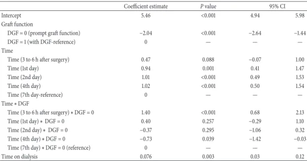
A linear mixed effects model was used to evaluate the longitudinal changes of the potential new biomarkers of early graft dysfunction over the first week after kidney transplantation and to identify the factors associated with these changes. The performance of the candidate biomarkers in the prediction of DGF was examined using receiver-operating characteristic (R.OC) curves. Survival analysis methods, including a survival analysis that accounted for competing risks were used to identify the predictive factors of long-term graft survival.
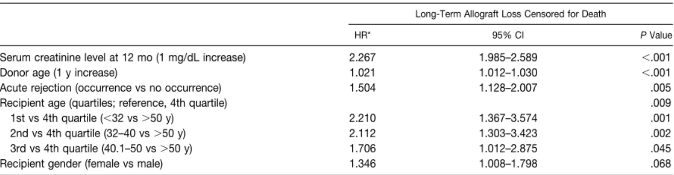
Results: Of the large number of variables that were considered, the SCr levels at 1, 6 and
12 months following kidney transplantation, as well as the changes between 1 and 6 months and between 6 and 12 months were independently associated with late graft failure.
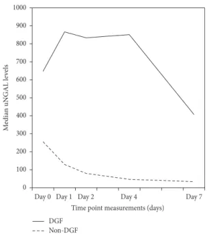
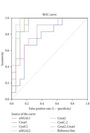
The R.OC curves identified urinary NGAL, MDA and CysC on the first postoperative day as moderately (NGAL) and highly (MDA and CysC) accurate in the prediction of DGF. Both urinary NGAL (at days 4 and 7) and MDA (day-7) were independently associated with one-year graft function, adjusting for variables that typically affect graft function, including acute rejection episodes and re-admissions during the first post-transplant year. Leptin at day-1 was slightly better than SCr in the prediction of the need for dialysis within the first week post-transplant, whereas adiponectin, SOD, GR, GPx and TAS were not. A triple-biomarker approach that used SCr, CysC, and MDA measured 8 to 12 h after kidney transplantation, was the most informative combination, which resulted in an increased ability (AUC=0.96) to distinguish patients with graft damage who would require dialysis within the first week. The application of a subdistribution regression model for competing risks indicated that DGF by itself and independent of acute rejection had a detrimental effect on long-term graft survival, but not on patient survival.
Conclusions: Independent of acute rejection, DGF per se was significantly associated
with poor-graft survival, but not with patient survival. Urinary NGAL and serum CysC and MDA were early, noninvasive, and accurate predictors of both the need for dialysis within the first week of kidney transplantation and one-year graft function. A triple-biomarker approach using SCr, CysC and MDA were highly predictive of DGF. Combining biomarkers from different pathophysiologic pathways appears to be a rational and reliable strategy to optimize sensitivity and specificity and obtain additive diagnostic and prognostic information.
Resumo
O transplante renal (TR) é considerado o melhor tratamento para a maioria dos doentes com necessidade de substituição da função renal. Apesar dos progressos alcançados, principalmente a nível da falência do enxerto nos primeiros seis meses após TR, a sobrevivência a longo prazo não tem acompanhado essa evolução. A ocorrência de atraso de função do enxerto (AF.E), nomeadamente por lesão provocada pela isquemia e
reperfusão associada ao transplante, condiciona a evolução do pós-transplante e tem um impacto negativo nos resultados imediatos e a longo prazo do TR. O desenvolvimento de intervenções eficazes na prevenção e/ou atenuação da agressão precoce no enxerto renal tem sido limitado pela ausência de marcadores precoces da lesão e disfunção renal. Os biomarcadores são substâncias ou “entidades” objetivamente quantificáveis, indicadores do curso de um processo biológico normal ou da ocorrência de uma lesão ou processo patológico, sendo usados na prática clínica para diagnóstico, monitorização terapêutica, estratificação de risco e previsão de eventos. Tendo em conta que os marcadores tradicionais de lesão e função renal, como a creatinina sérica (SCr), são tardios e insensíveis para o diagnóstico atempado de AFE, têm sido procurados novos biomarcadores capazes de identificar precocemente a disfunção renal e promover uma intervenção atempada e uma melhoria da sobrevivência a longo-prazo do enxerto renal. A análise dos fatores preditivos de disfunção e perda do enxerto renal a longo prazo, com ênfase na investigação de potenciais biomarcadores da disfunção precoce do enxerto, expressa pelo AFE, e do seu efeito na sobrevivência renal a longo prazo foi a principal motivação e objetivo desta tese.
Participantes e Métodos: O estudo envolveu três coortes: duas retrospetivas em que os
participantes foram selecionados a partir da base de dados do TR da Unidade de Nefrologia e Transplante Renal do Centro Hospitalar do Porto de 1983 a 2008 (para a primeira coorte retrospetiva) ou 2012 (para a segunda coorte retrospetiva) e uma coorte prospetiva de 40 doentes convocados para TR entre Dezembro de 2009 e Junho de 2010. A primeira coorte retrospetiva foi usada para validar a SCr observada durante o primeiro ano pós-TR como um marcador surrogate (substituto) da sobrevivência do enxerto renal a longo-prazo. A segunda coorte retrospetiva foi utilizada para avaliar o impacto do AFE (definida pela necessidade de diálise na primeira semana pós-TR) na sobrevivência do doente e do enxerto renal a longo prazo usando uma abordagem estatística baseada em eventos competitivos. Os estudos baseados na coorte prospetiva seguiram um desenho observacional longitudinal com início à data do transplante e pretenderam estudar nove potenciais biomarcadores para o diagnóstico precoce de AFE (um na urina e oito no sangue): a cistatina C (CysC), a lipocalina associada a gelatinase
capacidade antioxidante total (TAS)] e as adipocinas leptina e adiponectina. Foram colhidas 5 amostras por doente durante a primeira semana pós-TR: 3 a 6h antes do transplante; na manhã subsequente, aproximadamente 8 a 12 h após a reperfusão do enxerto renal (dia-1); e depois no segundo, quarto e sétimo dias pós-TR. A evolução longitudinal dos valores dos marcadores durante a primeira semana pós-TR e a identificação de fatores associados às alterações analíticas observadas nessa semana foram estudadas por modelos lineares de efeitos mistos. O estudo da performance dos biomarcadores no prognóstico de AFE foi efetuado com as curvas ROC. Métodos de análise de sobrevivência, incluindo a componente de eventos competitivos, foram usados para identificar fatores preditivos da sobrevivência do enxerto renal.
Resultados: Ajustando para os fatores tradicionalmente associados à perda a longo
prazo do enxerto renal, a SCr aos 1, 6 e 12 meses, assim como a diferença entre os valores de SCr entre primeiro e o sexto mês e entre o sexto e o primeiro ano associaram-se de forma significativa e independente à perda de enxerto renal a longo-prazo. As curvas R.OC revelaram que o NGAL urinário, o MDA e a CysC séricos no primeiro dia pós-TR foram moderadamente (NGAL) e fortemente (MDA e CysC) mais sensíveis no diagnóstico de AFE. Tanto o NGAL urinário (aos dias 4 e 7), como o MDA (ao dia-7) se associaram de forma independente à função renal observada no primeiro ano pós-TR, ajustando para os fatores que tradicionalmente afetam a função do enxerto. Os valores de leptina no primeiro dia pós-TR apresentaram uma performance ligeiramente melhor que a SCr para predizer o AFE, o que não ocorreu com a adiponectina, SOD, GR, GPx e TAS. Um multimarcador composto por SCr, MDA e CysC resultou da combinação de marcadores com melhor capacidade preditiva 8 a 12h após o TR (AUC=0.96) para identificar os doentes com lesão do enxerto renal e predizer a necessidade de diálise durante a primeira semana pós-TR. A aplicação de modelos de regressão de subdistribuição para eventos competitivos permitiu demonstrar que o AFE isolado e independentemente da rejeição aguda tem um efeito deletério na sobrevivência do enxerto renal, mas não na sobrevivência do doente.
Conclusões: O AFE por si só e independentemente da rejeição aguda associou-se a pior
sobrevivência do enxerto renal, mas não do doente. O NGAL urinário, o MDA e a CysC séricos são marcadores precoces e preditores da necessidade de diálise durante a primeira semana pós-TR e da função renal ao primeiro ano. Um marcador composto triplo com SCr, CysC e MDA foi altamente preditivo de AFE. A combinação de marcadores procedentes de diferentes vias patofisiológicas é uma estratégia racional para optimizar a
List of Publications included in the PhD Thesis
O
RIGINALA
RTICLESI. Fonseca I, Almeida M, Martins LS, Santos J, Dias L, Lobato L, Henriques AC,
Mendonça D. First year renal allograft function predicts long-term renal allograft loss. Transplant Proc. 2011; 43: 106-12
II. Fonseca I, Oliveira JC, Almeida M, Cruz M, Malho A, Martins LS, Dias L, Pedroso
S, Santos J, Lobato L, Henriques AC, Mendonça D. Neutrophil Gelatinase-Associated Lipocalin in kidney transplantation is an early marker of graft dysfunction and is associated with one-year renal function. J Transplant. 2013; 2013: 650123. doi: 10.1155/2013/650123. Epub 2013 Oct 31
III. Fonseca I, Reguengo H, Almeida M, Dias L, Martins LS, Pedroso S, Santos J,
Lobato L, Henriques AC, Mendonça D. Oxidative stress in kidney transplantation: Malondialdehyde is an early predictive marker of graft dysfunction. Transplantation 2014; 97: 1058-65.
IV. Fonseca I, Oliveira JC, Santos J, Martins LS, Almeida M, Dias L, Pedroso S,
Lobato L, Henriques AC, Mendonça D. Leptin and Adiponectin during the first week after kidney transplantation: Biomarkers of graft dysfunction?
(In Press, Accepted Manuscript in Metabolism – Clinical and Experimental)
V. Fonseca I, Reguengo H, Oliveira JC, Martins LS, Malheiro J, Almeida M, Santos J,
Dias L, Pedroso S, Lobato L, Henriques AC, Mendonça D. A Triple-Biomarker approach for the detection of delayed graft function using serum creatinine, cystatin C, and malondialdehyde. (Submitted) VI. Fonseca I, Teixeira L, Malheiro J, Martins LS, Dias L, Henriques AC, Mendonça D.
The Effect of delayed graft function on graft and patient survival in kidney transplantation: An approach using competing events analysis. Transplant
International 2015; 28: 738-50.
R
EVIEWA
RTICLEVII. Fonseca. I. Biomarkers in kidney transplantation: Translating to clinical practice
Table of Contents
Preface i
Acknowledgements iii
Abstract v
Resumo vii
List of publication included in the PhD thesis ix
Table of contents xi
Keywords xiii
List of abbreviations xiv
CHAPTER 1
1
General Introduction 1
Background and motivation for the work in this thesis 3
Aims of this dissertation thesis 6
Thesis outline 7
CHAPTER 2
9
Literature Review 9
Biomarkers 11
What is a biomarker? 11
Usefulness and advantages of biomarkers 12
Classification of biomarkers 12
Biomarkers versus Surrogates 13
Surrogates and biomarkers in kidney transplantation 14
Standard biomarkers for kidney damage 15
Why do we need new biomarkers for kidney transplantation? 15
Looking for new biomarkers of kidney graft dysfunction 16
Cystatin C 16
Neutrophil Gelatinase-Associated Lipocalin 19
Oxidative stress 22
Leptin and Adiponectin 24
CHAPTER 3
27
Material and Methods 27
3.1. Study Designs 29
3.1.1. Retrospective Cohort Studies (Studies I and VI) 29
Study samples 29
Data collection 30
Recruitment procedures 31
Biological sample collection, preparation and storage 31
Biomarker analyses/measurements 32
3.2. Clinical Definitions 34
3.3. Ethics 34
3.4. Statistical Analysis 35
Linear mixed effect model 35
Survival analysis considering competing risks 38
CHAPTER 4
43
Results 43
4.1. Study I 45
Serum creatinine as a surrogate endpoint for long-term graft survival 45
4.2. Study II 55
NGAL: a promising marker of graft dysfunction and a predictor of one-year graft function 55
4.3. Study III 69
Oxidative stress in kidney transplantation 69
4.4. Study IV 79
Leptin and adiponectin: biomarkers of graft dysfunction? 79
4.5. Study V 87
Combining biomarkers in kidney transplantation 87
4.6. Study VI 103
The impact of delayed graft function on graft and patient survival using a competing events approach 103
4.7. Review Article 121
Biomarkers in kidney transplantation: Translating to clinical practice 121
CHAPTER 5
131
Discussion 131
Discussion 133
CHAPTER 6
143
Conclusions and Future Directions 143
Concluding remarks 145
Future directions 148
REFERENCES
151
Keywords
Biomarkers Kidney transplantationDelayed graft function Long-term graft failure Cystatin C Leptin Neutrophil gelatinase-associated lipocalin
Oxidative stress Malondialdehyde
ANOVA Analysis of variance ANCOVA Analysis of covariance AUC Area under the curve ATP Adenosine triphosphate CIF Cumulative incidence function
CKD-EPI Chronic kidney disease epidemiology collaboration csHR Cause-specific hazard ratio
CysC Cystatin C
DGF Delayed graft failure
eGFR Estimated glomerular filtration rate EDTA Ethylenediaminetetraacetic acid GFR Glomerular filtration rate
GPx Glutathione peroxidase GLM Generalized linear models GR Glutathione reductase HLA Human leukocyte antigen H2O2 Hydrogen peroxide
KIM-1 Kidney injury molecule 1 KM Kaplan-Meier method
1-KM The complement of Kaplan-Meier estimator/estimates MANOVA Multivariate analysis of variance
MDA Malondialdehyde
MnSOD Manganese superoxide dismutase
NGAL Neutrophil gelatinase-associated lipocalin O2−· Superoxide
OH· Hydroxyl radical
ROC Receiver operating characteristic ROS Reactive oxygen species
sHR Subdistribution hazard ratio SCr Serum creatinine
SOD Superoxide dismutase TAS Total antioxidant status
Chapter 1
General Introduction
Content
Background and motivation for the work in this thesis Aims and objectives
B
ACKGROUND AND MOTIVATION FOR THE WORK IN THIS THESISKidney transplantation is considered the treatment of choice for almost all cases of renal failure, particularly because the quality of life and patient survival associated with transplantation are better than for chronic dialysis.1 Due to the new immunosuppressive drugs and consequent decrease in the rejection incidence, the short-term outcome of renal transplantation has improved substantially in the past 20 years. However, despite progress in short-term allograft survival, long-term survival has not paralleled that improvement.2-6
At present, late failure of kidney transplants is an important clinical problem and one of the leading causes of end-stage renal disease.7 The rate of chronic graft loss after the first year remains significant and the actual kidney allograft half-life showed only a marginal improvement over the past decade.2, 3, 6, 8 The reasons for this slight improvement remain unclear. It is possible that some important determinants of long-term graft survival may not have changed sufficiently to improve the overall outcomes of kidney transplantation.9 Patient death with a functioning allograft, mostly from cardiovascular disease, and chronic allograft failure are the two major causes of late transplant loss.10-12 The causes of chronic
allograft failure are multifactorial and are influenced by numerous immunological and non-immunological factors.2, 9 Generally, kidney transplants stabilize after recovering from the
stress of implantation until declining of graft function due to specific diseases or conditions, such as recurrent renal disease, antibody-mediated rejection or a common process involving interstitial fibrosis and tubular atrophy, which is encompassed by the previous descriptive term “chronic allograft nephropathy” and, more recently, “fibrosis / atrophy”.9, 13-15
Approximately half of deceased renal allografts are lost within 10 to 12 years after transplantation.16, 17 A patient submitted to kidney transplantation would wonder whether
his or her transplanted kidney will work well and how long it will last. There are no answers to these questions. Clinicians lack appropriate non-invasive methods to predict, diagnose and reduce the risk of graft failure in the years following renal transplantation. When will it be possible to identify valuable markers for distinguishing patients who are at an increased risk of graft dysfunction or of losing their transplant? Can biomarkers signal early transplant dysfunction, a process that is often undetectable? Can biomarkers help clinicians fine-tune their prognoses?
Many donor, recipient and immunologic characteristics are consistently associated with poor long-term outcomes, namely female gender, black ethnicity, prolonged pre-transplant dialysis time, older donor age, deceased donor source, delayed graft function (DGF), and acute rejection.11, 18-21 The association of human leukocyte antigen (HLA) matching and
panel reactive antibodies with the change in graft function suggests that immune mechanisms continue to have an effect on allograft function even among the long-term transplant recipients.22 Many of these factors coexist and act synergistically, and DGF is
one of them.
Delayed graft function is a well-known and the most common complication in the immediate post-transplantation period mainly in deceased renal allografts, almost invariably in the non-heart beating and in some live donor transplants.23-25 This condition
is a continuous spectrum of ischemia-reperfusion-related acute kidney injury and describes dysfunction of the kidney allograft immediately after transplantation.23, 26
Although not confirmed by some,27, 28 most studies have found associations between DGF and an increased risk for acute rejection and chronic allograft dysfunction,11, 29, 30 worse
graft survival21, 30-35 and higher mortality.26, 36-38
A range of factors could lead to DGF such as organ procurement (i.e. kidneys from non- heart-beating donors), donor characteristics (i.e. donors older than 55 years, donors with diabetes and/or high blood pressure), prolonged ischemia time, recipient factors (such as male gender, longer waiting time on dialysis, number of recipient’s previous transplants), renal toxicity, and ureteral obstruction, among others.39 With the present disparity between
supply and demand for organs, transplantation is proceeding with more marginal kidneys and therefore the problem of DGF is likely to increase in the future. Thus, DGF poses a significant challenge to clinicians in the context of kidney transplantation.
Ischemia/reperfusion injury after organ transplantation is a major cause of DGF, which is associated with prolonged hospital stay, additional invasive procedures, supplementary costs and greater risk of early and long-term graft loss.40 The association between DGF
and worse outcomes has led to increased efforts to better understand the mechanisms of ischemia-reperfusion injury and to develop interventions to reduce its occurrence and impact. This has included initiatives to discover and use biomarkers to stratify the risk of DGF, to diagnose dysfunction early and to target any intervention to those patients who will benefit most. This was the primary motivation for this thesis.
Thus, the major portion of this work focuses on one critical event after transplantation that is detrimental to the long-term graft and patient survival, DGF, as an expression of acute graft dysfunction. The following are some of the questions that this work tried to answer: a) Are there biomarkers that are significantly different between patients undergoing DGF,
versus those who are not?
b) Can a specific combination or panel of biomarkers work together and be potentially utilized for the diagnosis of DGF?
c) What can the identified biomarkers tell us about the underlying pathophysiology of this condition?
d) What is the impact of DGF on the patient and long-term graft survival over decades when using a competing risks approach?
A
IMS OF THIS DISSERTATION THESISThe analysis of predictive factors of graft dysfunction and long-term kidney allograft failure with a main focus on novel biomarkers was the major motivation for this work.
Thus, the general aim of this thesis was to investigate the potential of different biomarkers to reliably diagnose and predict early graft dysfunction and their effect on long-term kidney allograft failure as well as to gain insight into the underlying mechanisms of graft dysfunction.
The evaluation of the role of novel biomarkers on long-term graft failure requires a prospective approach that is impossible to achieve in a short-time period. On a short-term approach, alternative endpoints or short-term markers that can predict graft failure in the long-term may represent potentially useful surrogates and can be used in place of conventional endpoints.
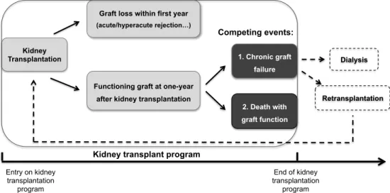
Thus, a phased approach was required to evaluate the clinical utility of novel biomarkers on long-term outcome, with the following design (Fig. 1) and specific aims:
- Develop a surrogate marker for long-term graft survival and evaluate the impact of early graft dysfunction, expressed by DGF, on the long-term graft loss and patient
survival. (Retrospective studies)
- Identify novel diagnostic biomarkers for early graft dysfunction by investigating the prognostic performance of the candidate biomarkers in the prediction of DGF and
one-year graft function. (Prospective studies)
DGF: delayed graft function; SCr: serum creatinine Fig. 1: Research design of this PhD thesis
Novel
Biomarkers Surrogate Endpoint Definitive Endpoint
e.g: SCr, DGF e.g: Graft Failure New potential biomarkers
T
HESIS OUTLINEThe present thesis is divided into six chapters.
Chapters 1 and 2 contain the scientific background and motivation for the research
performed in this thesis with respect to the investigation of novel graft dysfunction biomarkers.
Chapter 1 is a general introduction to some of the achievements and problems of kidney transplantation and summarizes the main objectives and outline of the thesis. Chapter 2 is as an introductory chapter to the field of biomarkers with relevant definitions and emphasizes the importance of novel and early markers of graft dysfunction in the immediate post-transplantation period, focusing on the biomarkers investigated in the current study.
The materials and methods are described in the original publications (I to VI), but some additional and more detailed information about the procedures and methodology are presented in Chapter 3.
Chapter 4 presents an overview of the results and the original research papers included
in this thesis as well as a review article. All published papers are reproduced with permission from the publisher.
Paper I aimed to develop a surrogate marker for long-term graft survival in our center.
Thus, factors associated with late kidney graft failure were identified and the predictive effect of serum creatinine (SCr) within the first year on long-term graft survival was examined.
In Papers II, III and IV the aims were to evaluate longitudinal changes of potential new biomarkers of early graft dysfunction over the first week after kidney transplantation and identify factors associated with these changes, to assess the performance of these candidate biomarkers in predicting DGF, and to appraise the long-term prognostic value of these biomarkers on kidney allograft function, evaluated by one-year SCr. These papers addressed the objectives of the prospective component of this work.
Paper V aimed to combine the studied biomarkers and develop a high sensitive
The aim of Paper VI was to evaluate the controversial impact of DGF on long-term graft loss and patient survival using a competing events approach.
Paper VII is an invited review article that presents a general discussion about
biomarkers in kidney transplantation, integrating some of the findings and biomarkers studied in this thesis.
Chapter 5 provides a general discussion of the main findings of the papers included in
this thesis and also considers aspects and reflections that were not included in the papers.
Chapter 6 provides a conclusion to the thesis and presents future perspectives.
Chapter 2
Literature Review
Content
Biomarkers
Surrogate markers
Surrogates and biomarkers in kidney transplantation Neutrophil gelatinase-associated lipocalin (N.GAL) Oxidative stress biomarkers
Cystatin C
B
IOMARKERS
A number of factors concerning donor, recipient and the peritransplant period influence the long-term graft outcome and have been widely discussed in the published literature. 41-46 Although the synergic action of immune and non-immune factors cannot be forgotten,
this literature review chapter will focus on biomarkers dependent of peritransplant kidney injury processes.
W
HAT IS A BIOMARKER?
Biomarker is a broad term that can be used to describe any indicator of a biological state. The term biomarker, or biological marker, was introduced in 1989 as a Medical Subject Heading (Me.SH) term and it was defined as “measurable and quantifiable biological
parameters (e.g., specific enzyme concentration, specific hormone concentration, specific gene phenotype distribution in a population, presence of biological substances), which serve as indices for health- and physiology-related assessments.” More recently, in 2001,
the definition was standardized by the Biomarker Definitions Working Group47, 48 as “a
characteristic that is objectively measured and evaluated as an indicator of normal biological processes, pathogenic processes, or pharmacologic responses to a therapeutic intervention.”
These definitions correspond to two concepts: first, the objective measurement of a parameter in a biological sample, and second, its application to classifying a patient. Although the term “biomarker” is relatively new, biomarkers have been used in preclinical research and clinical diagnosis for some considerable time. Body temperature is a long-standing and well-known biomarker for fever, for example.
Several types of objective biomarker measurements can be performed on patients. In fact, biomarkers appear in every form. They can be anatomic, physiologic, biochemical, or molecular parameters that are associated with the presence and severity of specific diseases and they are detectable by a variety of methods including physical examination, laboratory assays, and imaging. Serum creatinine (SCr) and blood glucose levels are biomarkers, as are blood pressure, enzyme levels, tumor size measurements obtained by imaging techniques, and the presence of a gene mutation or the expression level of mRNA. Unlike what is commonly believed, biomarkers are not only molecules. A biomarker is any type of measurable change that may have clinical relevance.49, 50 And
this list is far from exhaustive and countless measurements have been proposed as biomarkers.
U
SEFULNESS AND ADVANTAGES OF BIOMARKERSThe goal of a biomarker measurement is to make a useful prediction about the classification of the patient. In practice, biological markers are used to determine the disease status, monitor the efficacy of a treatment or predict the future outcome of a patient. In any case, using the biomarker values, one would like to split the patients into two classes according to the status of interest. Patients are typically labeled according to a known test that was assessed with certainty, called a gold standard. After identifying specific and accurate biomarkers, future patients will be classified without the need for the gold standard, usually more expensive and risky. Biomarkers are often cheaper and easier to measure than “true” endpoints.
Biomarkers usually provide information that is readily available and simple to interpret by clinicians.51 For example, a patient's blood pressure is easier to use than
echocardiography for measuring left ventricular function, and it is much easier to perform echocardiography than to measure morbidity and mortality from hypertension in the long term. Biomarkers can also be measured more quickly and earlier. Blood pressure can be measured today, whereas it takes several years to collect mortality data.
The usefulness of biomarkers is highlighted by the wide array of clinical settings, in which they are utilized, including disease diagnosis and prognosis. According to the Food and Drug Administration and regardless of the purpose for its use, biomarkers should be accurate, reproducible and standardized across different clinical settings. Ideally, a biomarker should be specific, sensitive, predictive, robust, simple, accurate, and inexpensive. In other words, it should be perfect and improve our understanding of a disease while providing new knowledge of pathological mechanisms, allowing for earlier diagnosis and the delivery of more efficacious and safer therapies possible.52, 53
C
LASSIFICATION OF BIOMARKERSPresently, it is not well established how biomarkers are categorized because they can be classified based on different parameters. Within the field of health care, biological markers are commonly classified based on the sequence of events from exposure to disease, including biomarkers of exposure, which are used in risk prediction, and biomarkers of
disease, which are used in screening, diagnosis and prognosis.51 Clinically, biomarkers
may be distinguished according to their uses; an early intervention biomarker is used for the early detection of disease to facilitate intervention, whereas a prognostic biomarker is
correlated with outcomes and is used to identify patients who may benefit from an intervention.54
The Biomarker Working Group further classified biomarkers based on their utility and this categorization is also commonly used in biomedical research:47, 48, 55
- Type 0 biomarker: A marker of the natural history of a disease that correlates longitudinally with known clinical indices such as symptoms over the full range of disease states;
- Type I biomarker: A marker that usually determines the biological effect of a therapeutic intervention according to the mechanism of action of that intervention (pharmacological, nutritional or any other), even though the mechanism might not be known to be associated with the clinical outcome.
- Clinical endpoint: An outcome that represents the target measures of a study. A characteristic or variable that reflects how a patient fares or functions, or how long a patient survives. In renal transplantation, for example, the standard clinical endpoints are graft failure and death for late outcomes.
- Surrogate endpoint biomarker (Type II biomarker): A marker that is intended to substitute a clinical endpoint. A surrogate endpoint is expected to predict the clinical benefit, harm, lack of benefit, or lack of harm on the basis of epidemiologic, therapeutic, pathophysiologic, or other scientific evidence. It is important to note that all surrogates are predictors, but not all predictors are surrogates.
Biomarkers versus Surrogates
It is important to distinguish, at the outset, the use of the term biomarker from that of surrogate endpoint or surrogate marker. The use of the term ‘surrogate marker’ in medicine dates from 1988,56 but it was preceded for some years by the term ‘biomarker’57
and was succeeded and replaced by yet another term, ‘surrogate endpoint’.58 Surrogate
endpoints may be a subset of biomarkers. Although all surrogate endpoints may be considered biomarkers, it is clear that only a few biomarkers will meet the requirements for achieving inclusion in this subset.
A surrogate endpoint is one that is measured in place of the biologically definitive or clinically meaningful endpoint, and it usually tracks the progress or extent of the disease. Investigators choose a surrogate endpoint when the definitive endpoint is difficult to obtain or inaccessible due to cost, time, or the complexity of measurement. As explained by Lachenbruch54 “a ‘surrogate’ variable is one that is used in lieu of the true endpoint, to
evaluate the outcome more rapidly, less expensively, and/or less invasively.” Some examples include CD4 counts in AIDS patients, tumor size reduction in cancer patients,
blood pressure in cardiovascular disease, intraocular pressure in glaucoma patients, and SCr in chronic kidney disease.
Since approximately 1989, biostatisticians have investigated approaches to evaluating whether a biological parameter that might serve as a substitute or “surrogate” for a clinical endpoint in the study of a particular therapy for a particular disease.48 A “perfect”
surrogate endpoint, as described by Prentice,59 can be measured simply and without invasive procedures, is related to the causal pathway for the definitive endpoint, yields the same statistical inference as the definitive endpoint, and should be responsive to the effects of treatments. The disease affects the surrogate endpoint, which in turn affects the definitive endpoints. This is more than a correlation between the surrogate and the true clinical point. For example, to accept a classification scheme for a biopsy score as a surrogate endpoint for graft survival requires that the biopsy score not only correlates with outcome but that changes in the outcome due to treatment or any other intervention are reflected in the biopsies.54 Accepting a biomarker as a surrogate for a clinically definitive
endpoint requires validation. To validate an endpoint as a legitimate surrogate endpoint, a meta-analysis is usually required because relationships presented in one study may not be generalizable to another. Then, for use in clinical practice, each center should test whether that surrogate works well in the local scenario because populations have different characteristics and therapeutic interventions and treatments are different across countries and centers.
S
URROGATES AND BIOMARKERS IN KIDNEY TRANSPLANTATIONOne of the concerns in transplant research is to obtain insight into the factors that are associated with long-term allograft survival and to identify early markers of chronic allograft dysfunction, as well as potential interventional pathways. Long-term graft survival is an ideal endpoint, but evaluating an outcome in the long term is usually difficult and time-consuming. For this reason, an easier approach is to identify alternative endpoints or short-term markers that can predict the long-term survival and therefore act as potentially useful surrogates. This approach is widely used in clinical research on cancer and cardiovascular disease and has recently been applied in the context of renal disease.60
Biomarkers used for screening or diagnosis also often represent surrogate manifestations of the disease or dysfunction. This is the case for oxidative stress markers in the process of renal ischemia-reperfusion injury following kidney transplantation. Both biomarkers and surrogates significantly contribute to early diagnosis, longitudinal prognoses, and outcome prediction. They often enable the detection of renal graft dysfunction when kidney injury is subclinical, allowing for faster evaluation of drug therapies, transplant techniques, and
patient care protocols. As a result, there has been a concerted effort within the transplant community to attain a diagnostic marker that may serve as a surrogate for eventual graft loss.
Standard biomarkers for kidney damage
Traditional non-invasive markers of kidney injury are insensitive and nonspecific in the detection of early stages of kidney injury. Standard biomarkers for kidney damage include the glomerular filtration rate (GFR), SCr and urea as well as several urine qualities such as proteinuria and hematuria. For decades, the increase in SCr has been the only detectable sign of a reduction in the GFR. At present, a decline in the SCr is still the traditional marker for detecting graft functional recovery after transplantation. However, this biomarker is an unreliable indicator of kidney function during an episode of acute injury.61 Serum creatinine changes are not specific for parenchymal damage and occur long after the event. It is estimated that more than 50% of kidney function is lost before the SCr rises, which makes SCr less sensitive to early kidney damage and the severity of dysfunction.61-63 As such, renewed efforts to improve long-term survival through enhanced
monitoring and diagnosis of short and long-term graft dysfunction have directed attention to the search of better biomarkers.
Why do we need new biomarkers for kidney transplantation?
In organ transplantation, initial graft dysfunction is one of the most important early postoperative problems, which is mainly due to the unavoidable ischemia-reperfusion injury that occurs in the transplanted organ. In kidney transplantation, ischemic injury of the renal allograft is a critical early insult that augments the risk of acute tubular necrosis and long-term graft loss.64, 65 The development of effective interventions is constricted by
the limited ability to detect graft dysfunction early. The delay period between initiation of injury and clinical and biochemical detection of renal damage calls for the use of more reliable and earlier markers of kidney graft damage. As previously stated, current clinical indicators of kidney injury, such as SCr, are inadequate for timely diagnosis and prognosis. Thus, the application of biomarkers in the field of kidney transplantation will allow for the detection of incipient graft dysfunction, refine diagnoses and enable more effective post-transplant management, potentially improving the short-term (e.g., delayed graft function, acute rejection) and long-term (e.g., allograft failure) outcomes.
L
OOKING FOR NEW BIOMARKERS OF KIDNEY GRAFT DYSFUNCTIONThe search for new biomarkers is expanding at an unprecedented rate. Recent efforts to identify biomarkers in kidney transplantation with early diagnostic and prognostic potential have yielded several candidates, including neutrophil gelatinase-associated lipocalin (N.GAL), kidney injury molecule 1 (KIM-1), cystatin C, interleukin-18, clusterin, kariopherins, glutathione S-transferase iso-enzymes, liver-type fatty acid binding protein, alpha-1-microglobulin, C-terminal agrin fragment and haptoglobin.66-77 Nevertheless, due
to the lack of evidence to support their use in routine, neither of these is currently used in clinical practice. And the search for an ideal marker continues.
In this dissertation, nine candidate biomarkers were studied and they are briefly reviewed in the following paragraphs.
C
YSTATINC
Cystatin C (CysC) is a monomeric, non-glycosylated polypeptide chain of 120 aminoacids with a low molecular mass of 13.3 kDa. This cystatin is produced at a constant rate by nearly all-human nucleated cells and it can be found in virtually all tissues and bodily fluids, preferentially in the cerebrospinal fluid, seminal plasma, and milk.78, 79 Its
concentration is the highest of all known low molecular weight cystatins in most of the extracellular fluids in humans at approximately 1–10 mg/L.78 Cystatin C is an endogenous
cysteine proteinase inhibitor belonging to the type 2 cystatin superfamily. Cysteine proteases are enzymes that are responsible for many crucial physiological processes, such as intracellular protein degradation, apoptosis, major histocompatibility complex class II immune responses, prohormone processing and bone remodeling.80 By inhibiting the function of several cysteine proteases, CysC participates in the regulation of the balance of catabolism and modulates many of these normal body processes.81 Other functions for CysC include a role in the atherosclerotic process,82 antigen presentation,83
defense against bacteria and viruses84 and as a growth factor for neural stem cells.85 The main catabolic site of CysC is the kidney; more than 99% of the protein is cleared from the bloodstream by glomerular ultrafiltration and tubular reabsorption. Because it is not secreted by the tubules, its concentration in urine in normal states is remarkably lower and approximately 0.1 mg/L (Lofberg and Grubb, 1979; Poulik et al., 1983). As CysC is
per se produced at a constant level, its concentration in the circulation remains nearly
stable when kidney function is normal.79, 86 Consequently, the rate at which CysC C is filtered at the glomerulus is the primary determinant of the blood CysC level. Due to these properties, CysC was first suggested as a marker of the glomerular filtration rate (GFR) in
1985.87 Since then, CysC has been extensively investigated in multiple clinical studies on
adults, children, and in the elderly. In almost all clinical studies, CysC demonstrated a better diagnostic accuracy than SCr in discriminating normal from impaired kidney function.88, 89
In fact, CysC fulfills many criteria that are set for an ideal endogenous biomarker of kidney function. As a low molecular weight protein, it is almost freely filtered through the glomerular membrane and then completely reabsorbed and catabolized by the proximal tubular cells.86, 90, 91 In studies performed with mice, the plasma clearance of CysC is 94%
that of 51Cr-EDTA and no secretion or reabsorption in the circulation occurs.86 Its plasma
or serum concentration is less dependent on the muscular mass, inflammatory diseases, gender or diet, and these properties make it a good measure of the GFR compared to the traditional measurement of the SCr.92, 93 As a result of this finding, several prediction
equations have been derived from both pediatric and adult patients to estimate the GFR from the serum CysC concentration.94, 95 Most of the studies that compared the CysC
levels or CysC-derived equations with gold standard methods found CysC to be superior or at least equivalent to SCr.93 Some studies on selected patient groups with either
reduced or rapidly changing muscle mass also demonstrated that CysC is a sensitive marker of the GFR independent of body composition.92
Renal transplant recipients are a target group for whom the precise determination of GFR is crucial. Allograft function following renal transplantation is commonly monitored using SCr. However, plasma creatinine is far from being an ideal marker of the GFR, despite its convenience and low cost. Since the first publication in 1998,96 quite a few original clinical
papers have addressed the question of the use of CysC in kidney transplantation. A good number of studies identified serum CysC (or CysC-based equations) as a promising, easily measurable marker to estimate the GFR with a higher diagnostic value than SCr (or creatinine-based equations) and 24-hour creatinine clearance for evaluating the GFR in the follow up of adult kidney transplant patients.97-99 Very recently, Masson et al99
validated both CysC-based CKD-EPI equations (2012) in 670 kidney transplant recipients and concluded that both performed better than the serum creatinine-based CKD-EPI equation (2009).
A drawback of the use of CysC in kidney transplantation is the routine use corticosteroids. Glucocorticoid medication can compromise the use of serum CysC in this population and it is important to take this into account when interpreting this serum marker. Glucocorticoid therapy is one of the few identified circumstances that have an impact on the production of CysC in a dose-dependent manner, leading to systematic underestimation of the GFR.100 Very large doses of glucocorticoids have been described to increase the
do not seem to alter the production of CysC.102, 103 However, underestimations of GFR
occur in some studies, e.g., with steroid dependent asthmatic patients.104 Hence,
moderate and high-dose glucocorticoids can limit the usefulness of CysC soon after kidney transplantation.105
For kidney transplant patients, early detection of decreased renal function is crucial so that measures to prevent further decreases in graft function can be taken. For this reason, the role of this marker in detecting post-transplant renal damage earlier than SCr has been investigated.106-110 During the early post-transplantation period, the serum CysC
decreases more rapidly than creatinine.107, 111 As previously stated, glucocorticoids
increase CysC concentrations and may lead to underestimation of the GFR; however, in stable renal graft recipients with low-dose immunosuppressive therapy, CysC is strongly correlated with the GFR and detects a GFR impairment earlier than SCr or creatinine-based eGFR.100, 106, 107, 111-115
In a prospective study of 30 consecutive patients with end-stage renal disease undergoing renal transplantation, Le Bricon and coworkers107 evaluated CysC as a marker of allograft function during the early postoperative transplantation period. Serum CysC was more sensitive than SCr for detecting decreases in the GFR and predicting DGF. Furthermore, a more prominent rise in the plasma CysC values allowed for a more rapid diagnosis of acute rejection or treatment nephrotoxicity with the potential for more timely intervention. A prospective study performed by Thervet et al 106 in another 30 renal transplant patients
also found that CysC allowed for earlier diagnosis of renal function recovery than SCr, particularly in patients with DGF. These findings were also confirmed by Hall and coworkers108 in a cohort of 78 deceased-donor renal recipients, which showed that CysC
outperformed SCr as a predictor of poor early graft function and the need for dialysis within the first week of kidney transplantation. Additionally, these authors demonstrated that CysC was a good prognostic marker of graft function at 3 months. In a recent article, Liu et al 110 evaluated the clinical value of CysC for the diagnosis of an acute rejection
episode after renal transplantation in 76 recipients and concluded that CysC can predict an acute rejection episode after renal transplantation. In a recent multicenter study,99
CKD-EPI formulae were compared for their accuracy in estimating the GFR, as determined by the gold standard, inulin clearance, in adult kidney transplant recipients (n=670) with stable graft function. This study used centralized, standardized assays for CysC and creatinine. Despite immunosuppressive treatment, formulae based on CysC and the combination of CysC and creatinine were less biased, more accurate and precise than the CKD-EPI-creatinine formula.
observed in a prospective multicenter study of 91 deceased-donor kidneys transplants.109 Serial urine samples were collected for 2 days following transplant and on the first postoperative day urine CysC was a predictor of DGF and of 3-month allograft function. In summary, CysC either in serum or urine displays several good characteristics that make it a practical and reliable biomarker for the early detection of DGF. Among the markers addressed in this review, serum CysC is likely the most commonly used biomarker as well as the closest to the clinical validation in kidney transplantation.
N
EUTROPHILG
ELATINASE-A
SSOCIATEDL
IPOCALINNeutrophil gelatinase-associated lipocalin is one the most promising and extensively studied biomarkers of acute kidney injury in a variety of acute clinical settings.116-129 Neutrophil gelatinase-associated lipocalin, also known as human neutrophil lipocalin or lipocalin-2, is a glycoprotein that belongs to the lipocalin family. Originally, NGAL was identified in neutrophils covalently bound to gelatinase, but this lipocalin is also expressed at low levels in other human tissues including the kidney, lung, liver and epithelial cells in response to various pathologic states.130 Human NGAL exists as a 25-kDa monomer, with
a 45-kDa homodimer and is conjugated to gelatinase as a 135-kDa heterodimeric form, which is normally the main cellular source of circulating NGAL.130
As stated by one of the main researchers of these molecules, “Lipocalins are small
proteins that cells send out to bind things and carry them back”.131, 132 Effectively, this
family comprises several proteins, such as α1-microglobulin, retinol-binding protein 4, prostaglandin D synthase, and nitrophorines, which are specialized in binding and transporting small hydrophobic molecules, such as vitamin A, free heme and heme groups that are complexed with nitric oxide.133-136
The main features of NGAL were described by Goetz et al 131, 137, who discovered that the
most important ligands of NGAL were siderophores, which are small iron-binding molecules. Bacteria produce siderophores to scavenge iron from the extracellular space and use specific transporters to recover the siderophore iron complex, ensuring their iron supply. These findings were consistent with the most important function attributed to this lipocalin, which is the inhibition of bacterial growth by the inhibition of iron-binding molecules that are important to specific bacteria.131, 137 Thus, NGAL behaves as a
bacteriostatic agent in acute infections and, under physiological conditions, bacterial infections represent the most common condition associated with marked increases in the NGAL levels.138 But, beyond its microbial effect NGAL seems to have more complex
activities.128, 139 Some systemic diseases that are not necessarily associated with infection
tissues may express and release NGAL as an acute-phase factor signaling a condition of sustained injury, which is the case for inflammatory processes involving skin, intestine and certain types of cancer, like adenomas and inflamed epithelia of the bowel, adenocarcinomas of the breast, and urothelial carcinomas.140-149 Renal tubular injury is another pathologic state that induces the expression of NGAL and that increases its levels by approximately 1000-fold, which is rapidly apparent in both the urine and serum.128, 139,
150 The relatively rapid time course of NGAL changes with respect to renal injury in
comparison to SCr levels is one of the main advantages of NGAL, which makes this a superior or complementary biomarker in the diagnosis and prediction of acute kidney injury.117, 151-158
The genesis and sources of serum and urinary NGAL in response to renal injury is a subject under study. It was demonstrated that NGAL exists in two separate body pools: the systemic and the renal pools. In the steady state, NGAL is normally expressed at very low concentrations in multiple cell types. Accordingly, in healthy individuals, NGAL is detectable in the systemic circulation only at low levels. In the kidney, circulating NGAL is filtered in the glomerulus and luminal NGAL is completely reabsorbed in the proximal tubule by a megalin-dependent pathway. Hence, only traces of NGAL are detectable in urine. During injury or inflammatory processes, NGAL is massively released from activated neutrophils and the urinary levels correlate with serum levels independent of the cause of increased NGAL production. However, when massive NGAL quantities are excreted in the urine this usually indicates injury and damage to the proximal tubular cells due to ischemia-reperfusion injury, hypoxia, nephrotoxins or chronic progressive changes.128, 150, 159 These kidney insults cause failure of absorption of the filtered NGAL
leading to particularly high NGAL levels in urine, which is potentiated by the increased expression and secretion of NGAL from the nephron epithelia and from distant organs mainly the liver and the lungs.126, 128, 150, 160-163
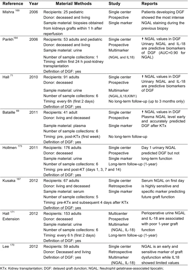
Ischemia-reperfusion injury is an inevitable consequence of the kidney transplantation procedure and can be considered as a form of post-transplantation acute kidney injury. For this reason, several studies have investigated the utility of NGAL for the diagnosis and prognosis of acute graft dysfunction following kidney transplantation, with promising results.69, 71, 72, 164-169 The values of NGAL collected shortly after renal transplantation were
shown to predict the dialysis requirement within the first week, preceding the postoperative peak in SCr levels that typically does not occur before two to four days.69, 71,
72, 164, 167, 170 Recently, the prognostic value of NGAL on graft function at one-year
post-transplantation was also examined.72, 171, 172 Different investigators reported consistent findings that NGAL may become one of the most important next-generation biomarkers in