CARLA MARIA DE CARVALHO BATISTA PINTO
PEROXISOMES IN BROWN TROUT (
Salmo trutta
f.
fario
):
REGULATION BY ESTROGENS
Dissertação de Candidatura ao grau de
Doutor em Ciências Biomédicas submetida
ao Instituto de Ciências Biomédicas Abel
Salazar da Universidade do Porto.
Orientador – Doutor Alexandre Manuel da
Silva Lobo-da-Cunha
Categoria – Professor Associado com
Agregação
Afiliação – Instituto de Ciências Biomédicas
Abel Salazar da Universidade do Porto.
Co-orientador – Doutor Eduardo Jorge
Sousa da Rocha
Categoria – Professor Associado com
Agregação
Afiliação – Instituto de Ciências Biomédicas
Abel Salazar da Universidade do Porto.
Co-orientador – Doutor Pedro Nuno
Simões Rodrigues
Categoria – Professor Associado
À minha mãe,
No cumprimento do disposto no nº 2 do Artigo 8º do Decreto-Lei nº388/70
(Decreto-Lei nº 216/92 de 13 de Outubro), declara-se que a autora desta
dissertação participou na concepção e na execução do trabalho experimental que
esteve na origem dos resultados apresentados, bem como na sua interpretação e
na redacção dos respectivos manuscritos.
Nesta tese inclui-se um artigo científico publicado numa revista internacional
resultante de uma parte dos resultados obtidos no trabalho experimental,
referenciado como:
Batista-Pinto C., Rodrigues P., Rocha E., Lobo-da-Cunha A., Identification and organ
expression of peroxisome proliferator activated receptors in brown trout (Salmo trutta f. fario),
Inúmeras foram as pessoas que, de algum modo ao longo destes anos, tornaram
possível ou contribuíram para a realização deste trabalho. A todos gostaria de
manifestar, desde já, o meu profundo reconhecimento. Alguns, no entanto,
merecem um agradecimento especial.
Reza a tradição que se agradeça aos orientadores. Embora goste de inovar, não
vou fugir a esta regra, pois, neste caso, a verdade está de mãos dadas com a
tradição. Reconheço que pertenço a um reduzido grupo de orientandos que
tiveram a sorte de ser realmente orientados por quem o deve fazer, tendo
recebido não só orientação teórica, como também uma verdadeira cooperação
experimental em trabalho de bancada. Também a convivência, assente numa
base de confiança, respeito e espírito de ajuda, foi facilitada pelo carácter dos
orientadores.
Assim, ao orientador desta tese, o Professor Doutor Alexandre Lobo da Cunha,
quero deixar expressa a minha profunda gratidão por me ter acompanhado de
perto ao longo dos últimos anos, fornecendo-me todas as ferramentas de
trabalho e meios técnicos necessários, bem como valiosos conselhos e rápido
auxílio na resolução dos problemas que sempre teimam em surgir. O bom
ambiente de trabalho foi, sem dúvida, favorecido pelo seu acutilante e sempre
bem-vindo sentido de humor. Estou também grata por me ter oferecido a sua
amizade, que muito estimo.
Não menos orientador, embora por imperativos legais apenas co-orientador, o
Professor Doutor Eduardo Rocha merece também o meu reconhecimento sincero.
Por ter sempre conseguido dispensar-me uma fatia do seu precioso tempo, pelos
sábios conselhos e sugestões, pela pertinência das suas críticas e amabilidade
dos elogios, pela valiosa ajuda experimental e paciência no tratamento dos
dados, pelo carinho e amizade, agradeço do fundo do coração.
O Professor Doutor Pedro Rodrigues, também meu co-orientador, merece
igualmente um agradecimento profundo. A ele devo os primeiros passos na área
da Biologia Molecular. O seu constante encorajamento e o hábil contorno dos
obstáculos após pequenos fracassos ajudaram a construir pequenas conquistas.
Agradeço com sinceridade os seus ensinamentos, a sua disponibilidade e o seu
por ter aceite pertencer à minha comissão de acompanhamento e por todas as
ocasiões em que tive oportunidade de escutar as suas sábias e bem-humoradas
palavras. Agradeço também a disponibilização do Laboratório de Histologia e
Embriologia, com meios técnicos e materiais essenciais ao desenvolvimento de
algumas etapas deste projecto.
Agradeço, ainda, ao Professor Doutor Jorge Azevedo, pela amabilidade que teve
em aceitar pertencer à minha comissão de acompanhamento e pelas sugestões
então feitas.
Ao Professor Doutor Carlos Azevedo, que foi o impulsionador da minha
actividade profissional na docência e o responsável pelo despertar para a
investigação, estarei sempre reconhecida pelas oportunidades que me deu, pelo
incentivo e pelos valiosos ensinamentos. Agradeço, ainda, a disponibilização do
Laboratório de Biologia Celular – incluindo o acesso ao Microscópio Electrónico de
Transmissão – para a realização de grande parte do trabalho envolvido neste
projecto.
Ao actual chefe do Laboratório de Biologia Celular, o Professor Doutor Mário
Sousa, agradeço as facilidades concedidas na utilização do mesmo. À Sra. D.
Laura Corral, bem como à Sra. D. Elsa Oliveira, estou grata pela agradável
companhia e por todos os conselhos e apoio técnico prestado. Pela esmerada
execução das fotografias de microscopia electrónica e pela disponibilidade na
resolução de pequenos problemas técnicos, agradeço amavelmente ao Sr. João
Carvalheiro.
No Laboratório de Histologia e Embriologia fiz também algumas amizades. Pela
ajuda bem-disposta durante as épocas de colheitas e pelo apoio pessoal,
agradeço à Filipa, à Nádia, à Paula e à Sra. D. Helena Galante.
Da vasta teia de cooperação estabelecida para o desenvolvimento do trabalho
fazem ainda parte outros laboratórios e serviços do Instituto de Ciências
Biomédicas Abel Salazar (ICBAS). Deste modo, gostaria de agradecer à Professora
Doutora Leonor Teles Grilo pelas facilidades concedidas na utilização do
Laboratório de Genética Molecular, à Eng.ª Carla Oliveira pelos conselhos técnicos
e ajuda prestada, e à Sra. D. Matilde Rocha pelo auxílio na esterilização do
material. À Sra. D. Teresa Barandela e à Lic.ª Cristiana Moreira agradeço o apoio
na área da Microbiologia. Agradeço também à Professora Doutora Berta Martins,
Baldaia estou grata pela amabilidade que teve em fazer a tradução do resumo
desta tese para francês. Ao Licº Rui Claro, do Departamento de Informática
agradeço todas as ajudas prestadas.
Também no Centro Interdisciplinar de Investigação Marinha e Ambiental (CIIMAR),
encontrei as condições adequadas para a realização das etapas finais deste
projecto. Ao Professor Doutor António Afonso devo um agradecimento muito
especial pela amável cedência do imprescindível espaço na sua arca congeladora.
À Professora Doutora Maria Armanda Henriques agradeço a utilização do
aparelho medidor de pH. Estou também grata à Professora Doutora Mª João
Rocha pela colaboração no doseamento de vitelogenina por ELISA e pela amizade
de longa data. Ao Eng.º Pedro Reis gostaria de agradecer a cuidada manutenção
dos aparelhos de uso comum. Pela ajuda na área informática, agradeço ainda ao
Eng.º Pedro Rodrigues.
À CESPU – Cooperativa de Ensino Superior Politécnico e Universitário – devo o
principal incentivo para a realização desta etapa, como parte integrante da minha
carreira docente e, acima de tudo, a oportunidade que me deu de fazer aquilo
com que sempre sonhei, num óptimo ambiente de trabalho. Na qualidade de
Director da CESPU, agradeço ao Professor Doutor Jorge Proença todas as
facilidades concedidas para a realização do doutoramento, nomeadamente a
possibilidade de dispensa do serviço docente e o alargamento do prazo
inicialmente acordado. Agradeço também a amável autorização de marcação de
férias em períodos alternativos. À Mestre Florinda Antunes agradeço a
disponibilidade e o precioso apoio prestado no Gabinete do PRODEP e no
Departamento de Recursos Humanos.
Como elemento fundamental para o desenvolvimento dos trabalhos, a Direcção
Regional dos Recursos Florestais - Circunscrição Florestal do Norte merece uma
referência especial pela disponibilização dos tanques necessários aos ensaios e
pelo fornecimento dos animais: os excelentes exemplares de truta fário cedidos
foram sempre transportados e mantidos em óptimas condições. Neste âmbito, a
preciosa cooperação do Eng. Augusto Maia merece o nosso maior apreço.
Gostaria também de deixar expressa a minha gratidão por alguém que deu um
empurrãozinho neste trabalho. Pela simpatia e disponibilidade, pelos conselhos
reconhecida ao Lic.º Eduardo Lopes, da Bio-Rad.
Ao Doutor José Manuel Costado Instituto Nacional de Saúde Doutor Ricardo Jorge
e à Mestre Paula Magalhães do Instituto de Biologia Molecular e Celular (IBMC)
agradeço a utilização dos respectivos aparelhos de PCR em Tempo Real. No IBMC
realizei ainda tarefas pontuais, pelo que agradeço a todos aqueles que, de algum
modo, me auxiliaram.
Ao meu designer favorito, Hélder Almeida, agradeço a fantástica criação da linha
gráfica do trabalho.
De entre todos os que me concederam algum do seu tempo, trabalho e paciência,
gostaria de agradecer de uma maneira muito especial ao Doutor Luís Filipe
Castro, à Lic.ª Graça Casal, à Lic.ª Fernanda Malhão e à Mestre Dolores Resende
que prontamente vestiram a bata por mim nas alturas em que eu não o pude
fazer sozinha e me proporcionaram um excelente ambiente de trabalho. O colega
e amigo Filipe, a quem estou profundamente grata pela realização das extracções
orgânicas prejudiciais ao meu estado de gestante, pela literatura partilhada, pelo
apoio científico e pelos ensinamentos, foi uma agradável surpresa. A elevada
capacidade científica desde logo sobressaiu do seu temperamento
despretensioso e humilde. Agradeço-lhe também a amizade e o sentido de
humor. Muito mais do que uma colega, a minha amiga Graça foi um apoio
precioso na leccionação das aulas e uma ajuda válida no laboratório,
demonstrando uma amizade verdadeira e constante. À minha amiga Fernanda
agradeço, além de uma bonita amizade, a sua boa-disposição, o apoio nas
colheitas, a ajuda técnica exaustiva e exímia nas áreas da microscopia e da
estereologia e a colaboração na leccionação de algumas aulas. Finalmente, à
minha grande amiga, colega de bancada e companheira de escrita Dolores quero
agradecer, antes de mais, a amizade sincera, o apoio, o companheirismo, a
generosidade, as palavras de carinho e incentivo, e o facto de ter tornado o peso
da minha protuberante barriguinha um pouco mais suave... No laboratório, estou
grata pela sua preciosa ajuda nas colheitas, nos doseamentos enzimáticos e em
inúmeras outras situações. Agradeço ainda a sua imprescindível colaboração na
minha actividade docente.
Para a minha maravilhosa mãe, Luísa, que, apesar da sua miopatia, conseguiu ser
o meu braço direito… e o meu braço esquerdo, as minhas mãos, as minhas
paciência e as horas extra que nos dedicou ao longo destes anos, num esforço
que reconheço como sobre-humano. A ti, Mamã, a quem eu dei quase nada em
troca de quase tudo, dedico esta centena e meia de páginas cheias de palavras
que não entendes mas que ajudaste a escrever.
Também os meus queridos sogros, Isabel e Agostinho, merecem uma palavra de
apreço e gratidão por todas as ajudas que me deram e, acima de tudo, pelo amor
que desde sempre me ofereceram.
Ao meu marido, Filipe, estarei sempre grata pelo amor e carinho constantes,
mesmo nos momentos mais difíceis. O seu sentido prático e racionalidade foram,
também, uma mais-valia: ao seu lado os problemas sempre encontraram uma
solução.
E aos meus filhos, Leonor e Henrique, devo o principal incentivo para a conclusão
deste trabalho: “Mamã, se tens mesmo que ir trabalhar… vai, trabalha muito,
A
BSTRACT17
R
ESUMO19
R
ESUME23
A
BBREVIATIONS27
1.
G
ENERALI
NTRODUCTION29
1.1. The brown trout 31
1.1.1. Life cycle 31
1.1.2. Liver metabolism and morphology 32
1.2. About peroxisomes 35
1.2.1. Discovery, morphology and biogenesis 35
1.2.2. Metabolic functions 41
1.2.3. Regulation by estrogens 49
1.2.4. Fish peroxisomes 50
1.3. Peroxisome proliferator activated receptors (PPARs) 51
1.3.1. Peroxisome proliferation 51
1.3.2. PPAR isotypes 53
1.3.3. Fish PPARs 57
1.4. References 59
2.
O
BJECTIVES79
Objectives 81
3.
I
DENTIFICATION ANDO
RGANE
XPRESSION OFP
EROXISOMEP
ROLIFERATORIntroduction 86
Materials and methods 87
Results 90
PPARs partial gene sequences 90
PPARs tissue expression 92
Discussion 94
Acknowledgments 97
References 97
4.
S
EASONAL ANDG
ENDERV
ARIATION OFP
EROXISOMEP
ROLIFERATORA
CTIVATEDR
ECEPTORSE
XPRESSION IN THEL
IVER OFB
ROWNT
ROUT101
Summary 103
Introduction 104
Materials and methods 105
Results 108
Fish morphometric parameters 108
Semi-quantitative RT-PCR 108
Quantitative real-time RT-PCR 110
Discussion 112
Acknowledgments 116
References 116
5.
C
ATALASE INB
ROWNT
ROUT:
G
ENEI
DENTIFICATION,
O
RGANT
RANSCRIPTIONP
ATTERN ANDS
EASONALE
XPRESSION IN THEL
IVER121
Summary 123
Introduction 124
Materials and methods 125
Tissue expression 128
Seasonal expression 129
Discussion 130
Acknowledgments 132
References 133
6.
K
INETICS OF THEM
ETABOLIC ANDM
ORPHOLOGICALA
LTERATIONS INB
ROWNT
ROUTH
EPATICP
EROXISOMES UNDERE
STRADIOLI
NFLUENCE137
Summary 139
Introduction 140
Materials and methods 141
Results 146
Enzymatic activities 146
Stereology of peroxisomes 146
Hepatosomatic index and plasma vitellogenin determination 151
Discussion 153
Acknowledgments 156
References 157
7.
C
ONCLUDINGR
EMARKS161
Concluding Remarks 163
References 168
Although the importance of peroxisomes in plants, fungi and protozoa has been recognized for long, the recognition of their relevance in mammal cellular metabolism was essentially based on the discovery of a class of severe human inherited diseases caused by peroxisomal deficiencies. Also the observation that many pharmaceutical and industrial compounds are able to induce proliferation of these organelles, and that prolonged treatment of rodents with most peroxisome proliferators origins hepatic tumours has been attracting researchers. On the other hand, recent evidence on the relationship between environmental pollutants and peroxisome proliferation phenomena is raising growing interest in the study of these organelles in aquatic organisms. Nonetheless, the information available on fish peroxisomes and the related PPARs (peroxisome proliferator
activated receptors) is, so far, scarce. As female brown trout (Salmo trutta f. fario)
has been shown to undergo hepatic peroxisome related seasonal variations, the purpose of this thesis was to provide a better understanding of the hypothesized regulation mechanisms of fish peroxisomes by estrogenic compounds, including initial approaches for studying the involvement of PPARs and estrogen receptor in the process. This work comprises a molecular component, involving the study of PPARs and peroxisomal enzymes genes, and a morphological and biochemical component, targeting peroxisomal morphofunctional parameters.
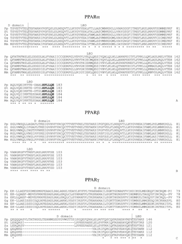
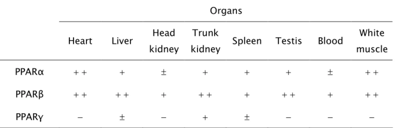
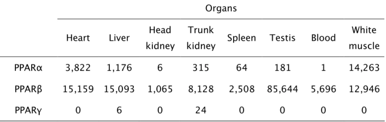
All PPAR isotypes genes were identified in brown trout through partial sequenciation and comparison with other species isotypes. For the first time, a parallel semi-quantitative and quantitative study of PPARs mRNAs was made in fish, showing the relative expression of these receptors in several organs and also their hepatic expression along the year in both genders. Among the organs
tested, PPARα was more expressed in white muscle, heart and liver. PPARβ, the
most strongly expressed isotype, was particularly abundant in testis, heart, liver,
white muscle and trunk kidney. With a much weaker expression, PPARγ mRNA
was only detected in trunk kidney, liver and spleen. PPARα expression in females
was higher in early vitellogenesis and lower in late vitellogenesis than in all other seasons. In early vitellogenesis, its expression was higher for females than for
males. PPARβ expression in males was higher in prespawning than in the other
postspawning than in all the other seasons.
The gene encoding for catalase was identified in the same way and its organ distribution pattern established by real-time RT-PCR, as well as its seasonal hepatic expression in both genders. It was more expressed in liver and blood, followed by testis, white muscle and trunk kidney. In females, hepatic catalase expression was higher in postspawning and early vitellogenesis than in late vitellogenesis and prespawning. Concerning gender differences, higher levels of expression were observed for males in prespawning.
The kinetics of morphological and metabolic alterations induced by waterborne estradiol in hepatic peroxisomes was followed within 30 days of exposure and 15 days after cessation of hormone treatment. Both catalase and urate oxidase activities were negatively influenced by estradiol, although exhibiting distinct behaviours. Catalase responded in a faster way to the treatment and to its suspension, and partially recovered its activity with a low dosage of the estrogen receptor inhibitor ICI 182,780 in the water (ICI:estradiol ratio of 1:9 in molarity). Urate oxidase showed a slower response in both cases and its activity was not affected by the estrogen receptor inhibitor under these conditions. Variations on peroxisome morphology under the same circumstances were less pronounced. Only the relative peroxisome volumes were negatively affected and just by the end of treatment. Likewise, waterborne ICI in this concentration did not have a significant effect on these parameters.
Both PPARα and catalase expressions in female brown trout liver followed the
same annual variation patterns as morphological and biochemical peroxisomal parameters, as well as an inverse pattern relatively to plasma estradiol levels previously determined. The effect of estradiol supply for a month on these peroxisomal enzymes of juveniles was similar to the effect of endogenous hormones on the same enzymes of late vitellogenic and prespawning mature females. These findings supported the baseline hypothesis that trout hepatic peroxisomes are directly or indirectly regulated by a mechanism involving
estradiol and further suggest that the estrogen receptor and PPARα play a role in
Embora a importância dos peroxisomas nas plantas, nos fungos e nos protozoários tenha sido desde cedo reconhecida, o reconhecimento da sua relevância no metabolismo celular dos mamíferos deveu-se, sobretudo, à descoberta de uma classe de doenças humanas hereditárias severas causadas por deficiências a nível peroxisomal. Também a constatação de que muitos fármacos e compostos industriais são capazes de induzir a proliferação destes organelos, e de que o tratamento prolongado de roedores com a maioria dos proliferadores peroxissomais origina tumores hepáticos malignos tem atraído investigadores. Por outro lado, o estudo de peroxissomas de animais aquáticos tem merecido maior atenção nos últimos anos devido às evidências que apontam para uma relação entre os poluentes ambientais e os fenómenos de proliferação peroxissomal. No entanto, a informação existente sobre peroxissomas e PPARs (receptores activados por proliferadores peroxissomais) de peixes é ainda limitada. Na sequência de estudos anteriores, que mostraram que as fêmeas de
truta fário (Salmo trutta f. fario) sofrem alterações sazonais nos peroxissomas
hepáticos, o objectivo desta tese foi contribuir para uma melhor compreensão dos hipotéticos mecanismos de regulação dos peroxissomas de peixes por compostos estrogénicos, incluindo uma primeira aproximação ao estudo do envolvimento dos PPARs e do receptor de estrogénios no processo. Este trabalho compreende uma componente molecular, abrangendo um estudo genético dos PPARs e de enzimas peroxissomais, e uma componente morfológica e bioquímica, direccionada para parâmetros morfofuncionais dos peroxisomas.
Os genes dos três isotipos de PPARs foram identificados na truta fário por sequenciação parcial e comparação com os isotipos de outras espécies. Foi, pela primeira vez, efectuado em peixes um estudo simultaneamente semi-quantitativo e quantitativo de mRNAs de PPARs, revelando a expressão relativa destes receptores em vários órgãos e ainda a sua expressão hepática ao longo do ano
em ambos os sexos. Entre os órgãos analisados, o PPARα mostrou maior
expressão no músculo branco, coração e fígado. O PPARβ, o isotipo com maior
expressão, revelou-se particularmente abundante no testículo, coração, fígado, músculo branco e rim posterior. Com uma expressão bastante mais fraca, o
mRNA do PPARγ foi apenas detectado no rim posterior, fígado e baço. A
da vitelogénese, a sua expressão foi maior nas fêmeas do que nos machos. A
expressão do PPARβ nos machos foi mais elevada na pré-postura do que em
todas as outras estações. Nas fêmeas, o PPARγ teve maior expressão durante a
pós-postura do que durante a vitelogénese avançada e a pré-postura. Em relação aos machos, a sua expressão mostrou-se mais elevada na pós-postura do que em todas as outras estações.
O gene da catalase foi igualmente identificado e o seu padrão de expressão nos órgãos estabelecido através de RT-PCR em tempo real, assim como a sua expressão hepática sazonal em ambos os sexos. A sua expressão revelou-se mais elevada no fígado e no sangue, seguida pelo testículo, músculo branco e rim posterior. Nas fêmeas, a expressão hepática da catalase foi mais elevada na pós-postura e no início da vitelogénese do que na vitelogénese avançada e na pré-postura. Diferenças entre sexos foram notadas durante a pré-postura, com níveis mais elevados para os machos.
A cinética das alterações morfológicas e metabólicas induzidas por estradiol exógeno nos peroxissomas hepáticos foi seguida durante 30 dias de exposição à água contendo a hormona e ainda 15 dias após a suspensão do tratamento. A catalase e a urato oxidase mostraram-se negativamente influenciadas pelo estradiol, embora exibindo comportamentos distintos. A catalase reagiu de modo mais rápido ao tratamento, bem como à sua suspensão, tendo recuperado parcialmente a sua actividade com a aplicação de uma concentração baixa do inibidor do receptor de estrogénios ICI 182,780 na água (razão ICI:estradiol de 1:9 em molaridade). A urato oxidase mostrou uma resposta mais lenta em ambos os casos e a sua actividade não foi afectada pelo inibidor, nestas condições. Nas mesmas condições, as variações na morfologia dos peroxissomas foram menos pronunciadas. Apenas os volumes peroxissomais relativos foram negativamente afectados e só no fim do tratamento. Do mesmo modo, o inibidor administrado na água nesta concentração não exerceu um efeito notório nestes parâmetros.
A expressão do PPARα e da catalase hepáticos nas fêmeas de truta fário seguiram
apoiam a hipótese inicial de que os peroxissomas de truta são regulados directa– ou indirectamente por um mecanismo envolvendo estradiol e sugerem ainda que
o receptor de estrogénios e o PPARα desempenham um papel no processo. Os
Bien que l'importance des peroxisomas dans les plantes, les fongus et les protozoaires a été depuis tôt reconnue, la reconnaissance de son importance dans le métabolisme cellulaire des mammifères se soit due, surtout, à la découverte d'une classe de maladies humaines héréditaires sévères causées par des insuffisances à niveau peroxysomal. Aussi la constatation que beaucoup de médicaments et composés industriels sont capables d'induire la prolifération de ces organites, et que le traitement prolongé des rongeurs avec la majorité de proliférateurs des peroxysomes donne lieu à des tumeurs hépatiques malignes a attiré les investigateurs. Par l’autre sens, l’étude des peroxysomes chez les animaux aquatiques mérite une plus forte attention ces derniers années, surtout, dû au fait des évidences suggérant un rapport entre les polluants environnementaux et la prolifération peroxysomal. Cependant, l’information sur les peroxysomes et les PPARs (récepteurs activés par les proliférateurs des peroxysomes) chez les poissons est encore limitée. A la suite des études
précédentes, montrant que chez les femelles de la truite fario (Salmo trutta f. fario)
les peroxysomes hépatiques souffrent des variations saisonnières, l´objectif de cette étude était de meilleur comprendre les mécanismes de l’hypothétique régulation des peroxysomes par les composés œstrogéniques chez les poissons, y compris une première approches à l’étude de l’engagement des PPARs et du récepteur de l’œstrogène dans ce processus. Ce travail comprend un abordage moléculaire par une étude génétique des PPARs et des enzymes peroxysomales et un abordage morphologique et biochimique vers les paramètres morpho-fonctionnels des peroxysomes.
Les gènes des trois isotypes de PPARs furent identifies chez la truite fario par une séquenciation partielle et comparaison avec les isotypes d’autres espèces. Une étude semi-quantitatif et quantitatif simultané des mRNAs des PPARs montrant l’expression relative de ces récepteurs dans plusieurs organes et aussi leur expression hépatique au cours de l’année chez les deux sexes, a été réalisée par la
première fois chez les poissons. Parmi les organes analysés, le PPARα a montré
une plus forte expression dans le muscle blanc, le coeur et le foie. Le PPARβ,
isotype le plus exprimé, c’est montré particulièrement abondant dans le testicule, le coeur, le foie, le muscle blanc et le rein postérieur. Montrant une expression
élevée pendant la pré-vitellogenèse et plus faible pendant la vitellogenèse avancée que dans les autres périodes. Pendant la pré-vitellogenèse son expression était
plus forte chez les femelles que chez les mâles. L’expression du PPARβ chez les
mâles était plus forte pendant la pré-ponte que ailleurs. Chez les femelles le PPARγ
était plus fort pendant la post-ponte que pendant la vitellogenèse avancée ou la pré-ponte. En ce qui concerne les mâles son expression c’est révélé plus forte pendant la post-ponte que ailleurs.
Le gène de la catalase fût aussi identifié et son sa forme d’expression dans les organes établi à partir de RT-PCR en temps réel, aussi bien que l’expression hépatique saisonnière chez les deux genres. Leur expression était plus forte dans le foie et le sang, moins dans le testicule, muscle blanc et rein postérieur. Chez les femelles l’expression hépatique de la catalase était plus forte dans la post-ponte et pré-vitellogenèse que dans la vitellogenèse avancée et la pré-ponte. Des différences entre genres furent observées pendant la pré-ponte, aux niveaux plus élevés chez les mâles.
La cinétique des modifications morphologiques et métaboliques induites par l’estradiol exogène dans les peroxysomes hépatiques fût suivie pendant les 30 jours d’exposition à l’eau contenant l’hormone et aussi pendant 15 jours après l’arrêt du traitement. Les activités de la catalase et de l’urato oxydase étaient négativement influées par l’estradiol, mais à des comportements distingués. La catalase a réagit de façon plus rapide au traitement et aussi à son stoppage, ayant récupéré partiellement son activité lorsqu’on applique une petite concentration de l’inhibiteur du récepteur d’estrogènes ICI 182,780 dans l’eau (à une raison de ICI:estradiol de 1:9 en molarité). L’urato oxydase a montré une réponse plus lente dans les deux cas et son activité n’a pas été affectée par le ICI, dans ces conditions. Les variations morphologiques des peroxysomes étaient beaucoup moins évidentes, dans ces mêmes conditions. Les volumes peroxymales relatifs furent les seuls négativement affectés et rien qu’à la fin du traitement. De même, le ICI ajouté à l’eau dans cette concentration n’a pas exercé un effet évident dans ces paramètres.
Les modèles de variations annuelles définies pour l’expression du PPARα et de la
enzymes peroxymales des juvéniles fût ressemblant à l’effet des hormones endogènes dans les mêmes enzymes observable chez les femelles en vitellogenèse avancée et en pré-ponte. Ces découvertes soutiennent l’hypothèse initiale dont les peroxysomes de la truite sont régulés direct ou indirectement par un mécanisme impliquant l’estradiol et suggèrent aussi que le récepteur des estrogènes et le
PPARα jouent un rôle dans le processus. Les normes saisonnières de l'expression
cDNA – complementary DNA
CoA – coenzyme A
DAB – 3,3’-diaminobenzidine
DNA – deoxyribonucleic acid
DNase – deoxyribonuclease
dNTP – deoxyribonucleotide
ELISA – enzime-lynked immunosorbent assay
FAD – flavin adenine dinucleotide
LBD – ligand binding domain
mRNA – messenger RNA
PCR – polymerase chain reaction
PPAR – peroxisome proliferator activated receptor
PPRE – peroxisome proliferator response element
RNA – ribonucleic acid
ROS – reactive oxygen species
RT-PCR – reverse transcription PCR
RXR – retinoid X receptor
SD – standard deviation
1.1. The brown trout
1.1.1. Life cycle
Depending on their natural habitat and life cycle, Salmo trutta species can be
found in distinct populations: the migratory trout, Salmo trutta morpha trutta or
sea trout, which divides the life period between rivers and seas, and the
freshwater trout or brown trout, which comprises Salmo trutta morpha fario and
Salmo trutta morpha lacustris, and spend all their lives in rivers and lakes, respectively (Elliott, 1994; Watson, 1999). Brown trout is widely distributed across the world, especially in Europe, from where it derived. It is also known as spotted trout because of its little coloured spots. The brown trout specimens used in this
study derived from the Portuguese freshwater resident Salmo trutta morpha fario
(or Salmo trutta f. fario) population, inhabiting the north region of the country. Its main economic interest is related to tourism, especially recreational fishing, and its culture has been assumed essential to the conservation of wild populations
(Laikre et al., 1999).
Brown trout prefer cool, unpolluted and well oxygenated streams with a swift flow in a stone and gravel bottom. In proper growth conditions, which include low population density, good food availability and an optimal temperature range from 13º to 18º C, they grow continuously with age. Depending on the habitat, a 4-year-old animal may weigh from 20 g to 1 Kg. Although they can live up to about
20 years, most of the specimens dye much younger (Reviewed by Klemetsen et
al., 2003).
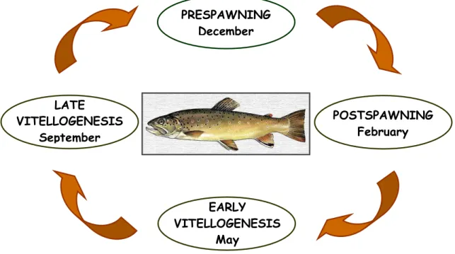
These trouts became sexually mature at an age between 1 and 10 years, which is indicated, in males, by the development of a hooknose. Spawning occurs during the following year, with females depositing about 3,000 eggs per kg in more than one digged and gravel covered nest. One large male usually fertilises the majority
of the eggs, after a strong competition (Klemetsen et al., 2003). Although minor
variations may occur, influenced by factors such as food availability, temperature and photoperiod, the major periods of the natural breeding cycle of this species are classified as follows (Fig.1): postspawning in late Winter, early vitellogenesis in Spring, late vitellogenesis in Autumn and prespawning in early Winter (Selman
Fig. 1 – The Portuguese brown troutbreeding cycle.
As sentinels of environmental pollution disturbances, aquatic species have been gaining attention in recent years, but the current knowledge on fish peroxisomes is yet restricted, which led us to chose a fish as a biological model to our study. Besides its economic interest and the fact of being a native species, the optimum handable size of the specimens, the not-too-long generation time and the availability to acquire good health status fishes, has made brown trout a good option for this study.
1.1.2. Liver metabolism and morphology
The liver can be considered the major gland of the complex organisms, having a vital role in the integration of diverse physiological and biochemical functions. This organ is involved in the metabolism and excretion of many compounds, including xenobiotics, digestion, accumulation of storage material, and also production of the yolk proteins. In this way, liver developed a peculiar morphology, with interrelating stromal and parenchymal components made of different cell types, all strategically positioned and organized. Since the middle of the late century, fish hepatic histology and cytology have been the subject of
LATE
VITELLOGENESIS September
POSTSPAWNING February PRESPAWNING
December
EARLY VITELLOGENESIS
numerous works (Elias and Bengelsdorf, 1952; Hampton et al., 1985; Beresford and Henninger, 1986; Hinton and Couch, 1998) and a crescent focus of interest.
Compared to the traditionally lobulated mammalian liver, fish liver has a somewhat different arrangement, often with no easily distinguishable afferent
versus efferent veins. Moreover, its structure is also variable from one fish species to another, even within the same family (Reviewed by Rocha and Monteiro, 1999). Over the years, several studies on salmonids liver structure and
ultrastructure have been undertaken. Data on the chum salmon, Oncorhynchus
keta, (Takahashi et al., 1977), coho salmon, Oncorhynchus kisutch, (Leatherland,
1982), rainbow trout, Oncorhynchus mykiss, (Chapman, 1981; Schär et al., 1985;
Hinton and Laurén, 1989), Sevan trout, Salmo ischchan gegarkuni Kessel,
(Kalashnikova and Kadilov, 1991) and Atlantic salmon, Salmo salar, (Robertson
and Bradley, 1991; 1992) are available. More recently, a great deal of information
on brown trout liver has also been released (Rocha et al., 1994a; 1994b; 1995;
1996; 1997; 1999; 2001a).
In brown trout liver, veins, arterioles and bile ducts can be observed randomly dispersed throughout the parenchyma. Additionally to these isolated elements, a three-dimensional engagement of venous-biliary-arteriolar tracts, venous-biliary tracts, venous-arteriolar tracts and biliary-arteriolar tracts is also present, as shown by serial sectioning analysis. The parenchyma is composed of branched, anastomosing and distorted tubular-like arrangements of the hepatocytes, as seen in Fig. 2, with segments of the biliary system as the axis of the “tubules”. A
network of sinusoidal capillaries encircles these structural units (Rocha et al.,
1995) whose exact three-dimensional structure is still under scrutiny.
Due to its metabolic functions, the hepatocyte is organelle rich and highly
organised, showing an apical biliar zone and a basal vascular region (Schramm et
al., 1998; Rocha and Monteiro, 1999). The centrally located spherical nucleus is
Fig. 2 – Brown trout liver semithin epoxy section, showing hepatocytes often in clear tubular associations, and surrounded by capillaries (C). E - erythrocytes. Fig. 3 – The hepatocytic ultrastructure. N – nucleus, Nu – nucleolus, RER – rough endoplasmic reticulum, Mi – mitochondria, Li – lipids, Di – dictyosome, Gl – glycogen.
3
N
Nu
Li
Mi
RER
Gl
Di
C
C
C
E
2
1
Although maintaining its general structure and components, liver is a pretty much dynamic organ, which can undergo morphological transformations to cope with metabolic demands. The relative liver weight of female salmonids varies along the annual breeding cycle, reaching highest values during late vitellogenesis
(Takahashi et al., 1977; van Bohemen et al., 1981). Stereological studies on
brown trout liver, in both genders and also along the year, were exhaustively
made (Rocha et al., 1997; 1999; 2001a). These works focused on liver
histological components, on hepatocytes themselves and also on hepatocytic organelles and structures, regarding their relative and absolute numbers, volumes and surfaces.
Because of the importance of peroxisomes in an array of biological processes, including the relationship with the development of hepatic tumours (Reddy and Lalwani, 1983) and the response to environmental water pollutants (Krishnakumar and Casillas, 1995), a special emphasis has been given to the study and quantification of this organelle, not only in the liver of healthy brown
trout (Rocha et al., 1999), but also in other fishes living under chemical pollution
conditions (Scarano et al., 1994; Oulmi and Braunbeck, 1996; Zahn et al., 1996).
1.2. About peroxisomes
1.2.1. Discovery, morphology and biogenesis
Peroxisomes were discovered and described for the first time in 1954, when Rodhin was observing mouse kidney proximal tubule cells at electron microscope level, during his PhD studies (Rodhin, 1954). Without any clue about their biochemical functions at that time, these new organelles were given the name “microbodies”. More than a decade later, de Duve and Baudhuin adopted the designation “peroxisome” for this hydrogen peroxide producer and catalase containing organelle (de Duve and Baudhuin, 1966). Due to their important metabolic functions, peroxisomes are ubiquitous in eukaryotic cells and have been described in many different kinds of species since then. These include mammals (Hruban and Rechcigl, 1969), birds (Shnitka, 1966), reptiles (Hruban and Rechcigl, 1969), amphibians (Hruban and Rechcigl, 1969), fishes (Veenhuis
(Lobo-da-Cunha, 1995), insects (St. Jules et al., 1989), fungi (Maxwell et al., 1975), protozoans (Lobo-da Cunha and Azevedo, 1993) and plants (Huang, 1983), among others. Sometimes a specific designation is used rather than peroxisome. It is the case of the “glyoxisome”, found in germinating seeds (Breidenbach and Beevers, 1967; Cooper and Beevers, 1969) and the “glycosome”, a related structure present in trypanosomes (Opperdoes and Borst, 1977; Hart and Opperdoes, 1984).
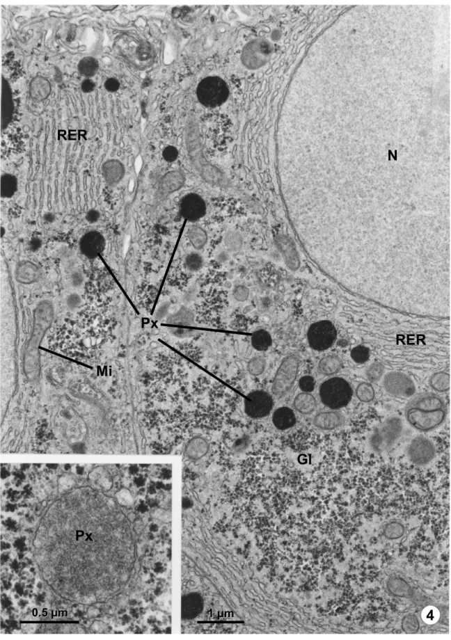
The development of the cytochemical technique based on the peroxidatic reaction of the peroxisomal enzyme catalase using 3,3’-diaminobenzidine (DAB) and hydrogen peroxide as substrate (Novikoff and Goldfischer, 1969) made it possible to unequivocally identify these organelles (Figs. 4 and 7). They are single membrane bounded, usually spherical or oval shaped, with a fine granular matrix, slightly denser than the cytosol (Fig. 4, inset). The peroxisomal approximate diameter – ranging from 0.05 to 3.0 µm – and its number may vary considerably depending on cell type, species, gender, metabolic conditions and stage of development. The rat hepatocytes are among the most peroxisome-rich cells, where they occupy about 1.5% of total cell volume (Beier and Fahimi, 1987).
Crystallized inclusions, named nucleoids or crystalloids, can be easily observed by conventional transmission electron microscopy in the peroxisomes of some species (Figs. 5 - 7). Peroxisome subfractionation (Hruban and Swift, 1964; Lata
et al., 1977) and immunocytochemistry (Usuda et al., 1988b) studies on the chemical nature of these crystalloids described them as being urate oxidase, a peroxisomal enzyme, in rat hepatocytes. In agreement with these results, it was verified that some peroxisomes with no nucleoid do not contain urate oxidase. This is the case of human tissues (Goldfischer and Reddy, 1984) and rat kidney
(Usuda et al., 1988a; Beard, 1990) peroxisomes. However, in fishes this enzyme
has been detected in hepatic peroxisomes with no visible nucleoids, in which it
behaves like a peroxisomal matrix soluble enzyme (Noguchi et al., 1979). On the
other way, reported nucleoids are not necessarily constituted by urate oxidase. One example of that is the formation of large crystalloids of alcohol oxidase
during the exponential growth of the yeast Hansenula polymorpha in methanol
(Veenhuis et al., 1981). Another peroxisomal enzyme – xanthine oxidase – was
also associated with rodent liver crystalloids (Angermüller et al., 1987). Although
appearing in peroxisomes of a great number of species, nucleoids are rarely
Fig. 4 – Ultrathin section of an S. trutta hepatocyte, with peroxisomes (Px) stained after DAB reaction for the detection of catalase, in the proximity of mitochondria (Mi), rough endoplasmic reticulum (RER) and glycogen (Gl). N – nucleus. Inset - Unstained peroxisome (Px) presenting a fine granular matrix with no visible nucleoid.
4
Px
Px
Mi
RER
RER
Gl
N
1
1µµmm 0
Figs. 5, 6, 7 – Gibbula umbilicalis digestive gland basophilic cells in ultrathin sections. Fig. 5 – Numerous peroxisomes (Px) are seen close to mitochondria (Mi), lipofuscin granules (L) and endoplasmic reticulum cisternae (arrows). Fig. 6 – These peroxisomes show a fine granular matrix (Ma) and a nucleoid (asterisk) with evident hexagonal crystalline structure. Fig. 7 – The product of DAB reaction accumulates in the peroxisomal matrix (Ma) but not in the nucleoid (asterisk). An endoplasmic reticulum cistern (arrows) is located at the peroxisome vicinity.
5
6
7
1 µm
1
1µµmm
0
Another kind of inclusion is sometimes seen strictly associated with the peroxisomal membrane, conferring an angular shape to the organelle (Gorgas
and Zaar, 1984; Zaar et al., 1991). These electron dense crystalline formations,
known as marginal plates, are usually flat or slightly curved and were identified as
L-α-hydroxyacid oxidase B (Zaar et al., 1991; Yokota and Hashimoto, 1995).
Some peroxisomes, like the rhesus monkey renal peroxisomes (Tisher et al.,
1968), present two marginal plates, appearing elongated and narrow. In the kidney cortex of beef and sheep (Zaar and Fahimi, 1991), the existence of multiple marginal plates was reported. Although mainly observed in renal cells, these structures are also seen in other cell types, such as glial tumour and sebaceous gland cells (Sima, 1980; Gorgas and Zaar, 1984).
Peroxisomes have frequently been observed in strict association with the endoplasmic reticulum. In 1964, Novikoff and Shin reported the existence of structural continuities between both organelles in rat liver, a feature that was later described in several tissues and species (Novikoff and Novikoff, 1972; Reddy and Svoboda, 1973; Zaar and Gorgas, 1985). Ever since these observations were reported, an attempt has been made by many investigators to establish if the peroxisomes originate from the endoplasmic reticulum. Indeed, morphological analysis of numerous electron microscopy pictures in several species suggested an endoplasmic origin of peroxisomes, a theory mainly defended by the above mentioned investigators, even after reports of cytochemical and biochemical studies pointing towards an opposite idea. Fahimi demonstrated, in a first approach, that rat hepatocytes endoplasmic reticulum lumen was negative to catalase activity (Fahimi, 1969). Subsequent studies based on serial sectioning analysis and cytochemical reactions indicated that peroxisomes and endoplasmic reticulum were distinct organelles, with catalase localized inside the former and
peroxidase inside the latter (Fahimi et al., 1976). This result was later
corroborated by the cytochemical localization of glucose-6-phosphatase activity in the endoplasmic reticulum and not in membrane structures associated with peroxisomes (Shio and Lazarow, 1981). Biochemical techniques reinforced this theory by proving that catalase and urate oxidase are synthesised in free ribosomes (Goldman and Blobel, 1978) and that catalase is translocated to peroxisomes without N-terminal cleavage (Robbi and Lazarow, 1982). Posterior studies led to the same conclusions for the remaining peroxisomal proteins,
either matrix or membrane proteins (Ozasa et al., 1983; Fujiki et al., 1984; Miura
1986; Köster et al., 1986; Suzuki et al., 1987; Imanaka et al., 1996). This theory is also supported by numerous ultrastructural studies in rat, fungi, yeast and crab, in which peroxisome matrix density maturation, peroxisome membrane
evaginations and peroxisome division were observed (Tsukada et al., 1968; Legg
and Wood, 1970; Osumi and Fukuzumi, 1975; Fahimi et al., 1993a; 1993b;
Lobo-da-Cunha, 1995). According to a model established for rat hepatocytes, peroxisomes form membrane loops (which were previously mistaken with endoplasmic reticulum extensions) capable of incorporating peroxisomal membrane and matrix proteins. These new compartments finally separate,
continuing to grow afterwards (Fahimi et al., 1993a; 1993b). Peroxisomal
membrane loops were also observed in brown trout hepatocytes, clearly
connected with the organelle (Rocha et al., 1999). In this way, the theory that
peroxisomes develop from fission of pre-existing ones, subsequently importing the necessary peroxisomal proteins, and implying at least one original peroxisome in each new cell after mitosis, has gained acceptation.
New controversy about this issue was shed after recent reports in both yeast and mammals of the appearance of new peroxisomes in cells which lacked these
organelles (Matsuzono et al., 1999; South and Gould, 1999; Sacksteder and
Gould, 2000) and the finding that some yeast peroxisomal membrane proteins
are only found in endoplasmic reticulum and peroxisomes (Titorenko et al., 1997;
Titorenko and Rachubinski, 2001; Faber et al., 2002). Hence, one of the current
believes is in the direction of a semi-autonomous origin of peroxisomes, with a contribution of the endoplasmic reticulum on their membrane formation (Geuze
et al., 2003). In this line, a rather revolutionary concept about peroxisome biogenesis was recently proposed, in which a group of peroxins enters the endoplasmic reticulum, concentrates in special regions and finally captures a piece of its membrane, banishing the resident proteins. The new compartment is
then detached to form pre-peroxisomes that undergo maturation (Tabak et al.,
2006). Despite being a conciliatory answer to an ancient question, this theory raises new ones. For instance, the mechanisms by which peroxins are directioned to the endoplasmic reticulum or how small vesicles are released from it.
Later on, this peroxisomal reticulum was also observed in vivo in cultured cells by fluorescent microscopy and shown to have a dynamic behaviour, with
peroxisomes transiently linked to each other (Schrader et al., 2000).
1.2.2. Metabolic functions
Since the first time an inherited disease was associated to a peroxisomal
dysfunction (Goldfischer et al., 1973), peroxisomes received a great deal of
attention in terms of metabolic studies. In mammals, more than 60 peroxisomal enzymes involved in important metabolic pathways (Fig. 8) were already identified (Reviewed by Singh, 1997).
Fig. 8 – Major metabolic pathways occurring in mammalian hepatic peroxisomes. The β -oxidation enzymes are shown in green. Shortened fatty acids can be further oxidized in mitochondria or used as substrates for the biosynthesis of ether glycerolipids in the smooth endoplasmic reticulum. Acetyl-CoA can enter the mevalonate pathway and give rise to dolichol, cholesterol and bile acids. Adapted from Fahimi et al., 1993a.
Dolichol
Acyl-CoA
oxidase Enoyl-CoA hydratase 3-Hydroxyacyl-CoA dehydrogenase 3-Ketoacyl-CoA thiolase
R-CH2-CH2-C-SCoA
=O
R-CH-CH-C-SCoA
=O
R-CH-CH2-C-SCoA
= O
OH
- R-CH-CH2-C-SCoA
= O
O
=
CH3-C-SCoA
=O HMG-CoA reductase Farnesyl pyrophosphate Cholesterol Bile acids
O2 H2O2 Catalase H2O + ½ O2
O2 O2
O2
D-Amin oacid o
xidase Urate oxidase α-Hy droxy acid oxida se
FAD FADH2
α-Ketoacid α-Hydroxyacid Uric acid Allantoin Ketoacid D-Aminoacid
H2O NAD+ NADH/H+ CoA
R-C-SCoA =O CoA DHAP Acyl-DHAP O-Alkyl-DHAP O-Alkyl-glycerol-3-phosphate DHAP-acyl transferase O-Alkyl-DHAP synthase O-Alkyl-DHAP oxidoreductase
Ether glycerolipid synthesis
CYTOSOL
SMOOTH ENDOPLASMIC RETICULUM
PEROXISOME M I T O C H O N D R I A Dolichol Acyl-CoA
oxidase Enoyl-CoA hydratase 3-Hydroxyacyl-CoA dehydrogenase 3-Ketoacyl-CoA thiolase
R-CH2-CH2-C-SCoA
=O
R-CH-CH-C-SCoA
=O
R-CH-CH2-C-SCoA
= O
OH
- R-CH-CH2-C-SCoA
= O
O
=
CH3-C-SCoA
=O HMG-CoA reductase Farnesyl pyrophosphate Cholesterol Bile acids
O2 H2O2 Catalase H2O + ½ O2
O2 O2
O2
D-Amin oacid o
xidase Urate oxidase α-Hy droxy acid oxida se
FAD FADH2
α-Ketoacid α-Hydroxyacid Uric acid Allantoin Ketoacid D-Aminoacid
H2O NAD+ NADH/H+ CoA
R-C-SCoA =O CoA DHAP Acyl-DHAP O-Alkyl-DHAP O-Alkyl-glycerol-3-phosphate DHAP-acyl transferase O-Alkyl-DHAP synthase O-Alkyl-DHAP oxidoreductase
Ether glycerolipid synthesis
CYTOSOL
SMOOTH ENDOPLASMIC RETICULUM
In animal cells, β-oxidation of fatty acids occurs in peroxisomes, as well as in mitochondria (Mannaerts and Van Velhoven, 1993). However, despite the similarity of the mechanisms in these organelles, there are significant differences between the two systems, mainly concerning the intervenient enzymes and the
nature of substrates. It is now clearly established that mitochondria are able to β
-oxidize medium and long chain fatty acids but the initial steps of the β-oxidation
of very long chain fatty acids and polyunsaturated fatty acids take place
preferentially in peroxisomes (Kondrup and Lazarow, 1985; Hiltunen et al., 1986;
Chance and McIntosch, 1994). It was also demonstrated that peroxisomes do not
act only as fatty acids chain-shorteners, but they also carry out the whole β
-oxidation pathway of very long chain fatty acids (>C22) in certain cell types like
hepatocytes, brain cells and skin cultured fibroblasts (Chance and McIntosch, 1994; Reddy and Mannaerts, 1994; Singh, 1997).
Fatty acids can only be β-oxidized in the form of their acyl-CoA derivatives. This
activation step is performed by different acyl-CoA synthetases, located in the peroxisomal membrane (Singh, 1992). Once activated, the acyl-CoA esters enter
the β-oxidation pathway to follow a sequence of four reactions by three different
enzymes (Fig. 8). Acyl-CoA oxidases dehydrogenate acyl-CoA esters to
2-trans-enoyl-CoA. These are hydrated to L-3-hydroxiacyl-CoA and then
dehydrogenated to 3-keto-acyl-Coa by the multifunctional enzymes acting both as enoyl-CoA hydratases and as L-3-hydroxiacyl-CoA dehydrogenases. Finally, 3-keto-CoA thiolases cut these molecules to free acetyl-CoA and an
acyl-CoA derivative two carbon atoms shorter than the original, which re-enters the β
-oxidation chain (Lazarow, 1978; Hashimoto, 1987; Reddy and Mannaerts, 1994; Singh, 1997).
In parallel with the β-oxidation of fatty acids, the chain-shortening of certain bile
acids intermediates occurs through a β-oxidation process with a distinct set of
enzymes located exclusively in peroxisomes (Kase et al., 1986; Björkhem, 1992;
Russel and Setchell, 1992). Moreover, it was recently shown that the entire β
-oxidation of 2-methyl branched-chain fatty acids, such as di- and trihydroxycholestanoic acid and pristanoic acid, is an exclusive peroxisomal task
(Ferdinandusse et al., 2001).
Peroxisomal β-oxidation is also essential for the metabolization of many other
Gregersen, 1986; Ferdinandusse et al., 2004), prostaglandins (Schepers et al.,
1988) and xenobiotics (Yamada et al., 1987; Yoshida et al., 1990).
Concerning the final metabolites and the controversial localization of some
enzymes, purine catabolism pathway (Fig. 9) is rather variable among species.
Nonetheless, some steps are known to take place in peroxisomes.
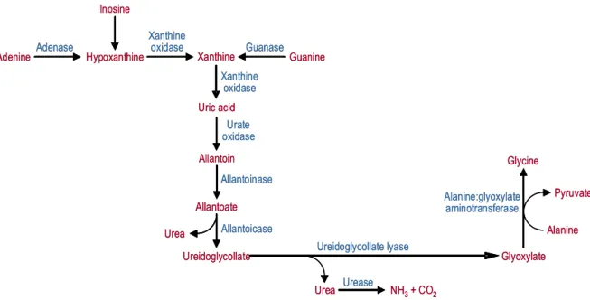
Fig. 9 – Route for the degradation of purines and reutilization of purine carbon skeletons in the liver. Adapted from Sakuraba et al., 1996.
Degradation of purines to uric acid is generally carried out in the cytosol, with few exceptions: xanthine oxidase was reported in hepatic and renal peroxisomes
of birds and amphibians (Scott et al., 1969) and rat hepatic peroxisomes
(Angermüller et al., 1987). During the evolutionary process, humans and some
hominoid primates have lost urate oxidase due to mutations in the corresponding
gene (Wu et al., 1992). In this way, uric acid is the final product of purine
degradation in these primates, as well as in birds, terrestrial reptiles, some insects and certain gastropods. Other mammals and several reptiles are able to catabolise uric acid to allantoin through a reaction catalyzed by urate oxidase
(Friedman et al., 1985; Usuda et al., 1988a). This enzyme has been largely
described in the peroxisome nucleoids of many species (Lata et al., 1977; Usuda
et al., 1988b; Usuda et al., 1994) and even in the peroxisomal matrix of some
fishes and crustaceans (Noguchi et al., 1979). Some teleost fishes and
Adenine Hypoxanthine Xanthine Guanine
Uric acid Xanthine oxidase Adenase Guanase Inosine Xanthine oxidase Glycine Glyoxylate Alanine Pyruvate Urea Allantoin Allantoate Ureidoglycollate Urate oxidase Alanine:glyoxylate aminotransferase Ureidoglycollate lyase Urea Allantoicase Allantoinase Urease
NH3+ CO2
Adenine Hypoxanthine Xanthine Guanine
Uric acid Xanthine oxidase Adenase Guanase Inosine Xanthine oxidase Glycine Glyoxylate Alanine Pyruvate Urea Allantoin Allantoate Ureidoglycollate Urate oxidase Alanine:glyoxylate aminotransferase Ureidoglycollate lyase Urea Allantoicase Allantoinase Urease
amphibians further degrade allantoin to allantoic acid through allantoinase, and allantoic acid to urea and glyoxylate by reactions catalyzed by allantoicase and
ureidoglycollate lyase, respectively (Scott et al., 1969). Although the intracellular
localization of allantoinase and allantoicase in some marine fishes and
crustaceans is attributed to peroxisomes (Noguchi et al., 1979; Hayashi et al.,
1989b), it has also been reported to be respectively mitochondrial and cytosolic
by immunocytochemical studies in frog liver and kidney (Yeldandy et al., 1995)
and, in some freshwater fishes, allantoinase is only found in cytosol (Fujiwara et
al., 1989). In marine invertebrates and crustaceans, urea is finally degraded to
NH3 and CO2 through the action of urease (Hayashi et al., 2000).
Peroxisomal enzymes are also implicated in the synthesis of cholesterol, dolichol
and other important isoprenoids playing a crucial role in the cellular homeostasis. Cholesterol is widely known as an essential constituent of cell membranes, being a major determinant of membrane fluidity. It is also a precursor of bile acids and
steroid hormones (Bloch et al., 1943; Payne and Hales, 2004). Although
ubiquitarily present in plant and animal tissues, dolichol has a more intriguing biological role. So far, it was only proven to be involved in protein glycosilation (Leloir, 1977; Burda and Aebi, 1999) and suggested to behave as a free radical
scavenger (Bizzarri et al., 2003).
Cholesterol and dolichol syntheses follow the same route until the formation of farnesyl pyrophosphate from acetyl-CoA through the mevalonate pathway (Goldstein and Brown, 1990) and then diverge (Fig. 10). This pathway is shared by cytosol, peroxisomes, endoplasmic reticulum and mitochondria (Mannaerts and
Van Velhoven, 1993; Aboushadi et al., 1999; Olivier et al., 2000). Peroxisomes
contain all the enzymes necessary for the conversion of acetyl-CoA to farnesyl pyrophosphate but the first two steps are duplicated in the cytosol and mitochondria, and the third one also occurs in the endoplasmic reticulum. The
rest of the pathway is thought to be exclusively peroxisomal (Olivier et al., 2000).
cholesterol implicated in bile acids synthesis and transport (Singh, 1997). For dolichol and other isoprenoids synthesis, farnesyl pyrophosphate is transferred to
the cytosol and further transformed (Aboushadi et al., 1999).
Fig. 10 – Current model of the subcellular compartmentalization of cholesterol biosynthesis. Conversion of acetyl-CoA to 3-hydroxy-3-methylglutaryl-CoA occurs in the cytosol, peroxisomes, and mitochondria. The further conversion of 3-hydroxy-3-methylglutaryl-CoA to mevalonate occurs both in endoplasmic reticulum and in peroxisomes. However, the conversion of mevalonate to farnesyl pyrophosphate occurs predominantly in the peroxisomes. The further metabolism of farnesyl pyrophosphate to squalene proceeds exclusively in the endoplasmic reticulum, and the final conversion of lanosterol to cholesterol occurs in the endoplasmic reticulum and may also be localized in peroxisomes. Adapted from Olivier et al., 2000.
Β-oxidation pathway
Long chain fatty acids
Acetyl-CoA Acetoacetyl-CoA HMG-CoA Mevalonate Mevalonatephosphate Mevalonatepyrophosphate Isopentenylpyrophosphate Dimethyallylpyrophosphate Geranylpyrophosphate Farnesylpyrophosphate Squalene Lanoesterol Cholesterol Acetyl-CoA Acetoacetyl-CoA HMG-CoA HMG-CoA Mevalonate Farnesylpyrophosphate Squalene Lanoesterol Cholesterol Cytosol ? Dolichol ? Acetyl-CoA Acetoacetyl-CoA HMG-CoA Mitochondria Peroxisome Endoplasmic reticulum
Β-oxidation pathway
Long chain fatty acids
Degradation of cholesterol to produce bile acids occurs in liver following two possible pathways, depending on species and enzyme availability (Chiang, 1998). The classic pathway requires the intervention of a set of enzymes located in cytosol, endoplasmic reticulum, mitochondria and peroxisomes (Pederson, 1993;
Chiang, 1998). Peroxisomes are responsible for the β-oxidation reactions that
shorten the cholesterol ring side chain of the bile acids intermediates 3α,7α
-dihydroxy-5β-cholestanoic acid and 3α,7α,12α-trihydroxy-5β-cholestanoic acid
to produce chenodeoxycholic acid and cholic acid, respectively (Kase et al., 1986;
Björkhem, 1992; Russel and Setchell, 1992; Pederson, 1993).
Among the peroxisomal pool of enzymes, stands a group of enzymes involved in
amino acids catabolism. D-Amino acid oxidase, responsible for the oxidation of the neutral and basic D-isomers, producing the corresponding ketoacids, ammonia and hydrogen peroxide, was the first to be discovered (de Duve and Baudhuin, 1966). An enzyme that oxidizes the acidic D-isomers, D-aspartate
oxidase, was also found in peroxisomes (Zaar et al., 1989), as well as L-α
-hydroxyacid oxidases A and B (Angermüller et al., 1986), alanine:glyoxylate
aminotransferase (Noguchi, 1987) and L-pipecolate oxidase (Wanders et al.,
1989). Since D-amino acids have no known metabolic meaning in mammals, the physiological roles of D-amino acid oxidase and D-aspartate oxidase remain unclear. Nevertheless, they are present in the peptidoglycans of bacterial cell walls and some results suggest that these enzymes might have a possible intervention in the catabolism of D-amino acids from the microbial intestinal
flora (Hoeprich, 1965; Konno et al., 1989). More recently, certain D-amino acids
have been found in nervous tissue from molluscs, amphibians and vertebrates, either in the free form or as residues of certain neuropeptides, where they seem
to participate in the modulation of important neuronal functions (Fujisawa et al.,
1992; Hashimoto et al., 1993; Yasuda-Kamatini et al., 1995; Zaar et al., 2002).
aspartate oxidase was suggested to be involved in the degradation of D-isomers of acidic dicarboxilic amino acids, which can behave as nonphysiological neurotransmitter ligands, and cause disturbances in the central nervous system neuronal function (Zaar, 1996). Another hypothesis was raised, suggesting that these oxidases use substrates other than D-amino acids, such as cysteamin, L-cysteine or L-cysteinylglycine, originating products possibly integrated in
intracellular messenger systems for several hormones, like insulin (Hamilton et