Instituto de Ciências Biomédicas Abel Salazar
Instituto de Investigação e Inovação em Saúde
Mestrado Integrado em Bioengenharia
Biotecnologia Molecular
Dissertação
Can osteoclast pre-conditioning enhance
osteogenic differentiation of MSC:
a 3D in vitro study with Fg scaffolds
By Ana Rita Sousa Caetano de Almeida
Supervisor:
Susana G. Santos, PhD
Co-supervisor:
Daniel Vasconcelos
iii
Acknowledgments
I would like to thank everyone that supported me during my thesis, in particular:
To Professor Mário Barbosa, for giving me the opportunity to perform my work in his group, the Microenvironments for New Therapies group, at i3S and INEB, and for often asking those hard questions that helped me improve my work.
To my supervisor Susana Santos, for giving me the chance to work on this subject, for always supporting my work, for her advice, for her patience, for always contributing to make my work better.
To my co-supervisor Daniel Vasconcelos, for the incredible patience and time spent helping me in the laboratory; for the constant advice, suggestions and technical tips, and for showing me many sides of the research world.
To Andreia Silva, José Henrique Teixeira, Maria Inês Almeida, Ana Paula Lima, João Brás and Ana Rita Ferreira, for always supporting me with a smile when I needed, sometimes without having to ask. Raquel Gonçalves, Graciosa Teixeira, Daniela Vasconcelos, Catarina Pereira, Filipa Lourenço and Maria Molinos, for their constant availability for helping me.
To my colleague and friend, Joana Rita Ferreira, for always giving me advice and help, for hearing me out whenever I needed, for transforming even the most boring chores in laughs and for improving my work with her questions.
To the whole Microenvironments for New Therapies group, for creating a good environment for working.
To all the researchers and technicians at i3S, for all their help.
To Serviço do Imunohemoterapia, at Centro Hospitalar de São João, for kindly donating the buffy coats.
To the ALM – Advanced Light Microscopy Unit, at i3S, and namely Paula Sampaio, for giving me training and helping me with all the technical problems, when performing confocal microscopy.
To CEMUP (Centro de Materiais da Universidade do Porto) and in particular Daniela Silva, for the help with the SEM.
To my sister Beatrice, for supporting when I am down, for pushing me to be better and for always caring about me.
Finally, to my parents, for all the love, support and inspiration. They already gave me more than I can repay in my whole life. Without their support this work would not be possible.
iv
To the project (NORTE-01-0145-FEDER-000012), supported by Norte Portugal Regional Operational Programme (NORTE 2020), under the PORTUGAL 2020 Partnership Agreement, through the European Regional Development Fund (ERDF) for funding.
v
Abstract
Bone tissue is a dynamic structure, constantly being remodeled to repair small injuries and maintain homeostasis. The regenerative capacity of bone may be impaired by clinical conditions such as bone disorders, and large or complex defects or fractures, which are particularly relevant in ageing populations. Research on the development of new biomaterials for bone repair tends to focus on osteoblasts, the bone forming cells, their metabolism and behavior towards other cells, drugs and implanted materials. On the other hand, osteoclasts, the bone resorbing cells, are much less explored, despite their interactions with other cells, including osteoblasts, and their remodeling activity playing crucial roles in bone repair and regeneration. As such, understanding osteoclast behavior must be one of the key factors to have into account when designing novel biomaterials for bone regeneration. Biological polymers, such as those made of proteins, have shown great potential in tissue repair and regeneration, thus the research interest in this kind of biomaterials is increasing. Fibrinogen is classically defined as a pro-inflammantory protein with pro-healing properties, and previous results have shown that it can be used in biomaterials, to promote bone regeneration.
The main aims of this work were to assess the differentiation of mature and functional osteoclasts in fibrinogen scaffolds (Fg-3D), and compare it to macrophages. Also, to evaluate their capacity to degrade Fg-3D and the effect of their paracrine factors on mesenchymal stem cells (MSC) osteogenic differentiation.
Fg-3D scaffolds were prepared by freeze-drying and characterized by scanning electron microscopy. Monocyte-derived macrophages were differentiated without addition of cytokines, while osteoclasts were differentiated in presence of RANKL and M-CSF. Cytotoxicity of Fg-3D extracts was inferred from cell morphology and survival, before cells were cultured directly on the scaffolds. Osteoclast differentiation in Fg-3D was evaluated by cell morphology and expression of enzymes cathepsin K and tartrate resistant acid phosphatase (TRAP). Conditioned media was collected along the cultures. Scaffold degradation was assessed through area measurement and D-dimer quantification. MSC were cultured on Fg-3D in presence of osteoclast or macrophage conditioned media, and osteogenic differentiation was assessed by alkaline phosphatase (ALP) production. TGF-1 was also quantified in conditioned media by ELISA.
Fg-3D extracts were found to be cytotoxic to primary macrophages and osteoclasts, while adsorbed fibrinogen was not. When cultured directly in Fg-3D cathepsin K and TRAP-positive multinucleated osteoclasts were formed. Macrophages remained largely mononucleated
vi
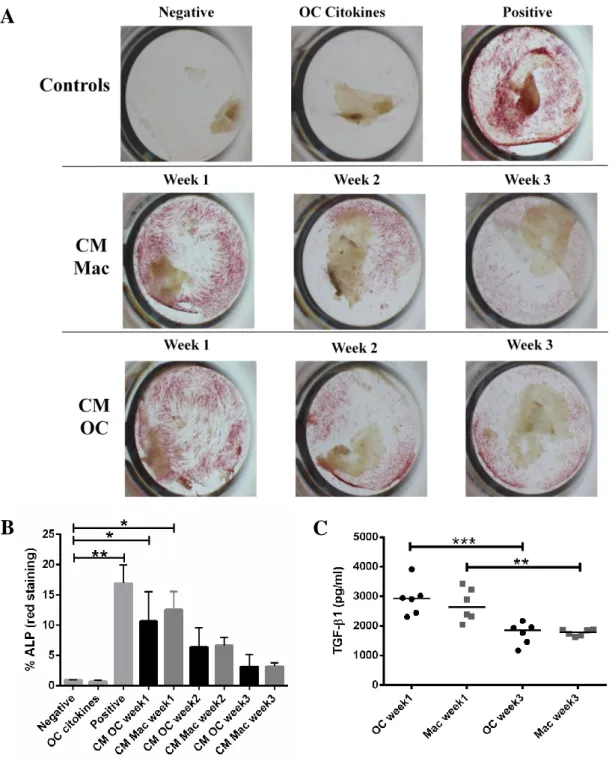
and expressed only TRAP. Although osteoclasts and macrophages were both capable of degrading Fg-3D over culture time, osteoclasts appeared more efficient. Remarkably, conditioned media from osteoclasts and macrophages at early differentiation stages promoted MSC osteogenic differentiation, to levels similar to the positive control. The levels of TGF-1 were also higher on the first week of differentiation, decreasing afterwards.
Taken together, our results suggest that both osteoclasts and macrophages are able to differentiate and are active in Fg-3D, having a positive influence on MSC osteogenic differentiation, potentially promoted by TGF-1.
vii
Resumo
O tecido ósseo tem uma estrutura dinâmica, sendo constantemente remodelado para reparar pequenas lesões e manter a homeostasia. Indivíduos saudáveis conseguem regenerar rapidamente pequenas lesões ósseas, mas doenças ósseas, assim como lesões maiores e/ou mais complexas, constituem problemas de saúde que se agravam em populações envelhecidas. A investigação sobre o desenvolvimento de novos biomateriais para reparação do osso tende a concentrar-se nos osteoblastos, as células formadoras de osso, o seu metabolismo e comportamento em relação a outras células, medicamentos e biomateriais. Por outro lado, os osteoclastos, células de reabsorção do osso, são muito menos explorados. No entanto, as suas interações com outras células, incluindo os osteoblastos, e a sua atividade de reabsorção desempenham um papel crucial em processos de reparação e regeneração óssea. Assim sendo, um melhor conhecimento do comportamento dos osteoclastos deve um dos principais fatores a ter em conta na concepção de novos biomateriais para regeneração óssea. Os polímeros biológicos, como os constituídos por proteínas, têm revelado grande potencial na reparação e regeneração dos tecidos, despertando assim o interesse crescente nestes materiais. O fibrinogénio é uma proteína pró-inflamatória, com um papel relevante na cicatrização e regeneração de tecidos. Trabalhos anteriores demonstram que o fibrinogénio pode ser usado em biomateriais, para promover a regeneração óssea.
Os principais objetivos deste trabalho foram a avaliação da diferenciação de osteoclastos funcionais em estruturas porosas de fibrinogénio (Fg-3D), em comparação com macrófagos, assim como a avaliação da sua capacidade para degradar as Fg-3D e do efeito de fatores parácrinos, secretados pelos osteoclastos, na diferenciação osteogénica de células estaminais mesenquimais (MSC).
As Fg-3D foram produzidas por liofilização e caracterizadas por microscopia eletrónica de varrimento. Macrófagos derivados de monócitos foram diferenciados sem a adição de citocinas, enquanto que os osteoclastos foram induzidos a diferenciar na presença de RANKL e M-CSF. A citotoxicidade de extratos de Fg-3D foi inferida a partir da morfologia celular e sua viabilidade, antes das células serem cultivadas diretamente em Fg-3D. A diferenciação dos osteoclatos cultivados em Fg-3D foi avaliada pela morfologia celular e pela expressão das enzimas catepsina K e fosfatase ácida resistente ao tartrato (TRAP). O meio condicionado foi recolhido ao longo da cultura. A degradação da estrutura de fibrinogénio foi determinada pela medição da área e quantificação de D-dímero. As MSC foram cultivadas em Fg-3D na presença de meios condicionados de osteoclastos e macrófagos e a sua diferenciação osteogénica foi
viii
avaliada pela produção de fosfatase alcalina (ALP). A quantidade de TGF-β1 nos meios condicionados foi determinada por ELISA.
Os extratos de Fg-3D foram citotóxicos para os osteoclastos e macrófagos primários, mas o fibrinogénio adsorvido não o foi. Quando cultivados diretamente em Fg-3D, formaram-se osteoclastos multinucleados e que expressavam catepsina K e TRAP. Os osteoclastos e os macrófagos foram capazes de degradar Fg-3D ao longo do tempo de cultura, mas os osteoclastos foram mais eficientes. Além disso, os meios condicionados de osteoclastos e macrófagos em fases de diferenciação precoces promoveram a diferenciação osteogênica das MSC em níveis semelhantes aos observados para o controlo positivo. Os níveis de TGF-1 também foram mais elevados na primeira semana de diferenciação, diminuindo ao longo do tempo de cultura.
Os resultados apresentados sugerem que tanto os osteoclastos como os macrófagos são capazes de se diferenciar e manter-se ativos quando cultivados em Fg-3D, tendo uma influência positiva na diferenciação osteogénica das MSC, provavelmente promovida por TGF-1.
ix
Index
Acknowledgments ... iii Abstract ...v Resumo ... vii List of figures ... xiList of abbreviations ... xii
1. Introduction ... 1
1.1. The bone system ... 1
1.2. Osteoblasts and bone formation... 2
1.3. Osteoclasts and bone remodeling ... 3
1.3.1. The relation between osteoclasts and macrophages ... 7
1.4. Coupling mechanisms between osteoblasts and osteoclasts ... 7
1.5. Bone diseases and therapies ... 9
1.5.1. Biomaterials for bone applications ... 12
1.5.1.1.The use of fibrinogen to promote bone regeneration ... 13
1.6. Osteoclasts in new biomaterials research ... 13
1.7. Aims ... 15
2. Materials and Methods ... 16
2.1. Production of fibrinogen 3D scaffolds (Fg-3D) ... 16
2.2. Monocytes isolation ... 16
2.3. Monocyte culture in 2D in the presence of fibrinogen or Fg-3D extracts ... 17
2.4. Monocytes seeding and differentiation into osteoclasts or macrophages on 3D fibrinogen scaffolds ... 17
2.5. MSC culture ... 17
2.6. Morphological analysis by SEM ... 18
2.7. Nuclei and cytoskeleton staining for confocal microscopy ... 18
2.8. Cathepsin K staining for confocal microscopy ... 18
2.9. Metabolic activity quantification on 3D fibrinogen scaffolds ... 19
x
2.11. TRAP staining ... 19
2.12. Measuring the scaffold area along time ... 20
2.13. MSC osteogenic differentiation on Fg-3D ... 20
2.14. TGF-β1 and D-dimer quantification... 20
2.15. Statistical analysis ... 21
3. Results ... 22
3.1. Fg-3D structure is modified by culture media ... 22
3.2. Fg-3D extracts are cytotoxic to primary macrophages and osteoclasts ... 23
3.3. Osteoclast and macrophage differentiate on Fg-3D scaffolds ... 24
3.4. Fg-3D are degraded by macrophages and osteoclasts... 26
3.5. Conditioned media from macrophages and osteoclasts differentiated in Fg-3D is capable of inducing osteoblastic differentiation ... 29
4. Discussion ... 31
5. Conclusions and future work ... 36
6. References ... 37
7. Annexes ... 46
Annex 1 ... 46
Annex 2 ... 47
xi
List of figures
Figure 1.1 The bone remodeling process. 4
Figure 1.2 Mechanism of osteoclast bone resorption. 6
Figure 1.3 Regulation os osteoclast differentiation and function by osteoblasts. 8 Figure 3.1 Structure of Fg-3D scaffolds is altered by incubation in culture media. 22 Figure 3.2 Fg extracts decrease viability of primary macrophages and osteoclasts. 23 Figure 3.3 Macrophages and osteoclasts differentiation on Fg-3D scaffolds. 25
Figure 3.4 Fg-3D scaffold degradation and cell ultrastructure. 27
Figure 3.5 Fg-3D scaffolds are degraded by macrophages and osteoclasts. 28 Figure 3.6 Conditioned media from osteoclasts and macrophages induce
osteogenic differentiation of MSC. 30
Annex 1 Cathepsin K staining is specific. 46
Annex 2 Cells adhered to Fg extract “structure”. 47
xii
List of abbreviations
3D
Three-dimensional
ALP
Alkaline phosphatase
BLM
Basolateral membrane
BMP
bone morphogenetic protein
BMU
Basic multicellular unit
BSA
Bovine Serum Albumin
CM
Conditioned media
CZ
Clear zone
DMEM
Dulbecco’s modified eagle's medium
ECM
Extracellular matrix
EDTA
Ethylenediamine tetraacetic acid
EGTA
Ethylene glycol-bis(beta-aminoethyl ether)-N,N,N',N'-tetraacetic acid
FBS
Fetal Bovine Serum
Fg
Fibrinogen
FGF
Fibroblast growth factor
HSC
Hematopoietic stem cell
IGF
Insulin-like growth factor
IL
Interleukin
M-CSF
Macrophage colony stimulating factor
M-CSFR
Macrophage colony stimulating factor receptor
MMP
Matrix metalloproteinase
MSC
Mesenchymal stem cell
NF-κB
Nuclear factor-kapa B
OCIF
Osteoclastogenesis inhibitory factor
OPG
Osteoprotegerin
P/S
Penicilin/Streptomycin
PBMC
Peripheral Blood Mononucleated Cell
PBS
Phosphate Buffered Saline
xiii
PDGF
Platelet derived growth factor
PEG
Poly(ethylene glycol)
PFA
Paraformaldehyde
PTH
Parathyroid hormone
PTHr
Parathyroid hormone receptors
RANK
Receptor activator of nuclear factor kapa-B
RANKL
Receptor activator of nuclear factor kapa-B ligand
RB
Ruffled border
RPMI
Roswell Park Memorial Institute
RT
Room temperature
SEM
Scanning electron microscopy
Src
Intracellular tyrosine kinase
TGF-β
Transforming growth factor beta
TNFRSF11B
Tumor necrosis factor receptor superfamily member 11B
TNF-α
Tumor necrosis factor alfa
TRAP
Tartrate resistant alkaline phosphatase
VEGF
Vascular endothelial growth factor
1
1. Introduction
1.1. The bone system
The bone system is formed essentially by bone and cartilage tissues, which come together in articulated joints that allow freedom of movement. Bone tissue is formed by cells and extracellular matrix (ECM) which includes an organic component, composed of collagen type I (up to 95% of bone organic composition), non-collagenous proteins (such as osteocalcin, osteopontin, osteonectin and bone sialoprotein) and proteoglycans (such as biglycan and decorin), and an inorganic component, mainly of hydroxyapatite crystals, formed from calcium and phosphorus, and placed into the organic matrix. The combination of these two components grants bone its characteristic rigidity and strength, while maintaining an appropriate degree of elasticity [1].
The main function of the bone system is to provide support to the body, as the skeleton forms a rigid structure to which softer tissues are attached, while also contributing for the movement of the body by working as a lever when muscles contract. Additionally, the bone system plays important roles in protecting vital organs, like the brain or the heart and lungs, protected by the skull and the ribcage, respectively; storage of minerals such as calcium and phosphate in hydroxyapatite crystals or magnesium and sodium; growth factors storage, for example bone morphogenic proteins; and hematopoiesis or blood cell formation (white and red cells, and platelets) in the bone marrow [1, 2].
The Harversian System, also known as osteon, is the structural unit of bone. Osteons resemble an elongated cylinder parallel to the bone axis and are composed of heavily packed collagen fibrils, called the lamellae. Lamellae’s function is to withstand torsion stresses, hence its fibrils orientation [2].
Bone tissue can have different structures, properties and functions. Cortical bone, also known as compact bone, constitutes an exterior layer, has dense bone matrix with passageways for blood, lymphatic vessels and nerves. Its main functions are mechanical support and protection of trabecular bone and organs. Unlike compact bone, cancellous or trabecular bone, also known as spongy bone, has a loosely organized matrix, with pores and trabeculae. The trabecular bone is where the bone metabolic functions are carried out. Some long bones (e.g. femur) have an additional compartment, the medullary cavity, filled with bone marrow [2].
Despite its appearance, bone is not a static tissue and is continually suffering alterations by remodeling, name given to the process of replacing old bone for new bone, in order to adapt to new stimuli from the environment, such as mechanical loads or necessity of mineral
2
homeostasis [3]. The remodeling process is performed by the bone cells: osteoblasts, responsible for bone formation, and osteoclasts, involved in bone resorption [2]. The deregulation of the remodeling process gives origin to several bone related diseases [3].
In the next sub-sections the biology of bone cells and their role in the process of bone remodeling are addressed, paving the way to review bone diseases and therapies, including the use of biomaterials for bone tissue repair and regeneration.
1.2. Osteoblasts and bone formation
Osteoblasts and osteoclast, the bone cells involved in remodeling, derive from distinct lineages, and have different life cycles and functions.
Osteoblasts are derived from mesenchymal stem cells (MSC) which have the potential to differentiate in the different cells of the mesodermal tissues, like osteoblasts, adipocytes, stromal cells, myoblasts, tenocytes and chondrocytes [1, 4]. MSC undergo several chances in metabolism and phenotype during osteoblastic differentiation. This process can be divided in three phases, with cells going through different stages of differentiation. In particular, MSC differentiate into osteoprogenitor cells, then into pre-osteoblasts and finally into mature osteoblasts [5]. Their differentiation is regulated by several molecular factors, including: bone morphogenetic proteins (BMPs), platelet derived growth factor (PDGF), fibroblast growth factor (FGF), insulin-like growth factors (IGFs), transforming growth factor β (TGF-β), interleukin 1β (IL-1β), interleukin 5 (IL-5) and tumor necrosis factor α (TNF-α) [5-7]. The Wnt signaling pathways have been reported to be involved in regulating gene transcription in osteoblast progenitor cells, and specially MSC, by stabilization of β-catenin [8].
The most explored mediators of osteoblast differentiation are BPMs, in particular BMP-2, BMP-4 and BMP-7, which up-regulate the transcription factors Cfba/Run-2. These BMPs are up-regulated through the activation of the Shh and Wnt signaling pathways and act on MSC, osteoprogenitor cells and pre-osteoblasts [5]. PDGF and TGF-β are reported to have an important role at early stages, recruiting MSC to the site of differentiation and/or stimulating proliferation of the osteoblast progenitors [5, 9]. Nonetheless, TGF-β has also been reported to inhibit later differentiation and mineral deposition by mature osteoblasts [5]. Later in the differentiation process IGFs up-regulate the transcription factor osterix, leading to late stage differentiation [5], and FGF reinforces the differentiation of already committed cells into the osteoblastic lineage, and also has a role in osteoblast apoptosis [7].
Mature osteoblasts express several transcriptions factors like Runx-2, osterix, Msx-2, Dlx-5 and the AP-family [5, 10], and several phenotypic markers that are important for bone formation [11], such as bone matrix proteins, like collagen type I, osteocalcin, osteopontin and bone sialoprotein. The ALP enzyme present at the osteoblast cell membrane and involved in bone mineralization, is greatly unregulated during bone formation phases, and usually
3
constitutes a hallmark of osteoblastic differentiation [12]. Parathyroid hormone/parathyroid hormone receptors (PTH/PTHr) that regulate calcium and phosphate ions homeostasis [1, 5], are also a osteoblastic products.
The average lifespan of an osteoblast is around one month, after it either undergoes apoptosis, being replaced in its role by new functional osteoblasts, or it is embedded in the bone matrix as an osteocyte, which is considered the last phase of osteogenic differentiation [5]. Osteocytes main functions are in maintaining the bone matrix and mechanotransdution, a mechanism that allows these cells to react to external stimuli, like stress and strain stimuli, without disrupting bone homeostasis. Several factors and pathways seem to be involved in mechanotransdution, such as the Wnt/β-catenin pathway [2, 13, 14]. When there is a microcrack in the bone matrix, osteocytes signal it by undergoing apoptosis and releasing osteoclastogenesis stimulation factors, thus promoting bone remodeling [14]. Additionally osteocytes can be involved in mineralization and phosphate metabolism, as they can produce fibroblast growth factor 23 (FGF23), a protein capable of controlling the reabsorption of phosphate in the kidney [15].
1.3. Osteoclasts and bone remodeling
Osteoclasts are derived from hematopoietic stem cells (HSC) that can give rise to the myeloid and lymphoid lineages. Osteoclasts are derived from the myeloid lineage, and share a common progenitor with monocytes, that differentiate into macrophages. The commitment of this progenitor into the osteoclast precursors occurs while the cells are still in the bone marrow, in the hematopoietic niche. Osteoclast precursors are then recruited to remodeling sites of the bone, where the differentiation into mature osteoclasts and fusion into multinucleated osteoclasts occurs [3]. Osteoclast differentiation is regulated by the interaction of receptor activator of nuclear factor κ-B (RANK), which is expressed on the cell membrane of osteoclast precursors and osteoclasts, and its ligand, RANKL (receptor activator of nuclear factor κ-B ligand). This interaction is influenced by macrophage-colony-stimulating factor (M-CSF), a cytokine that promotes the expression of RANK by these cells; and osteoprotegerin (OPG) (also known as osteoclastogenesis inhibitory factor (OCIF), or tumor necrosis factor receptor superfamily member 11B (TNFRSF11B)), a member of the TNF family, that acts as a decoy receptor for RANKL, thus decreasing its binding to RANK and inhibiting osteoclastogenesis [6, 16-18].
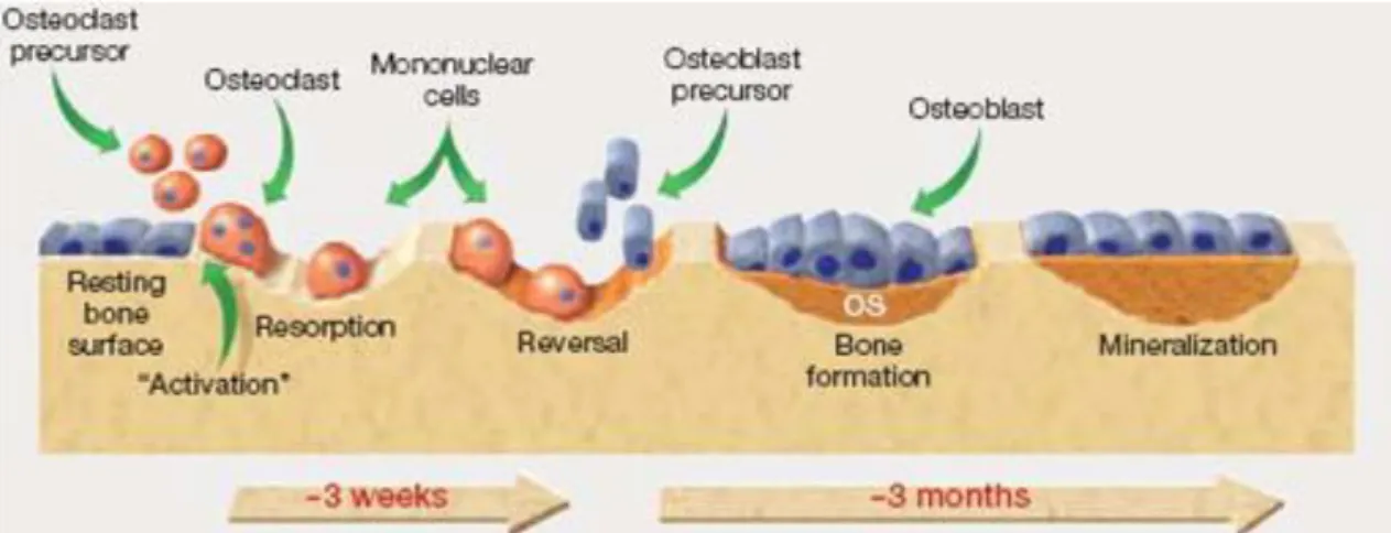
Bone homeostasis is maintained by bone resorption and formation, and there are tightly coupled mechanisms between the two, as for example inhibition of resorption suppresses bone formation [19, 20]. Approximately 5 to 25% of the bone surface is suffering remodeling at any given time [1]. The process of bone resorption takes about 3 weeks per site, while bone formation needs about 3 to 4 months (Figure 1.1) [21, 22]. Remodeling is important to repair
4
small injuries to the bone tissue, regulate the release of ions, such as calcium and phosphate [1], and to promote angiogenesis [23].
The remodeling process consists of three consecutive phases, initiation or activation phase, transition phase and termination phase [1, 24].
The initiation phase comprises the recruitment of osteoclast precursors and their differentiation into mature osteoclasts, capable of bone matrix resorption.
The transition phase occurs when osteoclasts activity ceases and coupling mechanisms recruit and activate osteoblasts so they can start to produce new bone. Osteoblasts synthesize proteins for the bone ECM to form the osteoid, an unmineralized matrix, that will later be mineralized due to the calcium binding capacity of said proteins [3].
It is considered that the bone remodeling entered the termination phase when bone formation by osteoblasts stops and the new bone is fully functional.
A group of osteoclasts and osteoblasts that cooperates to remodel bone is denominated Basic Multicellular Unit (BMU). BMUs are located at the leading edge of a cylindrical canal, in cortical bone. At one tip of the cylindrical canal, osteoclasts start bone resorption, giving rise to a tunnel, which will be filled by new bone produced by osteoblasts, with the exception of a channel in the center. This forms an osteon, or Haversian System, the structural unit of bone. In trabecular bone, BMUs are located on bone surfaces, such as the periosteum, forming a hemi-osteon [25].
To achieve bone resorption properties, osteoclasts merge together to form multinucleated cells with a large amount of mitochondria, to provide the support for the high metabolic activity required by the process. Osteoclasts also have well-developed endoplasmatic reticulum and Golgi apparatus placed around each nuclei, as well as a high number of vesicles, lysosomes and tubular lysosomes and vacuoles. The presence of this collection of organelles is necessary for energy production and protein synthesis for bone resorbing mechanisms [26].
Figure 1.1- The bone remodeling process. Recruited osteoclasts are responsible for bone resorption and later for recruitment of osteoblast, to initiate bone formation. OS – osteoid. Adapted from [22].
5
When fully active, mature osteoclasts express several characteristic molecules, such as RANK, a cell membrane receptor that activates the nuclear factor-κ B (NF-κB) pathway, activating c-jun terminal kinase and thus promoting the expression of osteoclastic proteins, leading to osteoclastogenesis [17, 18, 27]; macrophage-colony-stimulating factor receptor (M-CSFR), that binds M-CSF to up-regulate the expression of RANK [3]; integrin αvβ3, that helps the binding of the cell to the ECM [3, 28]; calcitonin receptor, which binds calcitonin, inhibiting osteoclast activity and also a unique marker of osteoblasts that helps differentiate them from macrophages [29, 30]; tartrate-resistant acid phosphatase 5b (TRAP-5b), involved in organic matrix degradation and present in the vesicles, golgi complex and ruffled border of active osteoclasts, this enzyme isoform is also sometimes used to distinguish macrophages from osteoclasts, as the formers only produce the TRAP-5a isoform and osteoclasts only express TRAP-5b [31, 32]; cathepsin K, that takes part in organic matrix degradation specially collagen I, having the same location as TRAP-5b [1]; and matrix metalloproteinase 9 (MPP-9), also involved in organic matrix degradation [1, 33].
In order to initiate bone resorption, osteoclasts must adhere to the bone matrix, through recognition of RGD sequences on bone matrix proteins, such as osteopontin and bone sialoprotein [26]. Recognition is performed by integrin αvβ3 and leads to activation of a pathway mediated by intracellular tyrosine kinase (Src). Src then acts on focal adhesion kinase Pyk2 [34] and proto-oncogene c-Cb1 [35], which contribute for the correct rearrangement of osteoclast cytoskeleton. Soriano et al reveled that Src-deficient mice are not capable of resorbing bone despite having a high number of osteoclasts [36], likely due to impaired osteoclast binding to the bone surface. The recognition by integrin αvβ3 seems to be sufficient for osteoclast activation, suggesting that matrix mineralization is not the determining factor for osteoclasts to identify their substrate [37]. In fact, a previous study demonstrated that osteoclasts can resorb untreated, unmineralized, anorganic or surface-demineralized mammalian dental tissues in culture [38], as well as “artificial” substrates like polymeric non-mineralized biomaterials [39]. On the other hand, impairment of integrin αvβ3 binding is enough to disrupt osteoclastic activity [40, 41].
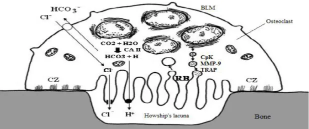
Fully active osteoclasts present well-marked cell polarity and characteristic membrane regions (Figure 1.2): clear zone (CZ), ruffled border (RB) and basolateral membrane (BLM). CZ receives its name from the fact that, on microscope images, it appears as a very light zone of the cell, due to the absence of cell organelles. The CZ cytoplasm has a network of actin filaments, concentrated on focal contacts, in a ring-like structure. The main functions of the CZ are the adhesion of the cell to the bone matrix and the total isolation of the bone resorbing compartment from the extracellular fluid and adjacent cells [26]. CZ also occasionally shows signals of receptor-mediated endocytosis, suggesting that CZ is involved in endocytosis of degraded bone matrix. Additionally, the presence of MT-MMP1 in this region implies that it
6
also has a function in the migration of the cell [42]. The CZ can be immune-identified by phalloidin staining of the actin ring [26].
The RB has as main goal the resorption of the bone matrix, by dissolution of mineral crystals and degradation of proteins. Several enzymes, produced by the complex organelle apparatus of osteoclasts, are involved in this process, and are present in vesicles in the RB [26]. Carbonic anhydrase, present in the cytoplasm, converts CO2 and H2O into HCO3
and H+ [43]. H+ is transported to the Howship’s lacuna, the space formed between the bone face and the osteoclast delimited by the RB, by active transport performed by H+ATPase in the RB membrane. The acidification of the Howship’s lacuna contributes for the dissolution of hydroxyapatite crystals and permits the action of enzymes that require acidic conditions [44], such as cathepsin K. The ionic balance in Howship’s lacuna is maintained thanks to the passive transport of Cl- through CIC-7 channels. Previous studies show that absence of either H+ATPase or CIC-7 channel is enough to impair bone resorption [45, 46]. Cathesin K, TRAP and MMP-9 are examples of enzymes responsible for the degradation of the bone organic component, all present in Howship’s lacuna. Cathesin K is an enzyme from the cysteine protease family and is capable of degrading the triple helix of collagen, the most abundant protein in bone, in acidic conditions, being helped by MMP-9 that degrades the segmented collagen fibrils [26].
It is believed that the BLM is responsible for the reception of stimulatory cues, such as cytokines. Additional functions are the communication with the osteoblasts through direct contact and secretion of factors, and transcytosis and exocytosis of degraded bone matrix [33].
Figure 1.2 - Mechanism of osteoclast bone resorption. The acidic environment of the Howship’s lacuna dissolves the inorganic component of bone, while enzymes like CpK, TRAP and MMP-9 degrade the organic component. The action mechanism is similar when degrading biomaterials instead of bone. CZ- clear zone; RB – ruffled border; BLM – basolateral membrane; CpK - Cathepsin K; MMP-9 - matrix metalloprotease 9; TRAP - tartrate-resistant acid phosphatase. Adapted from [26].
7
1.3.1. The relation between osteoclasts and macrophages
Macrophages and osteoclasts derive from the same multipotent precursor of the monocyte-macrophage lineage. So, these two cell populations share some characteristics and can be transdifferentiated into each other. In fact, osteoclasts can be differentiated in vitro from peripheral blood monocytes [47, 48]. Although their functions can differ greatly from each other, these cells display histochemical and functional similarities [49].
Macrophages are immune cells from the myeloid lineage with an important role in maintaining homeostasis, the inflammatory response, and aiding in tissue repair, though interaction with other immune cells and phagocytosis of unwanted foreign bodies or damaged cells. Macrophages are usually mononuclear cells, but in response to certain stimuli can fuse and form multinucleated cells just like osteoclasts, in an attempt to increase their phagocytic capacity [19]. Different tissues in our body contain a small population of resident macrophages that can act quickly in situations of stress or injury. However, for an efficient response the recruitment of blood circulating monocytes to the site of injury and their differentiation into mature macrophages is necessary [50].
The shared characteristics between macrophages and monocytes include expression of CD11b, CD68, TRAP (though different isoforms), MMP-9 and CD61, in slight different levels of expression [1, 31-33, 51]. Nevertheless, osteoclasts also display differences from these immune cells, such as the very low expression of MHC class molecules, CD14 and receptors for immunoglobulin Fc and complement [52, 53]. In addition, macrophages were never described to be capable of resorbing mineralized bone, while osteoclasts were never identified as antigen-presenting cells.
Recent studies suggest the existence of a myeloid population with specific functions in bone, called osteomacs. This special subset of macrophage residing in bone seems to have a phenotype with characteristics of both macrophages and osteoclasts, and seem to interact closely with the latter and osteoblasts. Absence of the cells nominated osteomacs reduced greatly mineralization and production of osteocalcin [54, 55]. Though osteomacs contribution for bone remodeling is already being studied, their interaction with biomaterials and even the bone cells is still poorly known with contradictory ideas emerging in the field [56].
1.4. Coupling mechanisms between osteoblasts and osteoclasts
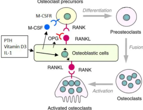
Coupling bone resorption and formation is regulated by a wide variety of molecules and signaling pathways. Osteoblasts (and osteocytes) are capable of recruiting osteoclasts and induce their differentiation (Figure 1.3) [57], and in turn, osteoclasts are able to recruit osteoblasts and induce their proliferation and differentiation.
Osteoblasts produce RANKL and M-CSF, important factores for osteoclatogenesis. Their presence is enough to induce osteoclast differentiation in vitro. Secreted M-CSF binds to
8 M-CSFR in osteoclast precursors’s membrane activating a pathway that will up-regulate the expression of RANK. Secreted RANKL acts by binding to RANK on osteoclast precursor membrane leading to pathways, mediated by NF-κB, activator protein-1 (AP-1) and nuclear factor of activated T cells c1 (NFATc1), that will end in osteoclastic differentiation [58]. The production of RANK by osteoblasts can be stimulated by tumor necrosis factor α (TNF-α), parathyroid hormone (PTH), interleukin 1 (IL-1), vitamin D3, prostaglandin E2, among some other cytokines/hormones/growth factors [1, 58], which means that these regulate osteoclast differentiation indirectly. Wnt5a signaling by osteoblasts is another way to promote osteoclastogenesis [59].
Besides producing factors that stimulate osteoclastogenesis, osteoblasts are also capable of producing one factor capable of limiting it, in order to preserve bone homeostasis, osteoprotegerin (OPG). OPG acts as a decoy receptor for RANKL, competing directly with it and so inhibiting its signaling on osteoclasts [1].
Moreover, osteocytes also regulate osteoclast differentiation, as dying osteocytes in damage sites send chemical signals to osteoblasts in order to produce more RANKL [60] and it seems that osteocytes themselves are capable of producing it [61].
Osteoclasts are, in turn, also involved in regulation of osteoblast differentiation, proliferation and function, especially during the transition phase. The degraded bone matrix releases factors such as TGF-β, IGFs and BMPs that have been shown to influence osteoblastic differentiation [1]. However, non-bone related signals produced by osteoclasts can also induce bone formation, as a previous study indicated that conditioned media from osteoclasts cultured on plastic surfaces, instead of bone surfaces, also had a significant impact on increasing mineralized matrix formation in vitro [62]. Recruitment of MSC or osteoblasts from other sites can also be promoted by osteoclasts. Osteoclasts produce PDGF-BB, suspected to have a role in osteoblasts chemotaxis [63]. Non-resorbing osteoclasts at the site of remodeling, instead of becoming active, secrete factors that will induce the up-regulation of osteogenic markers and recruitment of MSC [6]. A study by Zhao et al. showed that reverse signaling of EphrinB2, a ligand for Src receptor, in osteoclast precursors inhibited osteoclast differentiation and that forward signaling of EphB4, a receptor for Src, in osteoblasts induced osteogenic
Figure 1.3 - Regulation os osteoclast differentiation and function by osteoblasts. Osteoclast differentiation is induced by RANKL and M-CSF produced by osteoblasts. Factores like PTH, vitamin D3 and IL-1 up-regulate the production of RANKL, thus indirectly promoting osteoclast formation. Osteoblasts are also capable of decrease osteoclast differentiation and function thorugh the secretion of OPG. RANK - receptor activator of nuclear factor κ-B; RANKL – RANK ligand; M-CSF - macrophage-colony-stimulating factor; M-CSFR – M-CSF receptor; OPG - osteoprotegerin; PTH - parathyroid hormone; IL-1 – interleukin 1. Adapted from [57].
9
differentiation, proving the existence of a bidirectional signal between osteoclasts and osteoblasts [64].
Immune cells are capable of producing factors that interact with bone cells [65]. Macrophages and osteoclasts share the same precursor and some regulatory molecules (transcription factors, cytokines, membrane receptors) that are able to influence each other [20], suggesting that can exist cross-talk between these cells. A study reported that mice deficient in some immunomodulatory molecules also presented an abnormal osteoclast phenotype [66]. T-cells produce TNF-α and RANKL that up-regulate osteoclastogenesis and core binding factor 1, which is involved in osteoblast development. Because immune cells and osteoclasts share the same origin, start differentiating in the same environment and secrete molecules in the blood circulation, it is considered that they can influence each other both at the beginning of their life cycles and after differentiation [67]. Some other examples of molecules produced by immune cells that can interact with osteoclasts are IL-1, IL-4 and Interferon-γ [68]. Immune cells can also interact with osteoblasts. For example, macrophages are capable of promoting osteoblastogenesis by secretion of IL-18 [69]. As the immune system seems to have a close link with the bone system, deregulation of one of them can lead to deregulation of the other: osteopetrosis, a bone disease, can lead to decrease of the capacity to combat infection while auto-immune responses can lead to bone diseases such as arthritis rheumatoid [21].
Most studies regarding bone regeneration are focused on osteoblasts, as these are the cells responsible for new bone formation. However, considering the importance of bone resorption in bone regeneration, osteoclasts are likely as important to this process, as osteoblasts. Hence the growing number of reports studying the interaction between these two populations of cells, either by co-culture or culture with conditioned media [70]. Despite that, most studies continue to focus on the influence of osteoblasts on osteoclasts [71-74], with the opposite direction of signals, osteoclast to osteoblast [75], still being less explored.
It is necessary to uncover more about the interactions that osteoclasts have with others relevant cells, drugs and biomaterials, in order to develop more efficient new therapies to treat the increasing number of bone diseases.
1.5. Bone diseases and therapies
Bone diseases can be caused by a deregulation of the bone remodeling process, which can happen due to old age, hormonal alterations, physical activity changes, medication drugs, or as a consequence of disease. When bone resorption occurs at a higher rate than bone formation, the skeleton can present an overall fragility caused by lack of adequate structure. This can give rise to bone fractures, as a 10% decrease in bone mass is describer to double the risk of fracture [21]. The most common and widely studied bone fragility disorder is osteoporosis, which occurs most frequently in women after menopause, because of the decrease of estrogen due to the end
10
of the fertile life. Estrogen has receptors in both osteoblasts and osteoclasts and it is known to influence bone turnover. The low concentration of estrogen, along with the low concentration of some other steroids, induces an increase in the number of osteoclasts, resulting in unbalanced bone resorption. Decreased concentrations of these molecules have systemic effects, interfering with the function of other organs besides bone. So, it became common practice to give women in menopause an oral supplement of estrogen, to compensate for its absence, minimizing effects in the body, including on bone tissue preservation [76]. Osteoporosis can also be a consequence of other diseases, such as multiple myelomatosis, hyperparathyroidism or hyperthyroidism. In this case the best treatment is to treat the original condition, to eliminate the source of the problem [21].
Bisphosphonates have been used as a treatment for diseases related with excessive bone resorption for more than four decades. Bisphosphonates can inhibit osteoclast activity by entering the osteoclast and deregulating its metabolism, and potentially inducing apoptosis. It is a method that not only inhibits osteoclasts but also reduces their numbers, without being specific of any disease [77].
On the other hand, excessive bone formation can also result in disease, of which the most common example is osteopetrosis. In this condition, osteoclast failure leads to accumulation of bone, which in turns leads to complications such as reduction of bone marrow size (with a big impact on hematopoiesis, which can give rise to anemia and high susceptibility to infections) and compression of nerves and blood and lymphatic vessels (causing premature bone necrosis, infections and anemia). One of the treatments for this disease is the transplantation of HSC to both attenuate the symptoms and trying to increase osteoclast number and activity. However, this approach has a very low success rate [78].
Diseases of bone metabolism, where the activity of osteoblasts and osteoclasts is deregulated also result in bone problems. As an example, Paget’s disease is characterized by an excessive osteoclastogenesis with inherent increase in bone resorption that is followed by promotion of osteoblastic activity and bone formation, as the body’s attempt to compensate the bone loss. This ultimately results in the formation of defective enlarged bones (plexiform bone) that lacks the right structure and thus is susceptible to bowing, fractures and deformities. Paget’s disease’s cause seems to be a set of mutations that lead to excessive promotion of bone resorption caused by infection of virus from the paramyxovirus class [21, 79]. Renal osteodystrophy is another condition that leads to bone remodeling deregulation: it is a consequence of end-stage renal disease characterized by reduced bone mineralization and increased bone resorption, with implications on the whole bone system [80].
Some cancers have a tendency to form metastases in bone. Breast and prostate cancers are the ones that show a higher rate of bone metastases. In these cases, bone resorption is
11
enhanced to give the tumor more space to grow, which results in a formation of a structure that does not have the adequate characteristics to maintain bone functions [77].
Finally, inflammatory diseases can lead to bone remodeling imbalance, as expected from the large interactions that the immune cells are reported to have with bone cells. Rheumatoid arthritis is a disease where immune cells, like T lymphocytes, monocytes and macrophages become deregulated, increasing in number and secreting an excess of osteoclastogenesis stimulating factors, as the pro-inflammatory cytokine IL-17 or the osteclastogenesis promoter RANKL. The excessive osteoclastogenesis leads to augmented bone resorption that causes destruction of articular cartilage tissue and bone erosion by chemical and mechanical stresses [21].
Most treatments for bone diseases focus on the reduction of bone resorption by osteoclasts, the main common feature of most of these disorders. Along with the administration of estrogen supplements and bisphosphonates, injections of other osteoclast activity inhibitors are widely used. Calcitonin strongly inhibits bone resorption, however it can cause hormone-induced resistance, so it is not an ideal method [21, 29]. PTH administration seems to promote bone formation, so it is an alternative therapy focusing on counteract the diseases effects. Although it may seem strange that a hormone that actually promotes bone resorption is used to combat bone loss, studies show that intermittent and low doses are efficient in stimulating osteoblast activity, maybe due to cross-talk between these cells and osteoclasts [81]. The already mentioned osteoprotegerin (OPG) is another candidate for alternative therapies, since its original goal is the very inhibition of bone resorption. However, due to potential immune reaction and side effects related to repeated use, this complex protein is not one of the most promising options [21].
In 2008, a new approach was suggested by Bonnelye et al. that concluded that strontium ranelate has an influence in reducing bone resorption, by inhibiting osteoclast differentiation and function, and in stimulating bone formation, by promoting osteoblast differentiation. This means that this molecule has the potential to not only decrease the impact of excessive osteoclast activity, the cause of the problem, but also to promote healing of the damaged sites, by stimulating osteoblasts [82]. However, randomized placebo-control studies showed that this substance increased the risk of venous thromboembolism. During the post-marketing surveillance, DRESS syndrome, a severe dermatological reaction, and an increase in pulmonary embolism and myocardial infarction risks were reported. These side effects have limited the oral use of strontium ranelate to patients who do not respond to other medicines, and when the risk of bone fracture greatly overwhelms the cardio-vascular risk [83].
Many other molecules involved in the bone remodeling process have the potential to become drugs in new therapies. Both for these and for the ones already in use, directing the therapeutic molecule to the desired site instead of allowing its free circulation in the body, can
12
decrease side effects and potentiate the desired effect. Examples of approaches for directing molecules in vivo are the use of biomaterials and genetic therapies [21].
1.5.1. Biomaterials for bone applications
Extensive bone damage (e.g. non-union fractures, simple large fractures, bone defects or deformities) can arise from bone disease or trauma. While small defects or systemic conditions are usually treated using one of the therapeutic approaches discussed above, treatment of large defects usually involves surgery for providing bone or joint stabilization, or even replacement. The gold standard in bone implants is the autologous graft material (bone from the patient), as it has complete histocompatibility and all the properties of bone. Its main limitations are high donor-site morbidity and limited availability. Allografts are an alternative solution with the same bone characteristics. However, it is not completely histocompatible, is more expensive, has more processing, sterilization and storage problems, and can lead to disease transmission [84].
Nonetheless, the most used materials in orthopedic surgery are still the classic bone/joint replacement implants. Composed of metal alloys, ceramics and high density polymers, these biomaterials are designed to be “inert”, so as to avoid implant rejection. However, they present several drawbacks, like the generation of biomaterial-derived wear and corrosion debris, leading to a chronic inflammatory response, accompanied many times by the formation of a fibrous capsule around the implant. Accelerated bone resorption at the site of implant potentiated by the chronic inflammation then mostly leads to aseptic loosening and pain, requiring a revision surgery with implant substitution. This topic has been recently reviewed [85]. In order to overcome these current limitations, research is focused on the development of new biomaterials with or without pre-encapsulated cells (MSC or osteoblasts), capable of substituting or helping autologous grafts or allografts, in promoting bone regeneration [84].
In this context, biomaterials designed for bone repair should support and promote the bone remodeling process and combine good resorptivity with osteoconductivity (ability to promote vascular growth and allow free proliferation of bone cells) and osteoinductivity (ability to promote osteoblastic differentiation and new bone formation) [86-88].
New biomaterials currently being investigated for bone repair/regeneration applications include metals, ceramics, bioactive glasses, cements, composites and polymers The interaction of osteoclasts with these biomaterials was studied only to a limited extent, with most of the attention being devoted to osteoblasts [89].
13
1.5.1.1. The use of fibrinogen to promote bone regeneration
Fibrinogen (Fg) is a blood plasma protein, involved in blood coagulation. It is converted into fibrin by the action of thrombin in the presence of ionized calcium. Fg is synthetized in the liver by hepatocytes. Studies show that Fg is required for normal platelet function and wound healing [90]. Fg structure contains two RGD sequences, important for cell adhesion, as fibrinogen/fibrin forms a network in the site of injury that will act as a foundation for tissue regeneration [91]. Also, Fg promotes cytokine secretion by peripheral blood mononuclear cells (PBMC), suggesting that it can contribute to the inflammatory process [90].
It was reported that Fg may have an important role in tissue repair, by stabilizing wound fields and supporting local cell proliferation and migration of inflammatory, endothelial and stromal cells [92]. It may also have an important role in angiogenesis at injury site, as it has a great binding affinity to vascular endothelial growth factor (VEGF) [93]. Fg was previously used in tissue regeneration, for urinary tract regeneration [94] and reconstruction of blood vessels [95]. Fg or composite biomaterials containing Fg were also studied regarding cellular migration, proliferation and morphological and functional changes of several types of cells [96], such as smooth muscle cells [97, 98], MSC [99] and fibroblasts [100, 101]. Fibrin, a protein resulting from the action of thrombin on Fg, was already studied in the context of wound healing in the cardiovascular system [102-104] and in the nervous system [105, 106], as well as for MSC transplantation [99]. Scaffolds of fibrin were used in a rabbit bone model, suggesting its potential to help regenerate small bone injuries [107]. Another study envolving MSC and a composite biomaterial containing fibrin suggests that bone defects correction can be performed in a minimally invasive technique [108]. Adsorbed fibrin on polycaprolactone/ poly(glycolic acid) (PCL/PGA) hidrogels increased the mineralization and bone formation when compared with PCL/PGA hidrogels alone [109]. Importantly, a study by Peled et al. reported that poly(ethylene glycol) (PEG) conjugated with Fg materials have potential osteogenic properties in a tibial bone defect model [110]. A more recent study from our team shows that chitosan porous scaffolds modified with Fg lead to enhanced bone formation in vivo than chitosan-only scaffolds, while stimulating angiogenesis and correlating with differences of the systemic immune response [111]. Macrophages respond to adsorbed Fg by up-regulating angiogenic and osteogenic soluble factors [112]. A study showed that a Fg hydrogel supplemented with BMP-2 had a positive impact on bone regeneration [113].
1.6. Osteoclasts in new biomaterials research
Assessing in vitro the osteoclastogenesis process is possible, although it is still hard to compare quantitatively osteoclastic resorption in different models, due to lack of standardized systems to evaluate their behavior on biomaterials [89]. Dentine and cortical bone slices are
14
used as substrates for investigating the mechanisms of osteoclastogenesis in vitro, as well as for studying the therapeutic profiles of medication drugs [114, 115].
Several parameters of the biomaterials can influence osteoclastic activity [89], but biomaterial’s chemical composition is considered one of the most important factors. For example, the presence of RGD or other anchoring sequences is essential for adhesion to the substrate and initiation of resorption [26]. The chemical dissolution and/or cellular resorption rates been proposed to determine to a large extent the success of the biomaterial in bone regeneration, as they should be synchronized with bone formation [89]. The mineral density is inversely proportional to the rate of osteoclastic activity [116]. Surface characteristics, like roughness, pore size and interconnection and wettability, modulate osteoclasts [89]. Surface roughness plays an important role on osteoclast behavior, as it was reported that monocyte to osteoclast differentiation could be promoted by smoother surfaces, which can be explained by the enhanced possibility of cell spreading in low roughness surfaces [117]. Pore size and interconnection are important parameters that affect the biomaterial potential to promote cell migration and blood vessel formation for irrigation of new bone. So, macro-porous materials, or scaffolds, are believed to be the best structures to enhance bone repair [89]. Wettability, or surface energy, influences cell adhesion, with cells preferring high-energy surfaces, such as the one presented by hydroxyapatite crystals of bone matrix [118].
Although polymers, and specifically polymeric biological molecules (biopolymers), are reported to have great potential for tissue regeneration, due to their enhanced biocompatibility and biodegradability, research involving them and osteoclasts is scarce [89]. Collagen, naturally present in the body, was studied as a coating for polydimethylsiloxane, resulting in an increase of osteoclastic activity [119]. In another study, monocytes were allowed to differentiate into osteoclasts by stimulation with RANKL and M-CSF on fibroin, chitosan and poly(L-lactic acid) (PLLA) substrates. The number of TRAP-positive cells was higher on the first two substrates, suggesting that these promoted osteoclastogenesis more efficiently [120]. In a recent study, the formation of multinucleated osteoclasts was analyzed on chitosan and fibrinogen-modified chitosan surfaces. Both substrates showed multinucleated osteoclast formation, however the presence of adsorbed fibrinogen led to a significantly higher number of more multinucleated osteoclasts, and enhanced their activity, suggesting that fibrinogen-modified materials may be resorbed by osteoclasts [39]. Importantly, both soluble and adsorbed Fg seem to potentiate osteoclast fusion, leading to a higher number of mature osteoclasts available for bone regeneration [39, 121]. However, soluble Fg is correlated with a chronic inflammatory process [121, 122]. On the other hand, as discussed above adsorbed Fg on chitosan films leads to enhanced osteoclastogenesis and overall biomaterial performance, compared to chitosan-only [39].
15
Thus, in the current work the potential of Fg-only scaffolds for promoting bone regeneration will be investigated by examining their capacity to promote osteoclast differentiation and activity.
1.7. Aims
In the context of what was discussed above, the main aim of this work was to evaluate human primary monocyte-derived osteoclast differentiation and function in 3D Fg scaffolds, having monocyte-derived macrophages as a control.
To accomplish that aim, four specific objectives were delineated: 1- Produce and characterize the structure of Fg-3D scaffolds.
2- Culture monocyte-derived macrophages and OC in presence of FG-3D extracts and directly on the scaffolds, evaluating cell morphology and specific markers of osteoclast differentiation and activity.
3- Assess the capacity of macrophages and OC to degrade Fg-3D scaffolds.
4- Investigate the potential for conditioned media from OC, differentiated on Fg- 3D scaffolds, to promote MSC osteogenic differentiation.
16
2. Materials and Methods
2.1. Production of fibrinogen 3D scaffolds (Fg-3D)
For preparation of Fg scaffolds a protocol adapted from [111, 123] was used. Briefly, human Fg (fraction I, type III from human plasma; F3879 from Sigma) was dissolved in warm PBS at 60 mg/mL under magnetic agitation and cast in 48-well plates (800 µL per well). The plates were frozen at -20ºC and freeze-dried at -80ºC for 48 h. The scaffolds were removed from the wells and stored in the dark in a desiccator at room temperature until they were cut with appropriate cutters/molds into cylinders with 4 mm diameter and 2.5 mm height for use in the experiments. Before cell seeding, the scaffolds were neutralized, disinfected and hydrated, in sterile conditions and under vaccum, with a gradient of ethanol solutions (96% for 10 min, 70% for 30 min, 50% and 25% for 10 min each) and PBS (three times for 10 min). Prior to cell seeding, each scaffold was placed in the center of a well of a 96-well plate and allowed to incubate at 37ºC in complete cell medium for at least 1 h [124].
2.2. Monocytes isolation
Human monocytes were isolated by negative selection from buffy coats from healthy blood donors, kindly donated by Serviço de Imunohemoterapia, Centro Hospitalar de São João, Porto, using a method adapted from [125]. Briefly, the buffy coats were centrifuged at 1200 g for 35 min, with no acceleration or brake. Gradient separation formed three layers and the middle layer, enriched in PBMCs, was collected. RosetteSep™ Human Monocyte Enrichment Cocktail (StemCell™) were added, 67µL per mL of PBMCs, and incubated for 20 min at RT under orbital agitation. This suspension was then diluted 1:1 in 2% FBS in PBS, carefully transferred to a new tube, and overlaid on 1 volume of Histopaque and centrifuged at 1200 g for 35 min, with no acceleration or brake. Three new layers were formed and the middle layer, enriched in monocytes, was collected and washed with PBS and centrifuged at 700 rpm for 17 min, to remove platelets, until the supernatant was clear. The cell pellet was resuspended in complete medium (RPMI supplemented with 1% P/S, 1% glutamine and 10% FBS) and the number of viable cells was counted using the exclusion dye trypan blue.
17
2.3. Monocyte culture in 2D in the presence of fibrinogen or Fg-3D extracts
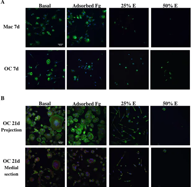
For the preparation of the Fg extract, 10 mg of Fg-3D scaffolds were incubated in 10 mL of α-MEM (1% P/S, without FBS) at 37ºC for 24 h, under agitation (120 rpm) (ISO 10993-5:2009 standard). After incubation time, supernatant was collected, filtered through 0.22 µm filters and stored at -20ºC until use. For obtaining the desired concentrations, Fg extract was diluted in α-MEM and 10% FBS was added. Prior to cells seeding, coverslips were placed in the bottom of 24 well plates and left uncoated or coated by adsorption of fibrinogen (100 µg/mL) for 1 h, before being washed twice with PBS. Monocytes (2.5x10⁵ cells per well) were cultured with RPMI supplemented with 1% glutamine, 1% P/S and 10% FBS, for macrophage differentiation; or α-MEM supplemented with 10% FBS, 50 ng/mL RANKL and 30 ng/mL M-CSF, for osteoclast differentiation [39]. Four conditions were studied: basal media only, fibrinogen adsorbed to the coverslip with basal media, and basal media with two concentrations of fibrinogen extract (50% and 25%). Media was changed twice a week. Cells were kept in culture for 7 days (macrophages and osteoclasts) and 21 days (osteoclasts).
2.4. Monocytes seeding and differentiation into osteoclasts or macrophages on 3D fibrinogen scaffolds
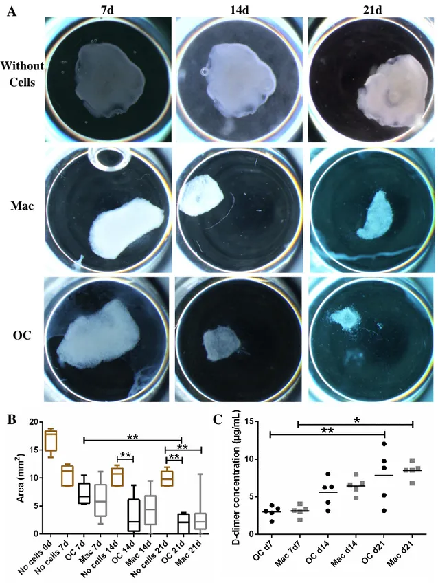
A total of 1x10⁶ cells, in 10 µL of complete medium, were seeded on each scaffold and incubated at 37ºC for 2 h. Monocytes were allowed to differentiate into macrophages, or differentiated into osteoclasts as described above, and in [39]. Media was changed twice a week and collected, as conditioned media. Conditioned media for each week (week 1 – up to day 7; week 2 – days 8 to 14; week 3 – days 15 to 21) of culture was pooled for each condition, centrifuged at 14000 rpm for 5 min and the supernatants were transferred to new tubes and stored at -20ºC until further use. Scaffolds without cells, but incubated in either media were used as controls.
2.5. MSC culture
Bone marrow MSC (mesenchymal stromal cell) had been isolated and characterized to follow the international stem cell society criteria, as described [126]. For MSC culture and maintenance, the cells were placed in flasks with DMEM (low-glucose with glutamax) supplemented with 10% FBS Hyclone and 1% P/S. Media was changed twice a week. Cells were passaged at 80% confluence. Medium was removed and the flasks washed twice with warm PBS. Trypsin EDTA was added to the flasks and allowed to act for 5 min at 37ºC. Trypsin was then inhibited with complete medium and cells were transferred to a tube and centrifuged at 300 g for 10 min. The viable cells were counted using trypan blue and plated at a density of 3000 cells/cm2.
18
2.6. Morphological analysis by SEM
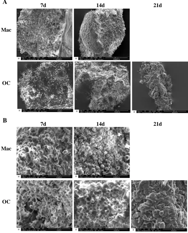
For observation of cell behavior on the fibrinogen structure, as well as the influence of the cells and time of culture in the scaffold itself, cells were fixated at 7, 14 and 21 days of culture. Scaffolds without cells were incubated in α-MEM or RPMI for 21 days. Media was removed from the wells, the scaffolds were washed twice with warm PBS for 5 min under orbital agitation and fixated with a solution of 2.5% glutharaldehyde in 0.1 M sodium cacodylate for 30 min under agitation. The scaffolds were washed three times with 0.1 M sodium cacodylate, dehydrated in a series of ethanol (50%, 60%, 70%, 80%, 90% and 99%, for 10 min each) under orbital agitation and stored in absolute ethanol at 4ºC until being critically point dried and mounted on an appropriate support using araldite glue. Dry and neutralized scaffolds were also prepared. Samples were then sputtered with a gold and palladium mixture, before being observed under a Scanning Electron Microscope (FEI Quanta 400FEG ESEM / EDAX Genesis X4M) at CEMUP (Porto).
2.7. Nuclei and cytoskeleton staining for confocal microscopy
Cells after 7, 14 and 21 days of culture were stained for f-actin and nuclei. Media was removed from the wells and the samples were washed twice with warm PBS for 5 min, fixed with 4% PFA (paraformaldehyde) at room temperature for 15 min and washed again twice with PBS (1X1 min, 1X15 min), always under orbital shaking. For cell membrane permeabilization, cells were incubated for 15 min with 0,2% Triton X-100 in PBS and washed three times with PBS for 5 min, under agitation at room temperature. For blocking non-specific binding, samples were incubated with 1% BSA at 37 ºC for 1 h under agitation and washed again with PBS for 5 min, twice. After that, the samples were incubated at 37ºC for 1 h with a 16:1000 solution of AlexaFluor 488 conjugated phaloidin in PBS (supplemented with 5mM of EGTA and MgSO4) and washed three times with PBS for 5 min under agitation. All incubation steps were performed in the dark. The samples were stored in Flouromount with DAPI at 4ºC until observation in the confocal microscope (Leica SP2 AOBS). Images were taken with Leica Confocal Software and processed with Fiji (ImageJ).
2.8. Cathepsin K staining for confocal microscopy
After 21 days of culture, the samples were fixed with 4% PFA, permeabilized and blocked as described in the previous subsection. Then the samples were incubated for 2 h with anti-cathepsin K rabbit polyclonal IgG antibody, 1:100 in PBS, in the dark and under agitation. After incubation, the samples were washed three times with PBS for 5 min under agitation and then incubated for 1 h with a solution of AlexaFluor-647 conjugated secondary antibody anti-rabbit 1:200 in PBS and AlexaFluor-488 conjugated phaloidin 16:1000 in PBS (supplemented with 5mM of EGTA and MgSO4). All incubation steps were performed in the dark. The
19
samples were stored in Flouromount with DAPI at 4ºC until observation in the confocal microscope (Leica SP2 AOBS). Images were taken with Leica Confocal Software and processed with Fiji (ImageJ).
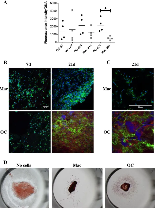
2.9. Metabolic activity quantification on 3D fibrinogen scaffolds
Macrophages and osteoclasts were differentiated on fibrinogen scaffolds as described before. At 7, 14 and 21 days, a resazurin assay was performed to quantify the metabolic activity of the cells present in the scaffold. Briefly, a dilution of 1:10 of a resazurin solution (0.1mg/mL) in appropriate culture medium for each type of cell was added to the wells and the samples were incubated for 2 h. After incubation time, the medium was collected, 100 µL was transferred to a well of a black 96-well plate (triplicates were made) and the intensity of fluorescence at 590 nm after excitation at 530 nm was read using a microplate reader (Biotek – Sinergy HT). Quantification of total DNA present in the sample was performed, as described bellow, in order to normalize the results by number of cells.
2.10. DNA extraction and quantification
After the resazurin assay, the media were collected and the scaffolds were washed twice with warm PBS. To collect the cells from the scaffolds, PBS was aspirated, trypsin EDTA was added and the plate was incubated at 37ºC for 30 min at 70 rpm. Then complete culture medium was added and the mixture was transferred to eppendorfs and centrifuged at 300 g for 5 min at 4ºC. The supernatant was discarded and the pellet was ressuspended in 50 µL Triton X-100 1% (v/v) in PBS and stored at -20ºC until quantification.
For DNA quantification, Quant-iT PicoGreen dsDNA Assay Kit (LifeTechnologies, Thermo Fisher Scientific) was used according to manufacturer’s instructions. The samples stored in Triton X-100 1% (v/v) at -20ºC were allowed to thaw at 4ºC for 1 h under orbital agitation (30 rpm). 450 µL PBS were added to each sample, homogenized and centrifuged at 10000 g for 15 min at 4ºC. The supernatant was transferred to new eppendorf tubes and 10 µL of the sample an 90 µL of TE Buffer were transferred to a black 96-well plate (duplicates were made). Then 100 µL of PicoGreen® solution was added to each well and the plate was incubated for 5 min in the dark. Fluorescence was read a fluorimeter (Biotek – Sinergy HT) (λexcitation = 480 nm, λemission = 520 nm) and the quantity of DNA was calculated through calibration curves.
2.11. TRAP staining
After 21 days of culture, the samples were assessed for the presence of TRAP positive cells using the Leukocyte Acid Phosphatase (TRAP) kit (from Sigma) according to the manufacturer’s instructions. After being fixed with a citrate/acetone solution for 30 s and