ON THE PREVENTION OF THE BLACK SPOT OF SHRIMP.:;'
I. EFFECT OF SOME CHEMICAL REAGENTS ON THE BLACK SPOT OF SHRIMP.lI. THE ISOLATION AND PURIFICATION OF THE ENZYME RELATED TO THE FORMATION OF BLACK SPOT.
CReceived in 29/4/1966)
YASUZO ITô
**
SYNOPSIS
The causes of the black discoloration of three species of shrimp commonly caught off the coast of the State of São Paulo, Brazil, are studied and measures for its prevention are advocated.
Landings of shrimp in Santos in 1964 equaled 13% of the total landing, by weight, while economically it is the principal fishery.
I - Blackening was effectively prevented by dipping the shrimp in cooled chemical solution for 30 minutes and then storing them at loW temperatures. L-ascorbic acid gave the best results, delaying discoloration up to 9 days at 0° to -~C and for a longer period at colder temperatures. The effect of
Sodium-thiosulfate and EDTA at different temperatures is aIs o discussed.
II - Tyrosinase was isolated and assayed by measuring the formation of Dopa-chrome from DL-Dopa at pH 6.8 an 37"C. Tyrosinase was obtained from the shrimp liver and was purified by approximately 25 times its activity from the initial extract by absorption chromatographY through Celite 545, as judged by the rate of dopachrome formation.
INTRODUCTION Since 1923, several authors (HARRISON & HOOD
1923; TANNER 1944; GREEN 1949; ALMEIDA 1955; DEGKWITZ 1954; BAILEY 1958; KAKIMOTO 1956; FIEGER 1956) reported on the black discoloration of shrimp and suggested various methods to prevent it.
tions; after that stored at low temperature (- 5°C ,-J - 10°C) ;
Earlier procedures may be summarized as follows:
1 - The fresh shrimp was boiled and then stored at low temperature (- 10°C ,-J - 15°C) ;
2 - The fresh shrimp was rapidly freezed and stored at low temperature (about -
lODC
,-J - 15°C);3 - The fresh shrimp's head was taken oH and the headless shrimp was stored at low tempera-ture (about - 10°C) ;
4 - The fresh shrimp was covered with ice wiLh added chemicals or dipped in cooled chemical
solu-• This work was accomplished while under contract as Vislting Researcher at the Instituto Oceanogrâflco da Universidade de São Paulo, on a Ford Foundation Grant. Present address: Hol<kaldo K,yoiku University, Hokkal-do, Japan.
Bolm Inst. oceanogr. S Paulo, 16(1):1-11, 1967
5 -
The fresh shrimp was stored with ice with added antibiotic;6 - The fresh shrimp was freeze dried and stored at low temperature.
In Brazil, people usually prefer to consume fresh
OI' dried shrimp. Therefore, it seems that we must
study according to procedures outlined under points 4, 5 and 6.
The shrimp landed in Santos is mainly , cama-rão-sete-barbas', and 'camarão-legítimo'. The amount of shrimp landed in Santos from 1962 to 1964 is shown in Table I.
As shown in Table I, in 1964 the total land-iugs of shrimp reached about 13% of the total landings of fish in Santos. Parallel to the increase of the catch of shrimp, the problem of the black spot of shrimp has become increasingly important also in Brazil.
In this connection, for a prevention of the black spot, fishermen have already used chemicals, called
T ABLE I - Landings in Santos from 1962 to 1964
~
1962Species Quantity (kg)
'Camarão-sete-barbas' 922,152 6 .8 %
'Camarão-rosa' 604,997 4 . 5 %
'Camarão-Iegítimo' 45,706 0.3 %
Total 1,572,955 11.6 %
I
% ratio of total landings
'pó-de-camarão', at Santos, they are sodium sulfite for 'camarão-rosa' and at São Sebastião, boric acid for ' camarão-sete-barbas'.
. Other chemicals usedor tested in other. cQun-tries were sodium thiosulfate, sodium bisulfite and a mixture of L-ascorbic and citric acids.
The fishermen
oI
V.S.A. boats fishing along the Guyana coast, dip shrimp in dilute sodium bisulfite solution at low temperature for a few minutes. After that, they cut oH the heads and stored the tails at low temperature. Moreover, since 1940 many researchers have studied the value of antibiotics to keep the quality of fish. However, from the view pointoI
food hygiene, the Ministry of Health and Welfare in the V.S.A., Cana da and Japan has per-mitted fishermen to use this procedure only on a few fishes (salmon, sea trout etc.) .On shrimp, DEGKWITZ (1954), FIEGER (1956), GREEN (1949), SOUTHéOTT et
alo
(1965) and MUCCIOLO & SCHNEIDER (1965) studied the effectoI
antibiotic to keep quality and to prevent black discoloration. Except for the result of MUCCIOLO & SCHNEIDER (1965) they reported that antibiotics kept the quality of shrimp but had no value in pre-venting black spot formation which is caused by tyrosinase.From these results it was observed that chemi-cals, such as sodium thiosulfate, sodium bisulfite and others were significant in preventing the black spot but did not contribute towards keeping the quality of shrimp. Therefore, shrimp treated with chemicals should be stored at low temperature.
It seems that the black spot of shrimp is caused by the action of the contained enzyme (tyrosinase) and of contaminating bacteria. Therefore, for a study of this problem, the author separated it into two parts: a study of the contained enzyme and a study of the contaminating bacteria.
The effect of chemicals in preventing discolo-ration by tyrosinase in shrimp was studied. It was observed again that reducing reagents were useful in preventing it. Furthermore, it was observed that a chelate reagent, such as EDTA, was similarly va-luable as a reducing agent.
The tyrosinase
oI
shrimp was aIs o studied. Bo-DINE & ALLEN (1941) reported on the presence of protyrosinase in blood of cray-fish and KAKIMOTOBolm Inst. oceanogr. S Paulo, 16 (1) :1-11, 1967
1963 1964
Qu a ntlty (kg) Qua ntlty (kg)
1 ,194,639 6.3 % 1,247,378 7.2 %
817,751 4.3 % 891,022 5.1 %
34,133 0.2 % 67,321 0.4 %
2,046,523 10.8 %
I
2,205,721 12 .7 %(1956) on the tyrosinase in blood, liver and other organs of Japanese spiny lobster. Hawever, no pur-ified enzyme has hitherto been obtained from marine animaIs .
The aulhor has undertaken the isolation and purification of the tyrosinase oI 'camarão-sete-barbas' for a study of its properties and obtained the enzyme purified approximately 25-times specific activity. In the present paper, the experimental results on the effect of chemicals in preventing black spot caused by a tyrosinase and a method of the isolation and purification of enzyme from the 'camarão-se te-bar-bas' liver are described.
EXPERIMENTAL PROCEDURE
I. On lhe effeet of some chemical reagents on lhe black spot of shrimp
MATERIAL - 'Camarão-sete-barbas' (Xyp hope-naeus kroyeri) samples were taken at the offing oI
Santos, from March to November, 1965. 'Camarão-rosa' (Penaeus aztecus) was bought at the central
market of Santos.
CHEMICALS - Sodium sulfate, L-ascorbic acid, Sodium thiosulfate, Ethylendiamin tetra acetic (EDT A), Acetic acid, n-Buthanol, Phenol, Acetone and Ninhydrine were used. AlI reagents were Merck A.G.
Organoleptic tests were done in two methods : A - Chemicals were a dded directly on shrimp and the treated shrimp were stored at low temperature. That is, at first, shrimp were separated into three masses (I, 11 and IH). Next, each mass was separ-ated in to three or five groups containing 10 or 20 shrimps. Then, each chemical (one
oI
tenth of weightoI
shrimp) was added to each mass. That is, sodium sulfa te was added to mass I, L·ascorbic acid to mass H and EDTA to mass IH, respectively. Then, shrimp treated were stored in the electric re-frigerator at O°C r-' - 2°C. B - ' Shrimp weredipped in each cooled chemical solution for 30 mi-nutes at low temperature and then stored at low temperature. That is, shrimp were separated on the same way as described above, and then, each mass was dipped in each cooled chemical solution
pared at O°C r-' - 2°C, for 30 minutes. After that,
shrimp were taken out from each solution and stored in the electric refrigerator at O°C r-' - 2°C
and - 5°C r-' 10°C. The concentration of various
chemicals solution 111 shown in Table 11.
TABLE II - Concentration of chemical solutions
Chemicals
Na2SO,
L-Ascorbic acid Na2S203
EDTA EDTA EDTA EDTA EDTA
Concentration ( % )
1 1 1 0.5 1 1.25 2 . 5 5
After the procedure described above, daily observations were made to see whether the shrimp in each group became black or noto
CHEMICAL TEST - a) The change of the free amino acids conten t in muscle of shrimp during storage at O°C r-' - 2°C was observed by means
of paper chromatography.
Preparation of sample for the paper
chromato-graphy - One gram of muscle was homogeneized
for a few minutes with 10 volumes of water. After 10 minutes, 5 ml of 20% trichloracetic acid were added and the precipitate was filtered oH. The fil-trate obtained was dried in a vacuum dessiccator, then, 0.1 ml of warm water (60°C) was added and the precipitate was '~ ompletely dissolved. At first, one third volume of the solution was applied to one dimension paper chromatography. Two third volu-mes of the remaining solution were applied to two dimension paper chromatography at room tempera-ture (about 25°C).
Solvent system - n-Buthanol - acetic acid
-water (4 :1:5 sup.) ar.d phenol--water (4:1) were used.
Development of colo r - 0.25% ninhydrin ace·
tone solution and special reagent for a few amino acids (isatin for proline, Pauly test for histidine and tyrosinase and Sakaguchi test for arginine) were used.
b) pH was measured with Metrohm A G Heri· sou Type E 148 C.
11. On the isolalion and purilicalion 01 tyrosinase
Irom shrimp liver
MATERIAL Shrimp (' camarão·sete·barbas',
Xyphopenaeus kroyeri) was captured at the oHing
of Santos, from March to November, 1965. Ali reagents used were analytical grade, unless other-wise indicated. Celite 545 was washed several times
Bolm Inst. oceanogr. S P a ulo, 16 (1) :1-11, 1967
by suspending it in distilled water and decanting. The slurry was poured into the column and equili· brated with 0.005 M sodium phosphate buffer, at pH 7.2 by running several hold-up volumes of the buHer through each column. Sephadex G·50 was prepared in the similar way as the Celite 545 except that 0.01 M sodium phosphate buffer was used for washing and equilibration.
Enzyme assay - Tyrosinase was assayed by
measuring the formation of 2.carboxy.2,3.dihydroin-dol-5,6-quinone (Dopa-chrome) from 3,4.dihydroxy-DL-phenylalanine (DL-dopa) at pH 6.8 and at 37°C. The assay system consisted of 0.2 ml of enzyme extract preparation, 1.8 ml of 0.1 M sodium phos-phate buffer at pH 6.8, and 0.1 mg of DL-dopa dissolved in 5 ml of the same buffer. The forma-tÍon of dopa·chrome at 37°C was measured colori-metrical1y using a Bausch & Lomb Spectronic 20 at 480 m,u.
Tyrosinase unit - According to the method of
HAROWITZ (1960) and FLlNG (1963), an amount of enzyme which produces an absorbancy increase of 0.1 at 480 m,u in the first 5 minutes, was adopted as a unit for the tyrosinase.
Protein determination - During the course of
the isolation, protein concentration was measured colorimetrically by the biuret method using a Bausch
& Lomb Spectronic 20 at 560
m,u
or 750 m,u.RESULTS
I. On the ellect 01 some chemical reagents on the
black spot of shrimp
The results of organoleptic methods are shown in Tables
UI·IV.
The value indicated in the Tables are shown as a ratio of blackening against total shrimp. Table111
shows the results of method A. From the results of Table111,
L·arcorbic acid was indicated as having a better effect than others, but in this case, the surface of a few shrimp wereTABLE III - Discoloration during storage (powder)
at
oDe
r-' -2°e
Storage
R e feren ce Chemicals period
\
I
% Na,SO, % L-Ascorbic EDTA
days acid % %
o o o o o
1 15 o o o
2 28 20 o 30
3 70 30 o 40
4 30 o 40
5 / / I
6 60 20' 50
7 80"
" to become y ellow
covered with a yellow color (that is yellow disco 10-ration) instead of blackening_ The results of method B are shown in Tables IV, V and VI. Tables IV and V show the results for ' camarão-sete-barbas' when storage temperature was
O°C
r-' -2°C
(Tab_IV) and -
5°C
r-' -10°C
(Tab. V), respectively.EDTA solution was indicated as having a effect than others. Table VI show the results for ' camarão-rosa'.
From the results of Table VI, it is evident that L-ascorbic acid strongly delayed black spot forma-tion. Sodium thiosulfate and EDT A delayed black
T ABLE IV - Discoloration during storage of 'camarão-sete-barbas' Csolution) at O"C r-' - 2°C
Storage
Reference perlod
L-Ascorblc %
days acid %
O O O
1 O O
2 15 O
3 40 10
4 55 40
5 100 /
6 50
TABLE V - Discoloration during storage of 'camarão-sete-barbas' Csolution)
at - 5°C r-' -lQ°C
Storage Referen ce Chemicals perlod
days % Na, 50. L-Ascorbic
I
EDTA% acld % %
O O
I
O O O1 O O O O
2 O O O O
3 O O O O
4 O O O O
5 O . O O O
6 O O O O
7 10 O O O
8 20 O O O
9 20 10 O O
10 20 10 O O
11 / / / /
12 / / / /
13 50 20 10 10
From the results of Tables IV and V, it was observed that the formation of black spot was strong-ly delayed at low temperature. In this case, shrimp didn't become black for about 6 days without chemi-cals, but after that, the black spot appeared rapidly on the shrimps. The author was unable to indicate organoleptic difference between these chemicals up to 8 days, but he could indicate the differences between three chemicals from pH value measured at 8th days.
The results of the pH value were shown in Table VII. At high temperature
(O°C
r-' -2°C),
dilutedBolm Inst. oceanogr. S Paulo, 16(1):1-11, 1967
Chemicals
EDTA %
0.5 %
I
1.25 %I
2.5 %I
5.0 %O O O O
O O O O
O 10 O 10
O 20 10 20
20 20 60 40
/ / / /
30 40 60 50
TABLE VI - Discoloration during storage of 'camarão-rosa' Csolution) at - 5°C
Chemicals
Sto,.~
perlod R"."n"Na,5,O.
%/
N 50 / L-Ascorbic lEDTA days %a, • % acid % %
O O O O O O
1 O O 10 O O
2 20 10 20 O 10
3 60 10 50 O 10
4 10 50 O 10
5 10 50 O 10
6 10 50 O 10
7 20 60 O 20
8 20 70 O 20
9 40 80 O 40
spot formation too, but their effects were not as strong as L-ascorbic acid, while, sodium sulfate had hardly any value in preventing black spot formation. At the 9th day, pH value of shrimp, which showed no black spot up to that day, was measured and the results obtained are shown in Table VII. The author was able to indicate slight differences between three chemicals, except sodium sulfa te.
The results of the chemical test are shown in Figures
1-5.
TABLE VII - pH value of each fraction during storage
'Camarão-sete-barbas'
I
'Camarão-rosa'-pH value
Chemlcals
- 50C '" - 100C
8th days
I
blackening After L-Ascorblcacid 6.80 7.70
0.5 % EDTA
-
-.,:: 1.25 % EDTA 7.20 7.80
.9
...
::s 2.5% EDTA
-
-Õ
<IJ 5.0 % EDTA
-
-Na,SO. 7.50 8.00
Na2S2Oa
-
-Na,SO.
-
-'"'
<li L-Ascorbic
'O
~
o acid
-
-P-< EDTA
-
-1) Concentration of EDTA ls 1 % 2) Yellaw color
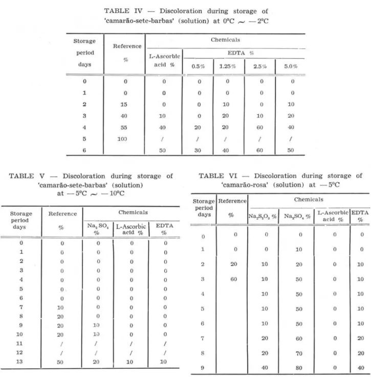
Figure 1 shows the results of one dimension paper chromatography.
11
o U
eX
c
n
U
;;Çfp
'~
j
~
~
(j
~
O
~
~
~
O
'
-:
,
,.
\(@; ~
,) I
~~~
~,
. "
24~
~~
O
~ ~
~
()~
~
()~
i
~ ()~
f
(J
~.-
~.,
""'
...
a ,g>.,.
36 48 h Tyr
Fig. 1 - One dimension papel' chromatogram of tyrosinase and shrimp muscle arter O, 24, 36 and 48 hours storage
time at OOC to - 20C.
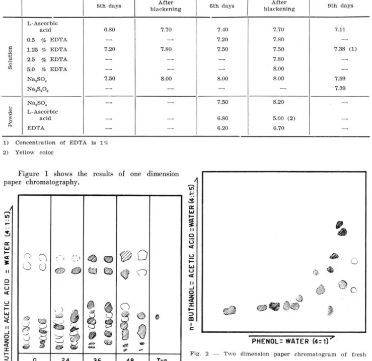
Figue 2 shows the results
oI
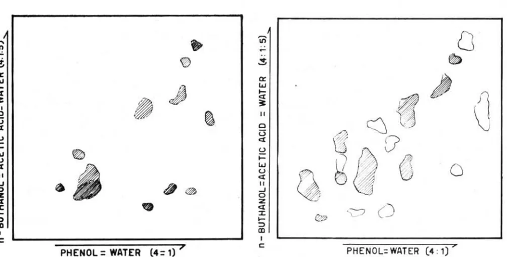
fresh material. Figure 3 shows the results after 24 hours of storage. Figure 4 shows the results after 36 hours. Figure 5 shows ·the results after 48 hours. After 48 hours,Bolm Inst. oceanogr. S P a ulo, 16 (1) :1-11, 1967
OOC", -20C - 50C '" - 100C
6th days
I
blackenin g After 9th daysI
7.40 7.70 7.117.20 7.80
-7.50 7 .50 7.38 (1)
-
7.80--
8.00-8.00 8.00 7.59
-
-
7.397 .50 8.20
-6.80 5.00 (2)
-6.20
I
6.70-~
...
S
a::
w
I-~
~
11
o
U
ct:
U
•
i=
O
w
~u ~
ct:
11
~
~O
-J
o
ã
$
x
~~~~
~
...
::::>ai
,
cPHENOL = WATER (4= 1)"
Fig. 2 - T wo dimension -paper chromatogram of fresh s hl'imp mus cle.
the surface of shrimp's neck beca me slightly black. From Figures 2, 3, 4 and 5, the author was able to indicate that some amino acids" (pro. arg. tyr. a
- amino butyric acid etc) has disappeared and an unknown substance was produced during storage at
O°C r-' - 2°C. The results of the change of free
amino acids in the muscIe of shrimps are shown in Table VIII.
"'"'
III....
~
a:
LrJ
I-e[
~
11 O
Ü
e[ U
I-~
LrJ u e[
II
...J
$
O z e[
:J: I-::l CII
I
c:
PHENOL
=
WATER (4= 1) "Flg. 3 - Two d lmenslon paper chromatogram of shrimp muscle after 24 hours of storage time at ooe to -20e.
11 O Ü
e[
(.) i=
LrJ u
ex:
11
5
zex:
:J:
I-;:)
CII
I C
PHENOL= WATER (4: 1) 7
Flg. 4 - Two dimension paper chromatogram of shrimp muscle after 36 hours storage time at 00 to -2°e.
The author again measured the pH as an indi-cator of autolysis. These results are shown in Table IX.
a: w
i
II o
Ü
ex: (.) i=
w u .
ex: 11
...J o z ex:
:J:
f-;:)
CD
c
Q
~
~
Jl
\j
o
PHENOL=WATER (4 : 1) >'
Fig. 5 - Two dimension paper chromatogram of shrimp muscle after 4S ·hours s torage time at ooe to - 20e.
11. On the isolaüon and purification of the enzyme
related to the formation of the black spot of shrimp
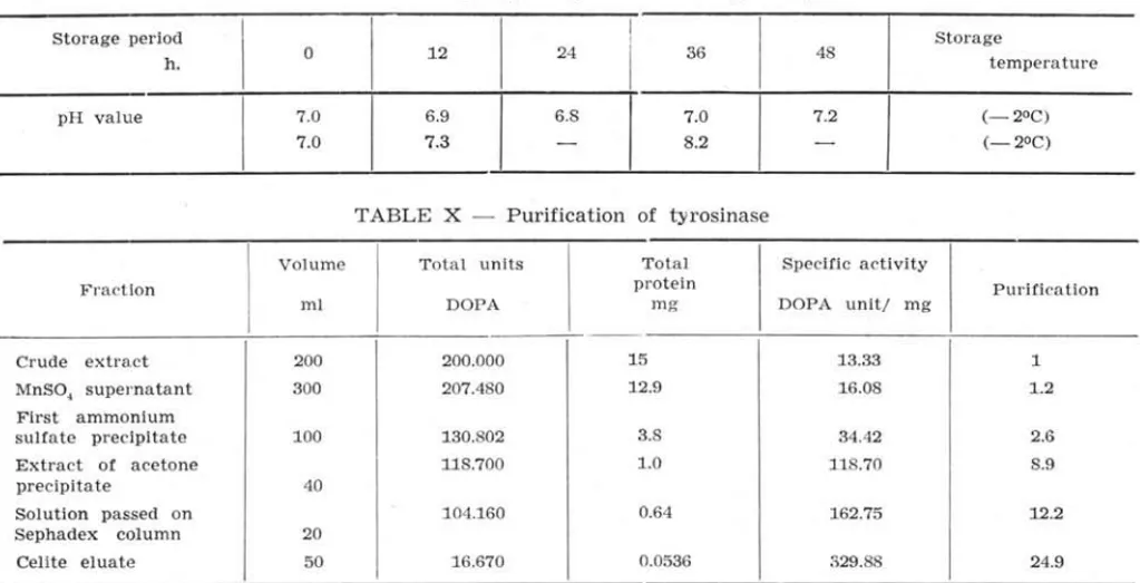
A summary of the isolation procedure of tyro-sinase from shrimp liver is shown in Figure 6 and Table X.
AlI the steps were carried out below 10°C. Unless otherwise indicated the buffer referred to be-low is 0.1 M sodium phosphate at pH 7.2.
Extration - Fresh liver of shrimp was mixed
with two volumes of cooled buffer and alIowed to stand for 24 hours; then the extract was filtered.
Preliminary purifícation - The filtrate was
treated with 50 ml of 0.2 M Manganous sulphate per !itero The pH value of this solution has to be kept at 7.2. After standing for 24 hours, the preci-pitate was removed by centrifugation and washed once with chilled water.
Ammonium sulphate precípitation - To the
mixture of the supernatant and the washing, solid ammonium sulphate was slowly added by sÍírring, to bring the concentration of ammonium sulphate to 60%, and then alIowed to stand overnight. The precipitated protein, containing the tyrosinase, was
TABLE VIII - Change of free amino acid in the muscles of 'camarão-sete-barbas'
Amino VaI <X -Amino
Phe Leu
+
Try Tyr - butyrlc Pro Ala Arg Thr Ser Gly Glu Aspacid Met acid
I
O
+
+
+
+
+
+
+
++ +++ +
+
++
+
+
24
+
+
+
±+
+
±++ ++
+
+
+
+
+
36
+
+
+
± ± ?-
+
±+
+
+
+
+
T ABLE IX - Change of pH value during storage
Storage period h.
pH value
o
7 .0 7.0
12
6.9
I
7.324 36
6.8
I
7.0
-
8.248
7.2
-Storage
tempe rature
(-2°C) (-2°C)
T ABLE X - Purification of tyrosinase
Volume Total units Fraction
ml DOPA
Crude extract 200 200.000
MnSO, supernatant 300 207.480 First ammonium
sulfate p recipitate 100 130.802 Extract of acetone 118.700
precipitate 40
Solution passed on 104.160 Sephadex column 20
Celite eluate
FRESH LIVER
FILTRATE
FILTRATE
PRE CIPITATE
PRE CIPITATE
PRECIPITATE
50 16.670
add. 2 volumes of 0.1 M sodium phosphate
buffer, pH 6.8 in the low t emperature, after 24 hours, filtered.
add. 0.2 M Mn-SO, (50 mVl) keep pH 7.2,
after 24 hours, filtered.
add. solid (NH,) 2 SO" at OOC, after 24 hours, the precipitate was collected by centrifugation at 2,000 r.p.m.
for 30 minutes.
dissolved in 0.1 M sodium phosphate buffer pH 7.2,
add. 2 volumes of acetone at - 10°C the precipitate was collected by centrifugation.
extraction with small quantities of buffer,
add. solid (NH,)2 SO" the precipitate was collected by centrifugation.
dissolved in buffer,
passed on Sephadex G-50 column, add. equal volume of chilled water.
CELITE CHROMATOGRAPHY
Fig. 6 - I solation or enzyme from 'camarão-sete-barbas'.
colIected by centrifu ga tion at 2,000 r.p.m. for 30 minutes.
Acetone preopaation - The precipitate was
dissolved in a mini mal amount of buffer and cooled to - 10°C, then two volumes of acetone were added
Bolm Inst. oceanog r. S Paulo, 16 (1) :1-11, 1967
Total Specific ac tivity
prot ein Purification
m g DOPA unit/ mg
15 13.33 1
12.9 16.08 1 .2
3 .8 34.42 2.6
1.0 118.70 8.9
0.64 162.75 12.2
0.0536 329.88 24.9
slowly by stirring. The suspension was centrifuged, the supernatant of which was discarded. The tyro-sinase was taken out of the precipitate by extracting
it several times with smalI amounts of buffer and was precipitated from lhe combined extracts by addition of solid ammonium sulphate.
Procedure on Sephadex C-50 - The ammonium
sulphate precipitate from the preceding step was dissolved in buffer. The solution was made free from ammonium sulphate by passing it through Sephadex C-50 column (2.5 x 8 or 2.5 x 15 cm) which had been equilibrated with 0.01 M buffer.
eelite chromatography - The ammonium-free
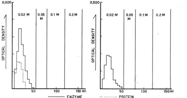
eluate from the preceding was diluted with two volumes of water and placed on a column (2.5 x 8 or 2.5 x 15 cm) of Celite 545 which had been equilibrated with 0.005 M buffer. Stepwise elution of the column was employed for the chromatographic separation of the enzyme. The effluent volume of each tube was controlIed for 5 ml in 30 minutes. About 70% of the activity originally placed on the column was recovered in the 0.02 M buffer. These results are shown in Figure 7.
DISCUSSION
The author studied the effect of chemicals against shrimp blackening with organoleptic and chemical methods.
Fishermen except for those operating from São Sebastião did not use chemicals for the prevention of black spot formation in 'camarão-sete-barbas'. Therefore is valuable to use ' camarão-sete-barbas' for studies in the laboratory on the effect of chemi-cals on the prevention of the black spot formation.
From the results of Table
IH
and V, the author observed that L-ascorbic acid had very strong value in preventing black spot formation.o.soo
>-
t-Vi Z
W
o
..J
ct o
;::::
Cl.
o
0.02 M
.. \. ..
l
50
0.05 0.1 M 0.2M M
100 150 ml. ENZYME
0.500
>-
t-üi
Z
W
o
..J
ct o
t-Cl.
o
0.02 M 0.05 0.1 M
M
50 ... ... PROTEIN
.,
100
0.2M
150ml
Fig. 7 - Elution of tyrosinase from Celite 545 column.
If the phenomenon of blackening is caused by the action of tyrosinase in shrimp, it should be inhibited when copper ion is removed from the shrimp muscles, because it is subject to a~tivation
by tyrosinase. It is known that copper lOns are contained in the muscles of shrimp, therefore a chelate reagent (EDTA) was used to remove it. And it was observed that the effect of EDT A was much the same as that of L-ascorbic acid when dilute solution of EDTA was used.
'Camarão-rosa' treated with chemicals (sodium sulfate) were used. From Table VI, the author observed that L-ascorbic acid, sodium thiosulfate anil EDT A were valuable in preventing black spot for-mation caused by tyrosinase. L-ascorbic acid and sodium thiosulfate were reducing reagents and strongly delayed black spot formation. The fact ao-reed well with the results of other authors. o
Sodium thiosulfate, sodium sulfate and sodium bisulfite have various uses, are widely used and their price is reduced. But, when we use these rea-gents, we must keep within the limits allowed by legislation on the hygenie of food. While L-ascorbic acid and EDT A are harmless their price is high. The author lhinks that this is the difficulty to the widespread adoption of these chemicals as preser-vatives.
In this experiment, the author observed that reducing reagents and cheIa te reagents are valuable in preventing black spot formation in shrimp. From these results, the author is also able to conclude that blackening occurs by the action of tyrosinase in shrimp.
The author recommends the following method for the prevention of. the black spot formation of shrimp as follows:
Procedure - The fresh shrimp are dipped in
the cooled chemical (reducing reagent or EDT A) solution for a few minutes. After that, treated shrimp are put in a box and stored at low temperature for
Bolm Inst . oceanogr. S Paulo, 16 (1) :1-11, 1967
the prevention of the evaporation of moisture from shrimp and to inhibit the action of contaminated bacteria.
Moreover, the author studied the change of the free amino acids in shrimp muscles when stored at about - 2°C. At this temperature, black spot appeared on the surface of the shrimp within 48 hours. The fresh shrimp have many free amino acids in the muscle as shown in Figure 2. This fact agreed well with the results of ALMEIDA (1954) and CAMIER et ql. (1951).
On paper chrom~togram, the author did not identify some substances that would have occurred during storage. As shown in Table IX it was observed that autolysis occurred in shrimp. The author was unable to clear the relation of blackening and free amino acids in muscle, but he thinks that part of amino acids will be used as substrate of tyrosinase, beca use tyrosinase and tryptophane dis-appeared at once.
Moreover, the author studied the isolation and purification of the e~zyme related to the discolora-tion of shrimp.
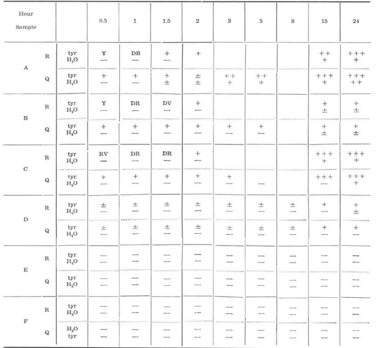
In our preparatory experiment, it was studied wheter tyrosinase present or not in various organs of 'camarão-sete-barbas' and on some solvents for the extraction of tyrosinase from the 'camarão-sete-bar-bas' liver. The results obtained were shown in Table XI.
From the results of Table XI, tyrosinase was extracted from ' camarão-sete-barbas' liver with phos-phate buffer, at pH 6.8 r-' 7.2. Also the activity
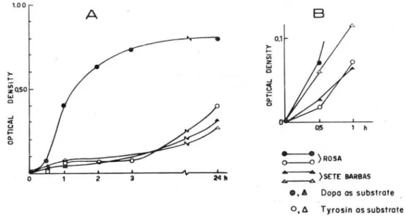
of tyrosinase of three sorts of shrimp (' camarão-sete-barbas', 'camarão-rosa' and ' camarão-legítimo') was compared. The results obtained were shown in Fi-gures 8 and 9.
From the results shown on Figures 8 a, b, and 9, it may be seen that the activity of tyrosinase from these shrimps indicated the same activity, after 24 hours.
TABLE XI - The enzymatic activity of the various organs from 'camarão-sete-barbas'
Hour
0.5 1 1.5 2 3 5 8 15 24
Sample
tyr Y DB
+
+
+ +
+ ++
R
H.o
+
+
A ... ... . 0 . 0 0 . 0 • • 0 . 0 0 . 0 • • ' ... .. • • . • • • • , • • I • • ., ...• .. , ...
tyr
+
+
+
±+ +
+ +
+ + +
+ ++
Q H.o ± ±
+
+
+
+ +
tyr Y DR DV
+
+
+
R
H.o ± ±
B .. ... ... .' .• . •. .• , • • • • • • ••• oI
tyr
+
+
+
+
+
+
+
+
Q H.o ± ±
-tyr RV DR DR
+
+ + +
+ ++
R
H,0
+
+
C ... ... ... ... " .. .... ...
tyr
+
+
+
+
+
+ + +
+ + +
Q H,O
+
tyr ± ± ± ± ± ± ±
+
+
R
H.o ±
D ... .. . " ... • .. ... . ...
t~'r ± ± ± ± ± ± ±
+
+
Q H,O
-R Htyr
20
E ... ... ... . ...
Q H,O tyr
R H.o tyr
F ... .. ... .... ... . ...
Q H.o tyr
Sample A - extract solution by physiologlcal salto
B - extract solution by phosphate buffer, pH 6.8.
C - extract solution by 30% acetone.
D - liver; E - muscle a nd F - intestine. D, E and F were extract solution by
phosphate buffer, pH 6.8.
R == raw m aterial; Q == heated material ; Y == yellow; DE == d a rk brown; DR dark reeI;
DV == dark violet; RV == redish violet;
+,
black (darkeing), -, no black.As shown in Table X, the tyrosinase was purifi-ed, by approximately 25 times its activity from the initial extract, by means of absorption chromato-graphy through Celite 545, as judged by the rate of the formation of dopachrome.
Recently, lhe purification of tyrosinase from
Bolm Inst. oceanogr. S Paulo, 16 (1) :1-11, 1967
various sources such as mushroom, hamster and Neu-raspara was published (HAROWITZ 1960; FLING 1963; BOUCHILLOUX et alo 1963; POMERANTZ 1963). The
author was unable to compare his results with the present ones, beca use of the lack of more detailed data on the physical and chemical properties of this enzyme, which will be reported elsewhere.
1.00
>-
o-~o.50
I&J
o
o
A
2 3
B
=)ROSA
2411
..,.:.---:.0.
)SETE BARBAS_, • Dopo os substrote
0.6 Tyrosin os substrote Fig. 8 - Enzyme activity of 'camarão-sete-barbas' and 'camarão-rosa'.
0.500
>-~
(/) z w Cl
...J
c:t C,.)
~
a.. o
400 500
DOPA AS SUBSTRATE
TYROSINE
} AS SUBSTRATE
-
0----0-,
r
ROSA•
.f'r---i.::,4 ]
LE G I TI MO600
Fig. 9 - Enzyme activity of 'camarão-rosa' and 'camarão-legitimo'.
ACKNOWLEDGEMENTS
The author wishes to express his sincere thanks to Dra. M. V ANNUCCI for her support and also wishes to thank Miss D ULCINDA RODRIGUES DA SILVA and Mr. LUIZ SANCHES for their helpful assistance.
RESUMO
Foram estudad'as, no presente trabalho, as causas do enegrecimento do camarão, tendo-se verificado que o mesmo é causado, fundament1:!lmente, pela ação da
BoIm Inst. oceanog r. S Paulo, 16 (1) :1-11, 1967
tirosinase, produzida no hepato pâncreas do crustá-ceo. O isolamento e a purificação da enzima foram realizados. A purificação foi feita, até se obter uma atividade 25 vêzes maior do que o extrato inicial, por meio de cromatografia de absorção, através de Celite 545 e avaliada através da velocidade de for-mação de dopacromo.
O uso de diferentes drogas também foi tratado experimentalmente, tendo-se verificado que o melhor inibidor do processo de enegrecimento é o ácido L-arcórbico que evita a formação da mancha preta durante 9 dias à temperatura de O a - 2°C e du-rante tempo mais longo em temperaturas mais bai-xas. Tiosulfato de sódio e EDTA também têm boa ação inibidora. As técnicas empregadas são descri-tas detalhadamente e é apresentada discussão com resultados obtidos por outros autores. Ao que se sabe, esta é aprimeira vez que tirosinase é isolada de um invertebrado.
REFERENCES
ALMEIDA, M. E. W. de
1955. Aminoácidos livres em camarões variações decorridas durante a decom-posição. Revta Inst. Adolfo Lutz, voI. 15, n.o único, p. 158-167.
BAILEY, M. E.
1958. Biochemical of melanin formation in shrimp. Ph. D. Thesis. Louisiana State Univ., Baton Rouge, Louisiana.
BODINE , J. H. & ALLEN, T. H.
1941. In: The proteins, ed. by Hans Neurath.
New York, Academic Press, vaI. 2, part A, p. 149.
BOUCHILLOUX, S. et alo
1963. The multi pIe forms of mushroom tyro-sinase. J. bioI. Chem., voI. 238, p. 1699-1707.
CAMIER, et alo
1951. Nonprotein amino acids in muscle and blood of marine and fresh water crus-tacea. J. bio!. Chem., vol 193, p. 881-885.
DEGKWITZ, E. et alo
1954. Untersuchungen uber die Ursachen der Schnillen Verderblichkeit des Krabben Fleisches. Arch. Fischereiwiss., voI. 5, p. 35-42.
FIEGER, E.
1956.
FLING, M.
1963.
GREEN, M.
1949.
HAROWITZ,
1960.
HARRlSON,
1923.
A. et alo
Objective tests applicable to quality studies of ice stored shrimp. Fd Res., voI. 21, p. 611-620.
et alo
The isolation and properties of crystal-line tyrosinase from Neurospora. J . bioI. Chem., voI. 238, p. 2045-2053.
Bacteriology of shrimp.
r.
Introduction and development of experimental pro-cedures.n .
Quantitative studies on freshly caught and iced shrimp. Fd Res., voI. 14, p. 365-371, p . 372-383. N. H. et aloGenetic det ermination and enzymatic induction of t yrosinase in Neurospora. J. molec. BioI., voI. 2, p. 96-104.
E. G. et alo
The red discoloration of cured cod fish. Trans. R . Soc. Can., vol. 3, 11.° 16, p.
101-152.
Bolm Inst. oceanogr. S Paulo, 16 (1) :1-11, 1967
KAKIMOTO,
1956. D. Studies lobster. voI. 22,
011 the black discoloration of
Bull. Jap. Soc. sciel1t. Fish., p. 471-479.
M UCCIOLO,
1965. P. Eficiência da clortetraciclina na con-& SCHNEIDER,
r.
S. servação do pescado. Revta naco Pesca S. Paulo, voI. 6. n .o 46, p. 14-15.POMERANTZ, S . H.
1963. Separation, purification and properties of two tyrosinase from Hamster m ela-noma. J. biol. Chem., vol. 238, p. 2351-2357.
SOUTHCOTT,
1965 .
TANNER, F.
1944 .
B. A. et alo
Tetracycline antibiotics in shrimp pre-servation. J . Fish. Res. Bd Can., voI. 22, p. 117-129.
W .
The microbiology of foods. 2nd ed. Champaign, Ill. , Garrard Press.