Vasopressin Intravenous Infusion Causes Dose Dependent Adverse
Cardiovascular Effects in Anesthetized Dogs
Luiz Cláudio Martins, Maricene Sabha, Maria Ondina Paganelli, Otávio Rizzi Coelho, Silvia Elaine Ferreira-Melo, Marcos
Mello Moreira, Adriana Camargo de Cavalho, Sebastião Araujo, Heitor Moreno Junior
Faculdade de Ciências Médicas da Universidade Estadual de Campinas - Unicamp - Campinas, SP - Brazil
Abstract
Background: Arginine vasopressin (AVP) has been broadly used in the management of vasodilatory shock. However, there are many concerns regarding its clinical use, especially in high doses, as it can be associated with adverse cardiovascular events.
Objective: To investigate the cardiovascular effects of AVP in continuous IV infusion on hemodynamic parameters in dogs.
Methods: Sixteen healthy mongrel dogs, anesthetized with pentobarbital were intravascularly catheterized, and randomly assigned to: control (saline-placebo; n=8) and AVP (n=8) groups. The study group was infused with AVP for three consecutive 10-minute periods at logarithmically increasing doses (0.01; 0.1 and 1.0U/kg/min), at them 20-min intervals. Heart rate (HR) and intravascular pressures were continuously recorded. Cardiac output was measured by the thermodilution method.
Results: No significant hemodynamic effects were observed during 0.01U/kg/min of AVP infusion, but at higher doses (0.1 and 1.0U/kg/min) a progressive increase in mean arterial pressure (MAP) and systemic vascular resistance index (SVRI) were observed, with a significant decrease in HR and the cardiac index (CI). A significant increase in the pulmonary vascular resistance index (PVRI) was also observed with the 1.0U/kg/min dose, mainly due to the decrease in the CI.
Conclusion: AVP, when administered at doses between 0.1 and 1.0U/kg/min, induced significant increases in MAP and SVRI, with negative inotropic and chronotropic effects in healthy animals. Although these doses are ten to thousand times greater than those routinely used for the management of vasodilatory shock, our data confirm that AVP might be used carefully and under strict hemodynamic monitoring in clinical practice, especially if doses higher than 0.01 U/kg/ min are needed. (Arq Bras Cardiol 2010;94(2): 213-218)
Key words: Arginine vasopressin; cardiac output; hemodynamic / drug effects; drug toxicity; dogs.
Mailing address: Heitor Moreno Jr. •
Departamento de Farmacologia, Faculdade de Ciências Médicas da Universidade Estadual de Campinas - UNICAMP - 13083-970, Campinas, SP - Brazil
E-mail: [email protected]
Manuscript received November 13, 2008; revised manuscript received June 19, 2009; accepted July 01, 2009.
Introduction
Vasopressin is a neuropeptide consisting of nine amino acids with antidiuretic and vasoconstrictor effects1-7. Its
powerful effect on vascular smooth muscle increases blood pressure and systemic vascular resistance2-4. Vasopressin
is synthesized in neurons located in paraventricular and supraoptic nuclei of the hypothalamus, and it is stored in the posterior pituitary1,5,6. The vasopressin release is complex,
being increased by hyperosmolality, hypotension and hypovolemia1. There are three types of vasopressin receptors:
V1, V2 and V3. Vasopressin V1 receptors present in blood vessels
are responsible for vasoconstriction, vasopressin V2 receptors present in renal collecting duct cells are mainly responsible for the antidiuretic effectsand vasopressin V3 receptors present in the adenohypophysis are responsible for ACTH secretion8.
Vasopressin is essential for cardiovascular homeostasis, acting via the kidney to regulate water reabsorption, on the vasculature to regulate smooth muscle tone, and as a central neurotransmitter, modulating brainstem autonomic function1.
Although it is massively released in response to stress or shock states, a relative deficiency of vasopressin has been found in prolonged vasodilatory shock, such as is seen in severe sepsis9,10. In this circumstance, exogenous vasopressin has
marked pressor effects, even at doses that would not affect blood pressure in healthy individuals. These two findings provide the rationale for the use of vasopressin in the treatment of septic shock11,12.
In the last decade, vasopressin has been broadly used as an adjunct vasopressor agent for the management of catecholamine-resistant vasodilatory shock9,11-13, and is
also recommended to increase peripheral vascular tone during cardiopulmonary resuscitation as an alternative to epinephrine14,15. Despite considerable research attention, the
mechanisms for vasopressin deficiency and hypersensitivity in vasodilatory shock remain unclear9,12,13,16-20. Moreover, the
effects in continuous infusions with progressive doses has been limited. For this reason, we investigated the cardiovascular effects of vasopressin on hemodynamic parameters when used as continuous infusion and at progressive doses in anesthetized healthy dogs.
Methods
Ethical aspects: all procedures were approved by our institutional Ethical Animal Care Committee and the experiments were carried out in accordance with the guidelines published by the National Institutes of Health and the European Community Guidelines for use of experimental animals.
Setting: Cardiovascular Pharmacology Laboratory at the Pharmacology Department of the Faculty of Medical Sciences, University of Campinas (UNICAMP), São Paulo, Brazil.
Population: Sixteen adult healthy mongrel dogs, both sexes, weighing 15 ± 1kg.
Animal handling and experimental model preparation: the animals were prepared as described by Tanus-Santos21. After
a fasting night with free access to water, the animals were anesthetized with a loading dose of sodium pentobarbital (10 mg.kg-1, IV) and an adequate level of anesthesia was
maintained by a continuously IV infusion of the same drug (2-4 mg.kg-1.h-1). The dogs were tracheally intubated and
mechanically ventilated with room air using a volume-cycled respirator (Dual Phase Control Respirator; Harvard Apparatus, Boston, MA, USA). The tidal volume was 15 mL/kg, and the respiratory rate was adjusted to maintain a baseline physiologic PaCO2 (around 35-40mmHg, as demonstrated by end-tidal CO2 monitoring).
A fluid-filled catheter was placed into the left femoral artery for mean arterial pressure (MAP) monitoring, via a pressure transducer (AS-3 Datex- Engstrom, Helsinki, Finland). Another plastic catheter was placed into the left femoral vein for fluid administration. A 7F balloon-tipped Swan-Ganz thermodilution catheter was placed in the pulmonary artery via the right femoral vein, and its correct location was confirmed by detection of typical pressure wave of this artery. The catheter was connected to a pressure transducer (AS-3 Datex-Engstrom, Helsinki, Finland) to allow monitoring of the mean pulmonary artery pressure (MPAP), central venous pressure (CVP), and pulmonary capillary wedge pressure (PCWP). The transducers were zeroed at the level of the right heart and recalibrated before each set of measurements.
Cardiac output was measured in triplicate by a bolus injection of 5 mL of normal saline solution, and the results were recorded and stored in a computerized system (Datex-Engstrom, Helsinki, Finland). Body surface area (BSA) of each dog was calculated according to the following formula: Km .BW0.67.100-1 [where Km for dogs = 10.1; body weight (BW)
is measured in kg; and BSA is expressed in m2]22, and cardiac
index (CI), systemic vascular resistance index (SVRI), and pulmonary vascular resistance index (PVRI) were calculated by using standard formulas. Heart rate (HR) was measured using a surface electrocardiogram (lead I).
The experiment: the animals were maintained with a
continuous IV infusion of 0.9% NaCl solution (5mL.kg-1.h -1) throughout the whole experiment. After the end of the
intravascular catheterization procedures, a stabilization period of 20 minutes was observed and baseline (BL) hemodynamic data were firstly recorded. Subsequently, the animals were randomly assigned to two equal groups: CONTROL (saline-placebo; sham-group; n=8) and VASOPRESSIN (n=8). The CONTROL group received continuous infusions (20 mL) of 0.9% NaCl for 10 minutes, at 20-minute intervals, for three times. The VASOPRESSIN group was infused with AVP (Arginine Vasopressin- Acetate Salt, Sigma Chemical Co. USA) for three consecutive 10-min periods at logarithmically increasing doses (0.01; 0.1 and 1.0U/kg/min) diluted in 20 mL of 0.9% NaCl solution, at 20-min intervals. Heart rate and hemodynamic data were recorded just after the end of each AVP dose (or placebo) infusion in both groups.
Statistical analysis was performed by applying Student’s t-test for unpaired observations or analysis of variance (ANOVA) for repeated-measures, followed by Dunnett’s multiple comparison test. A value of p<0.05 was considered significant. All statistical calculations were carried out using Sygma Stat for Windows (Jandel Scientific, CA, USA).
Results
The results are expressed as mean ± SEM. No statistically significant alterations in the hemodynamic parameters were observed during vasopressin infusion at the lowest dose (0.01 U/kg/min) compared to the CONTROL group or to the baseline values (p = NS).
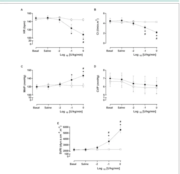
At the intermediate dose (0.1 U/kg/min), vasopressin induced significant decreases in cardiac index (CI) and heart rate (HR), when compared to both CONTROL group and baseline values. Additionally, increases in MAP and SVRI were verified at the end of the 10-minute drug infusion period (p < 0.05) (Figure1).
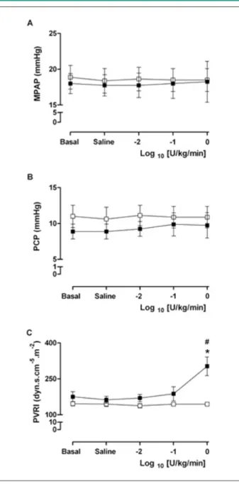
After the infusion of the highest vasopressin dose (1.0 U/ Kg/min), the previous changes observed in CI, HR (decrease) and in MAP and SVRI (increase) were exacerbated (p <0.05) (Figure 1). Additionally, at this dose, the calculated pulmonary vascular resistance index showed a statistically significant increase (p < 0.05), mainly due to the greater decrease in CI, rather than a significant increase in MPAP (Figure 2).
Discussion
This study showed that the continuous infusion of vasopressin for 10 minutes at a “low dose” (0.001U/kg/min) had no appreciable effects on HR, MAP, MPAP and CI in healthy anesthetized dogs. However, at “moderate” (0.1 U/kg/min) and “high” doses (1.0 U/kg/min), it increased MAP, SVRI and the PVRI. These doses also significantly decreased HR and CI. However, no statistically significant effects were observed in CVP, MPAP or PCWP.
The pressor effect of vasopressin is due to its action on the V1-receptors in vascular smooth muscle, and it is more predominant in the peripheral systemic arteriolar vasculature than in venous or pulmonary circulation13,18. Additionally, vasopressin leads to
Figure 1 -A) Heart Rate (HR), B) Cardiac Index (CI), C) Mean Arterial Pressure (MAP), D) Central Venous Pressure (CVP) and E) Systemic Vascular Resistance Index
(SVRI) in basal (bas), after saline (sal) and after injection, as continuous infusion, of 20 ml of NaCl 0,9% for three times in the control group (□); and after injection, as continuous infusion, of 20 ml of 0.01 U/kg/min, 0.1 U/kg/min and 1.0 U/kg/min doses in log10 of -2, -1 and 0 in the vasopressin group (■). The results are expressed as mean ± SEM. *p <0.05 vs. basal. #p <0.05 vs. control group.
muscle23,24. Vasopressin also inhibits nitric oxide production in
the vascular smooth muscle19 and acts on K
ATP channels 20. Both
of these actions lead to vasoconstriction, which, in conjunction with the effect on V1-receptors, result in MAP increase.
In 1895 Oliver & Schaefer first reported the effects of the posterior pituitary extract on blood pressure25, and more
recently its has been broadly used in situations that need MAP increase, such as in septic shock11,12 and cardiopulmonary
resuscitation (CPR) 14,15.
The use of vasopressin as an adjunct drug for catecholamine-dependent or refractory vasodilatory shock has been suggested, as there is an inappropriate autonomic response and an excessive
inflammatory vasodilation in this condition26-28. However, its ideal
doses29, as well as its safety during short and long-term use in this
condition remain a matter of controversy30.
The recommended doses of vasopressin to use in shock cases are relatively very low (0.01-0.04 U/min, or 0.00014-0.0006 U/kg/min), and aim at elevating the arterial pressure, as well as reducing the need for high doses of catecholamines11,12.
Figure 2 -A) Mean Pulmonary Artery Pressure (MPAP), B) Pulmonary Capillary
Pressure (PCP) and C) Pulmonary Vascular Resistance Index (PVRI) in basal (bas), after saline (sal) and after injection, as continuous infusion, of 20 ml of NaCl 0.9% for three times in the control group(□); and after injection, as continuous infusion, of 20 ml of 0.01 U/kg/min, 0.1 U/kg/min and 1.0 U/kg/min doses in log10 of -2, -1 and 0 in the vasopressin group (■). The results are expressed as mean ± SEM. *p <0.05 vs. basal. #p <0.05 vs. control group.
fact that these dogs have a normal operating baroreceptor reflex system, which blunts the hemodynamic effect of low vasopressin doses31, or even of high doses32.
Otherwise, in baroreceptor-denervated dogs, as shown by Cowley et al33, the dose-response (blood pressure) curve
for vasopressin is displaced to the left by a factor of 60-100 when compared to the curve for intact dogs, in whom the baroreceptor reflex was allowed to compensate. Additionally, in decapitated animals, this factor of displacement for vasopressin was 8,000 for doses that caused a 50-mmHg rise in
systemic arterial pressure33. Consistent with these experimental
findings is the observation that the dose-response curves for vasopressin in patients with idiopathic orthostatic hypotension (Shy-Drager’s syndrome) were markedly displaced to the left when compared to those of normal subjects34,35. Similar
findings have also been reported in brain-dead patients36.
Therefore, in patients with severe sepsis/septic shock, an abnormal baroreceptor reflex system function, due to critical illness polyneuropathy or to excessive inflammatory response, has been postulated as a possible mechanism for their high sensitivity to low-dose vasopressin in increasing blood pressure13.
The observed MAP increase, caused by vasopressin use, reinforces its use in vasodilatory shock. However, a SVRI increase may lead to decreased tissue perfusion and severe adverse events in patients needing high doses of continuous vasopressin infusion37. Indeed, in a recent publication,
Westphal et al38, studying the effects of vasopressin on healthy
and septic sheep, reported a reduction in the CI and an increase in the SVRI and PVRI, suggesting that these collateral effects may limit the use of this drug as the only vasopressor during septic shock. However, the simultaneous use of AVP and norepinephrine, considering their combined beneficial effects and reduced adverse events, could represent a useful therapeutic option in septic patients38.
In our study, “high-dose” vasopressin (1.0 U/kg/min) led to a significant increase in PVRI, but this was mainly due to the decrease in the CI and no significant increase was observed in the mean pulmonary artery pressure (MPAP) or in the pulmonary wedge pressure (PWP), as shown in Figure 2. Leather et al39, studying the effect of vasopressin on the right
ventricle function in a dog model of experimental pulmonary hypertension, concluded that vasopressin causes pulmonary vasoconstriction and an important negative inotropic effect in the right ventricle, suggesting that vasopressin should be used cautiously when the right ventricle function is compromised.
Study limitations
The present study has some limitations. Firstly, we have measured only global hemodynamic parameters, and no regional or metabolic effects of vasopressin infusion were evaluated. Secondly, we used vasopressin doses that were ten to one thousand times higher than those routinely used in clinical practice for the management of vasodilatory shock. Thirdly, the time of vasopressin infusion was very short, and probably not long enough to elicit the full activation of the normal cardiovascular compensation mechanisms. And, finally, healthy animals were studied, and obviously the obtained data cannot be directly extrapolated to those expected to occur in septic human patients.
Conclusion
References
1. Vincent JL, Su F. Physiology and pathophysiology of the vasopressinergic system. Best Pract Res Clin Anaesthesiol. 2008; 22: 243-52.
2. Cartheuser CF, Komarek J. Effects of vasopressin on the circulation, myocardial dynamics, and left ventricular oxygen consumption in the anaesthetized dog. Basic Res Cardiol. 1980; 75: 668-82.
3. Évora PRB, Pearson PJ, Rodrigues AJ, Viaro F, Schaff HV. Effect of arginine vasopressin on the canine epicardial coronary artery: experiments on V1-receptor-mediated production of nitric oxide. Arq Bras Cardiol. 2003; 80: 489-94.
4. Hollenberg SM, Ahrens TS, Annane D, Astiz ME, Chalfin DB, Dasta JF, et al. Practice parameters for hemodynamic support of sepsis in adult patients: 2004 update. Crit Care Med. 2004; 32: 1928-48.
5. Holmes CL, Patel BM, Russell JA, Walley KR. Physiology of vasopressin relevant to management of septic shock. Chest. 2001; 120: 989-1002. 6. Katori E, Ohta T, Nakazato Y, Ito S. Vasopressin-induced contraction in the
rat basilar artery in vitro. Eur J Pharmacol. 2001; 416: 113-21.
7. Kusano E, Tian S, Umino T, Tetsuka T, Ando Y, Asano Y. Arginine vasopressin inhibits interleukin-1 beta-stimulated nitric oxide and cyclic guanosine monophosphate production via the V1 receptor in cultured rat vascular smooth muscle cells. J Hypertens. 1997; 15: 627-32.
8. Maybauer MO, Maybauer DM, Enkhbaatar P. Physiology of the vasopressin receptors. Best Pract Res Clin Anaesthesiol. 2008; 22: 253-63.
9. Landry DW, Oliver JA. The pathogenesis of vasodilatory shock. N Engl J Med. 2001; 345: 588-95.
10. Lin IY, Ma HP, Lin AC, Chong CF, Lin CM, Wang TL. Low plasma vasopressin/ norepinephrine ratio predicts septic shock. Am J Emerg Med. 2005; 23: 718-24.
11. den Ouden DT, Meinders AE. Vasopressin: physiology and clinical use in patients with vasodilatory shock: a review. Neth J Med. 2005; 63: 4-13. 12. Russell JA, Walley KR, Singer J, Gordon AC, Hébert PC, Cooper DJ, et al.
Vasopressin versus norepinephrine infusion in patients with septic shock. N Engl J Med. 2008; 358: 877-87.
13. Barrett LK, Singer M, Clapp LH. Vasopressin: mechanisms of action on the vasculature in health and in septic shock. Crit Care Med. 2007; 35: 33-40. 14. Wenzel V, Krismer AC, Arntz R, Sitter H, Stadlbauer KH, Lindner KH.
A comparison of vasopressin and epinephrine for out-of-hospital cardiopulmonary resuscitation. N Engl J Med. 2004; 350: 105-13. 15. Gueugniaud PY, David JS, Chanzy E, Hubert H, Dubien PY, Mauriaucourt P,
et al. Vasopressin and epinephrine vs. epinephrine alone in cardiopulmonary resuscitation. N Engl J Med. 2008; 359: 21-30.
16. Malay MB, Ashton RC Jr, Landry DW, Townsend RN. Low-dose vasopressin in the treatment of vasodilatory septic shock. J Trauma. 1999; 47: 699-705. 17. Maxime V, Siami S, Annane D. Metabolism modulators in sepsis: the abnormal
pituitary response. Crit Care Med. 2007; 35 (Suppl): S596-S601. 18. Obritsch MD, Bestul DJ, Jung R, Fish DN, MacLaren R. The role of vasopressin
in vasodilatory septic shock. Pharmacotherapy. 2004; 24: 1050-63. 19. Patel BM, Chittock DR, Russell JA, Walley KR. Beneficial effects of short-term
vasopressin infusion during severe septic shock. Anesthesiology. 2002; 96: 576-82.
20. Rozenfeld V, Cheng JW. The role of vasopressin in the treatment of vasodilation in shock states. Ann Pharmacother. 2000; 34: 250-4.
21. Tanus-Santos JE, Gordo WM, Udelsmann A, Cittadino MH, Moreno H Jr. Nonselective endothelin-receptor antagonism attenuates hemodynamic changes after massive pulmonary air embolism in dogs. Chest. 2000; 118: 175-9.
22. Hand MS, Thatcher CD, Rimillard RL, Roudebush P (eds.). Small animal clinical nutrition. 4th ed. Marceline (MO): Walsworth; 2000.
23. Bartelstone HJ, Nasmyth PA. Vasopressin potentiation of catecholamine actions in dog, rat, cat, and rat aortic strip. Am J Physiol. 1965; 208: 754-62.
24. Commarato MA, Lum BKB. Cardiovascular interaction of amphetamine and ephedrine with norepinephrine and with vasopressin. Eur J Pharmacol. 1969; 7: 127-34.
25. Oliver G, Schäfer EA. On the physiological action of extracts of pituitary body and certain other glandular organs. J Physiol (London). 1895; 18: 277-9. 26. László FA, László F Jr, De Wied D. Pharmacology and clinical perspectives of
vasopressin antagonists. Pharmacol Rev. 1991; 43: 73-108.
27. Tayama E, Ueda T, Akasu K, Oda T, Fukunaga S, Akashi H, et al. Arginine vasopressin is an ideal drug after cardiac surgery for the management of low systemic vascular resistant hypotension concomitant with pulmonary hypertension. Interact Cardiovasc Thorac Surg. 2007; 6: 715-9.
28. Wakatsuki T, Nakaya Y, Inoue I. Vasopressin modulates K+-channel activities of cultured smooth muscle cells from porcine coronary artery. Am J Physiol. 1992; 263: H491-6.
29. Holmes CL, Walley KR. Arginine vasopressin in the treatment of vasodilatory septic shock. Best Pract Res Clin Anaesthesiol. 2008; 22: 275-86. 30. Ertmer C, Rehberg S, Westphal M. Vasopressin analogs in the treatment of
shock states: potential pitfalls. Best Pract Res Clin Anaesthesiol. 2008; 22: 393-406.
31. Montani JP, Liard JF, Schoun J, Möhring J. Hemodynamic effects of exogenous and endogenous vasopressin at low plasma concentrations in conscious dogs. Circ Res. 1980; 47: 346-55.
32. Gaskill III HV, Sirinek KR, Levine BA. Hemodynamic effects of vasopressin: can large doses be safely given? Arch Surg. 1983; 118: 434-7.
33. Cowley AW Jr, Monos E, Guyton AC. Interaction of vasopressin and baroreceptor reflex system in the regulation of arterial blood pressure in the dog. Circ Res. 1974; 34: 505-14.
34. Wagner HN, Braunwald E. The pressor effect of the antidiuretic principle of the posterior pituitary in orthostatic hypotension. J Clin Invest. 1956; 35: 1412-8.
Although these doses are ten to one thousand times higher than those routinely used for the management of human vasodilatory shock, our data confirm that vasopressin should be used carefully and under strict hemodynamic (and metabolic) monitoring in clinical practice, especially if doses higher than 0.01 U/kg/min are needed.
Potential Conflict of Interest
No potential conflict of interest relevant to this article was
reported.
Sources of Funding
This study was funded by FAPESP.
Study Association
35. Möhring J, Glänzer K, Maciel JA Jr, Dasing R, Kramer HJ, Arbogast R, et al. Greatly enhanced pressor response to antidiuretic hormone in patients with impaired cardiovascular reflexes due to idiopathic orthostatic hypotension. J Cardiovasc Pharmacol. 1980; 2 (4): 367-76.
36. Iwai A, Sakano T, Uenishi M, Sugimoto H, Yoshioka T, Sugimoto T. Effects of vasopressin and catecholamines on the maintenance of circulatory stability in brain-dead patients. Transplantation. 1989; 48: 613-7.
37. Luckner G, Dünser MW, Jochberger S, Mayr VD, Wenzel V, Ulmer H, et al. Arginine vasopressin in 316 patients with advanced vasodilatory shock. Crit
Care Med. 2005; 33: 2659-66.
38. Westphal M, Stubbe H, Sielenkamper AW, Ball C, Van Aken H, Borgulya R, et al. Effects of titrated arginine vasopressin on hemodynamic variables and oxygen transport in healthy and endotoxemic sheep. Crit Care Med. 2003; 31: 1502-8.