Electrocardiographic Profile and Muscle Glycogen Content of Rats
Treated with Nandrolone
Carlos Alberto da Silva, Adriano Cesar Rocco Pardi, Talita Moral Gonçalves, Sergio Henrique Borin
Universidade Metodista de Piracicaba - UNIMEP, Piracicaba, SP - BrazilMailing address: Adriano Cesar Rocco Pardi •
Rua Haldumont Campos Ferraz, 118 - Castelinho - 13403-052 - Piracicaba, SP - Brazil
E-mail: [email protected]
Manuscript received December 01, 2008; revised manuscript received September 05, 2009; accepted October 09, 2009.
Abstract
Background: We considered both the indiscriminate use of steroids by top athletes and by physically active individuals.
Objective: To evaluate the effects of nandrolone decanoate on the electrocardiographic profile, glycogen content and total-protein profile of skeletal and cardiac muscles, as well as the plasma albumin concentrations.
Methods: The drug was administered subcutaneously, at a concentration of 5 mg/kg, twice a week for three weeks, to animals in the treated group. Once a week, the rats were anesthetized with sodium pentobarbital (50 mg/kg, ip) and they underwent an electrocardiogram (ECG). After the trial period, samples of the cardiac muscle (left ventricle - LV), soleus muscle (S), white gastrocnemius muscle (WG), red gastrocnemius muscle (RG), pectoral muscle (P), intercostal muscle (IC) and diaphragm muscle (D) were promptly collected and analyzed. An analysis of variance (ANOVA) and then a Tukey test (p>0.05) were carried out to assess the data (mean ± sem).
Results: There were changes in the following parameters of rats in the treated group: QRS interval, QTc interval and heart rate, characterized by an increase in these parameters, with the peak being reached in the period between the pre-treatment week and the first week. There was an increase of 127% in glycogen reserves in the LV. In relation to the total-protein amount, the significant difference was found in S, RG and D. As for the hematocrit and biochemical profile, it was possible to notice an increase in the percentage of erythrocytes.
Conclusion: The study shows that major cardiac changes are triggered at an early stage, which indicates a hierarchy in the sequence of changes that compromise the homeostasis of the body. (Arq Bras Cardiol 2010;95(6):720-725)
Keywords: Decanoates/adverse effects; nandrolone/adverse effects; steroids/contraindications; electrocardiogram pattern; rats.
at catabolic processes manifested in specific situations and diseases, such as chronic infections, extensive surgery, testosterone deficiency, malnutrition, aplastic anemia, male impotence (caused by testicular failure), delayed male puberty, eunuchism (castration), menopause, in people with AIDS (decreased degradation of muscle and maintenance of muscle mass), treatment of hereditary angioedema, hypogonadism and decreased dehydroepiandrosterone and DHEAS, which typically affects the elderly, treatment of patients for weight gain, after serious trauma or continuous infection, besides enabling, in animals, the regeneration of tissues such as blood, cornea, among others2,3.
In the 70s, an increasing number of people became interested in the topic and studies were carried out with the purpose of maintaining and reestablishing the muscular strength of young or elderly people4. After that, a study
carried out by Ryan5 demonstrated that during World War
Two, AAS were widely used to restore the positive nitrogen balance in malnourished victims that had been subjected to forced fasting.
Developed by laboratory Organon and introduced in the market in 1962, nandrolone decanoate or Deca-Durabolin™, whose active ingredient is nandrolone, is one of the AAS most
Introduction
High-performance athletes try to maximize the results at any cost and, to do that, they often associate physical training with the use of substances that are illegal in the sporting world, such as androgenic anabolic steroids (AAS). Such substances are natural or synthetic compounds, made up of testosterone and its derivatives, and they are divided into two groups, namely: esterified derivatives and alkaline derivatives. The first ones are represented by testosterone propionate, testosterone enanthate and testosterone cypionate. They are administered preferably via intramuscular route and remain active for days and weeks. The second group, however, is administered orally1.
widely used in the world6. It is available commercially as an
injectable anabolic preparation and its action lasts for up to three weeks, after intramuscular administration in humans7.
Compared to testosterone, nandrolone shows more anabolic action and less androgenic activity8.
All steroids that are considered anabolic are compounds that derive from testosterone. When these anabolic steroids work on androgen receptors, they modulate, in an inseparable way, both the anabolic effects and the androgenic effects. These substances vary in relation to the androgenic anabolic activity and there is a hierarchy with respect to their ratios. Thus, methandrostenolone is 2-5 times more potent than testosterone, and the ratios of oxymetholone, oxandrolone, nandrolone and stanozolol are 9, 10, 10, 30 times higher, respectively. On the other hand, no drug currently available is able to trigger only anabolic effects9.
AAS have attracted the attention of health researchers because athletes have been using this drug without prescription and at high doses, with the purpose of increasing muscle mass, for aesthetic purposes or to improve physical performance10. This use has several side effects, such as
atrophy of testicular tissue, prostate tumors11, changes in lipid
metabolism12 emotional changes13 and the development of
psychotic symptoms14.
In addition, there are studies that associate the abuse of AAS with cardiovascular changes, such as a predisposition to hypercoagulability, increased platelet aggregation, decreased fibrinolysis15, increase in interventricular septum thickness,
but with preservation of normal systolic and diastolic functions16, ventricular thrombosis, systemic embolism17,
dilated cardiomyopathy, acute myocardial infarction caused by blockage of the left anterior descending artery and sudden death caused by left ventricular hypertrophy18.
For some time, energy metabolism, mainly in relation to the muscle glycogen content, has formed the basis for the research of many scientists. All of such scientists agree that the endurance time for a given exercise is related to the amount of muscle glycogen available.
Thus, high contents refer to improvement in performance, and low reserves are directly related to exhaustion19. As the
exercise progresses, the muscle glycogen reserves decrease progressively, and part of the energy spent in the effort starts to be provided by triglycerides, by glucose and free fatty acids (FFA) circulating in plasma20,21.
This study was based on the assessment of the effects of nandrolone at a dose of 5 mg/kg/week, which is equivalent to the dose mentioned in the literature. This dose is regarded as excessive and it is usually administered to athletes at the beginning of sports practice6. In the proposal presented herein,
the evaluation was directed at the electrocardiographic profile, glycogen content total-protein content of skeletal muscles and heart muscles, as well as plasma albumin concentrations.
Material and methods
Three-month-old Wistar rats, purchased from company ANILAB™, were used. The animals were housed in collective cages with no more than four animals per cage. The cages
were kept in a temperature-controlled room (23 ± 2°C) with 12/12h light/dark cycle and received water and feed ad libitum. All procedures adopted in this experiment were in line with the standards of COBEA (Brazilian College of Animal Experimentation) and the Guidelines of the Department of Comparative Medicine at the University of Toronto22.
The animals were randomly divided into two experimental groups, which were named “Control” (n = 10) and “Treated with Nandrolone Decanoate” (n = 13) (Deca-Durabolin™; 5 mg/kg) with n=23. The rats were anesthetized with sodium pentobarbital (50 mg/kg, ip) and their cardiac electrical activity (ECG) was evaluated in the week before the treatment and in the three subsequent weeks, with the device ECG 98 - HEART WARE™. Subcutaneous injections of nandrolone decanoate were administered to the treated group twice a week for three weeks, between 10 a.m. and 10:30 a.m., and injections of PBS (phosphate buffer solution) were administered to the control group on the same days and at the same time. After the trial period, samples of the cardiac muscle (left ventricle - LV), soleus muscle (S), white gastrocnemius muscle (WG), red gastrocnemius muscle (RG), pectoral muscle (P), intercostal muscle (IC) and diaphragm muscle (D) were promptly collected and the glycogen content was evaluated, as proposed by Siu et al23, with the values being expressed in mg/100 mg
of wet weight. The muscle samples were also used to evaluate the amount of total proteins (g/dl), and blood samples were used for evaluating the glycemia and concentration of free fatty acids24. An analysis of variance (ANOVA) and a Tukey test
(p>0.05) were carried out to evaluate the data (mean ± sem).
Results
Initially, we direct the evaluation to the behavior of the following electrocardiographic parameters: QRS duration, QT interval, QTc and resting heart rate (HRest).
When we evaluated the electrical behavior of cardiac muscles of rats treated with nandrolone, we saw that there was an increase in the QRS interval between the week before the treatment and the second week of treatment, as shown in Table 1. With respect to the QT interval, there was homogeneousness in the behavior of such interval, as shown in the same table. On the other hand, the QTc interval reached the acrophase in the first week of treatment, getting to values that were 19% higher, and then where was a reduction of 10% and 17% respectively. In the analysis of the heart rate, it was possible to note a significant increase of approximately 10% in the period between the pre-treatment week and the first week of treatment (p > 0.05). After that, there was a small decrease of 2%, which continued in last week (Table 1).
Then, we evaluated the muscle glycogen reserves, and it was possible to see that the control group had a pattern of glycogen reserves that was similar to that mentioned in recent publications of the literature25-27. With respect to the group
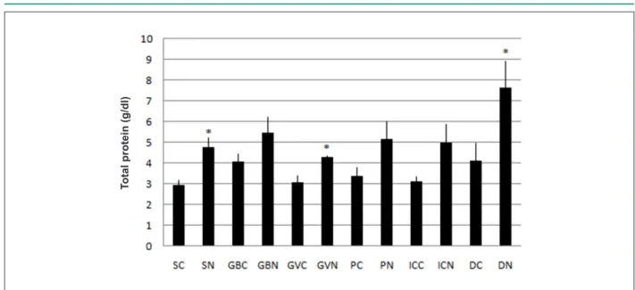
the soleus muscle, red gastrocnemius muscle and diaphragm muscle. However, in the white gastrocnemius muscle, pectoral muscle and intercostal muscle, no significant difference was found (Figure 2).
Finally, we assessed the plasma biochemical profile and hematocrit profile. With respect to changes in the metabolism, there was no difference in blood glucose levels and in the plasma concentration of free fatty acids. Similarly, there was difference neither in body weight nor in the following structures: heart, prostate or epididymal fat. However, in the hemacrocrit evaluation, it was possible to see that the percentage of erythrocytes in the treated group was 18% higher than in the control group. However, it is important to highlight that the other parameters evaluated were not statistically significant (Table 2).
Discussion
The classic therapeutic indication of AAS is intended to address androgenic deficiencies or pathological conditions that may cause deficiency in the protein metabolism10. It is
known that, in and out of sports, nandrolone is widely used. This steroid is administered at high doses with the purpose of making aesthetic changes and improving performance.
However, together with this practice, the literature shows an increasing rate of mortality among users of this substance28.
The electrocardiographic evaluation revealed changes in the electrical pattern of the cardiac muscle represented by an increase in the time to propagate the signal in the ventricular chamber, as the QRS became longer. This fact was accompanied by a similar event that was observed in the QTc interval, which was also high and indicated a delay in the processes that are inherent in the re-polarization of ventricular chambers.
A point to consider is that the presence of estrogen receptors has been demonstrated in a variety of tissues, including in the cardiovascular system29. In particular, in relation to muscle
tissue, the expressed receptors are of the ERa type. This expression has already been characterized in mice, rats, cattle and humans, and its action is linked to the regulation of cellular energy metabolism that can act both through the nongenomic, cytosolic pathway and through the genomic pathway30. As these
receptors act on metabolism, the fact that the action of the nandrolone is multifactorial and represented by the capacity to modify the expression of androgen receptors, the affinity and activity of post-receptor pathways. Moreover, there is a direct relationship between changes in adrenergic sensitivity of the sinoatrial node and the presence of steroids. This may indicate
Table 1 -Electrocardiographic parameters and heart rate of rats in the group treated with nandrolone (Deca-Durabolin™; 5 mg/kg). The values correspond to the mean ± sem, n=23
Weeks QRS (ms) QT (ms) QTc (ms) HR (bpm)
Pre-treatment 92.57 ± 5.42 210.14 ± 8.55 341.09 ± 15.06 186.46 ± 8.53
Week 1 94.46 ± 7.62 210.3 ± 8.47 408.1 ± 1.9* 203.69 ± 4.43*
Week 2 128.36 ± 9.53* 200.3 ± 6.65 367.07 ± 1.2* 199.61 ± 4.08
Week 3 106.18 ± 8.25 196.92 ± 5.4 338.75 ± 1.79 199.83 ± 8.72
* Signiicant (p> 0.05), compared to the pre-treatment week.
Figure 1 -Glycogen content (mg/100 mg) of soleus muscle (S), white gastrocnemius muscle (WG), red gastrocnemius muscle (RG), pectoral muscle (P), intercostal muscle (IC), diaphragm muscle (D) and left ventricle (V) of rats in the control group (C) and in the group treated with nandrolone (N, Deca-Durabolin™; 5 mg/kg). The values correspond to the mean ± sem, n=23. * p<0.05, compared to control.
G
ly
c
o
g
e
n
(m
g
/1
0
0
m
g
Table 2 - Body weight, biochemical and hematological parameters of rats in the control group and the group treated with nandrolone (Deca-Durabolin™; 5 mg/kg). The values correspond to mean ± sem, n=23
Control Nandrolone
FFA (mmol/l) 0.41±0.1 0.47±0.07
Blood glucose (mg/dl) 139.42±11 139.42±9.1
Epididymal fat (g) 6.32±0.49 7.11±0.8
Prostate (g) 0.64±0.06 0.57±0.04
Albumin (g/dl) 1.69 ± 0.07 1.93 ± 0.05
Body weight (g) 404.46 ± 9.3 433.68 ± 6.3
Heart weight (g) 1.382 ± 0.4 g 1.430 ± 0.4
Hematocrit (%) 43.9 ± 1.6 51 ± 2.4*
* Signiicant (p> 0.05) compared to controls.
Figure 2 -Content of total proteins (g/dl) in the soleus muscle (S), white gastrocnemius muscle (WG), red gastrocnemius muscle (RG), pectoral muscle (P), intercostal muscle (IC) and diaphragm muscle (D) of rats in the control group (C) and the group treated with nandrolone (N, Deca-Durabolin™; 5 mg/kg). The values correspond to the mean ± sem, n=23. * p<0.05, compared to control.
T
o
ta
l
p
ro
te
in
(g
/d
l)
that in the presence of a supraphysiological dose of nandrolone, there may be changes in the sensitivity of the heart, which can be expressed by the increase in the population of adrenergic β receptors, thereby causing super-sensitiveness in the cardiac pacemaker, as suggested by Norton et al31. It is important to note
that heart rate also rose, indicating a change in the sensitivity/ activity interface of the sinoatrial node.
If we consider that nandrolone may increase metabolic patterns, another option here is directed at a cardiotropic action with a biochemical base, because the nandrolone can stimulate the activation of the glucose-6-phosphate, 6-phosphogluconate dehydrogenase and isocitrate dehydrogenase enzymes that are present in the cardiac muscle32. At first, this change in the biochemical pattern of
cardiac fibers causes an increase in the generation of NADPH and consequently, a change in the time to convey the electrical signal, thereby overloading the cardiac function and increasing
the systole time of the ventricular chamber.
When we evaluated the glycogen content of the cardiac muscles, we observed that in the presence of nandrolone, there was an increase in reserves. This increase in glycogen content may reflect the action of the steroid, which is capable of changing the tissue responsiveness to other hormones, such as the IGF, which is the insulin-like growth factor. This shows some potent action on the glycogenic pathway, thereby favoring the formation of this reserve of metabolizable substrates. Thus, we suggest that, besides the increase in the functional requirement and metabolic activity of the cardiac muscle, changes occur in the functional relationships that can be characterized by an increase in the uptake and metabolism of substrates, with consequent increase in energy use, as suggested Falkenberg et al15. In other words, nandrolone acts
on the cardiac muscle and stimulates a rise in heart rate and metabolic rate, but in contrast, it may increase energy reserves, causing the heart to have a greater intake of metabolizable substrates and implementing a metabolic status that meets the largest demand. In this sense, a sign that suggests a rise in energy needs may be the rise in hematocrit, indicating that it is part of the responsiveness that is enabled in the early moments.
One needs to consider that the anabolic action, which is expressed by an increase in weight of some organs and which is inherent in nandrolone, has not been verified and it is possibly due to the experimental observation period, since it deals with actions related to nuclear codes expressed in the long term. This fact suggests that the effects that are inherent in the action of steroids may have a time relationship.
Conclusion
the relationship between nandrolone and the genesis of these changes, which indicate overloading of cardiac muscles, which is something that can cause arrhythmia and sudden death.
Potential Conflict of Interest
No potential conflict of interest relevant to this article was reported.
Sources of Funding
There were no external funding sources for this study.
Study Association
This article is part of the thesis of master submitted by Adriano Cesar Rocco Pardi, from Universidade Metodista de
Piracicaba.
References
1. Snyder PJ. Androgens. In: Hardman Limbird JGLE, Goodman & Gilman A. (eds). The pharmacological basis of therapeutics, 10th ed. New York: McGraw Hill; 2001. p. 1635-48.
2. Gebara OCE, Vieira NW, Meyer JW, Calich ALG, Tai EJ, Pierri H, et al. Efeitos cardiovasculares da testosterona. Arq Bras Cardiol. 79 (6): 644-9.
3. Larsson CE, Farias MR, Andrade SF, Brito AF. Terapêutica tópica e sistêmica: pele, ouvido e olho. In: Andrade SF. Manual de terapêutica veterinária. 2ª ed. São Paulo: Roca; 2001. p. 116-78.
4. Simonson E, Kearns WM, Enger M. Effects of methyl testosterone treatment on muscular performance and the central nervous system of older men. J Clin Endocrinol. 1974; 4 (11): 528-34.
5. Ryan AJ. Anabolic steroids are fool´s gold. Fed Proc. 1981; 40 (12): 2682-8.
6. Cunha TS, Tanno AP, Marcondes FK, Perez SEA, Selistre-Araujo HS. A administração de nandrolona não promove hipertrofia do músculo sóleo em ratos. Arq Bras Endocrinol Metab. 2006; 50 (3): 532-40.
7. Kutscher EC, Lund BC, Perry PJ. Anabolic steroids: a review for the clinician. Sports Med. 2002; 32 (5): 285-96.
8. Wilson JD. Androgen abuse by athletes. Endocr Rev. 1988; 9 (2): 181-99.
9. Lise MLZ, Gama e Silva TS, Ferigolo M, Barros HMT. O abuso de esteróides anabólicoandrogênicos em atletismo. Rev Ass Med Bras São Paulo. 1999; 45 (4): 364-70.
10. Creutzberg EC, Schols AMWJ. Anabolic steroids. Curr Opin Clin Nutr Metab Care. 1999; 2: 243-53.
11. Yesalis CE. Anabolic steroids in sport and exercise. Champaign, (IL): Human Kinetics; 1993. p. 325-9.
12. Kuipers H, Wijnen JA, Hartgens F, Willems SM. Influence of anabolic steroids on body comosition, blood pressure, lipid profile and liver functions in body builders. Int J Sports Med. 1991; 12 (4): 413-8.
13. Gruber AJ, Pope HG Jr.. Psychiatric and medical effects of anabolic-androgenic steroid use in women. Psychother Psychosom. 2000; 69 (1): 19-26.
14. Pope Jr HG, Katz DL. Affective and psychotic symptoms associated with anabolic steroid use. Am J Psychiatry. 1988; 145 (4): 487-90.
15. Falkenberg M, Karlsson J, Ortenwall P. Peripheral arterial thrombosis in two young men using anabolic steroids. Eur J Vasc Endovasc Surg. 1997; 13 (2): 223-6.
16. Di Bello V, Giorgi D, Bianchi M, Bertini A, Caputo MT, Valenti G. Effects of anabolic-androgenic steroids on weight-lifters´ myocardium: an ultrasonic videodensitometric study. Med Sci Sports Exerc. 1999; 31 (4): 514-21.
17. McCarthy K, Tang ATM, Dalrymple-Hay MJR, Haw MP. Ventricular thrombosis and systemic embolism in bodybuilders: etiology and management. Ann Thorac Surg. 2000; 70 (2): 658-60.
18. Ferrera PC, Putnam DL, Verdile VP. Anabolic steroid use as the possible precipitant of dilated cardiomyopathy. Cardiology. 1997; 88 (2): 218-20.
19. Baldwin J, Snow RJ, Gibala MJ, Garnham A, Howarth K, Febbraio MA. Glycogen availability does not affect the TCA cycle or TAN pools during prolonged, fatiguing exercise. J Appl Physiol. 2003; 94 (6): 2181-7.
20. Romijn JA, Coyle EF, Sidossis LS, Gastaldelli A, Horowitz JF, Endert E. Regulation of endogenous fat and carbohydrate metabolism in relation to exercise intensity and duration. Am J Physiol. 1993; 265 (3): E380-91.
21. Lima-Silva AE, Fernandes TC, De-Oliveira FR, Nakamura FY, Gevaerd MS. Metabolismo do glicogênio muscular durante o exercício físico: mecanismos de regulção. Rev Nutr. 2007; 20 (4): 417-29.
22. Chan CB, De Leo D, Joseph JW, McQuaid TS, Ha XF, Xu F, et al. Increased uncoupling protein-2 levels in beta-cells are associated with impaired glucose-stimulated insulin secretion: mechanism of action. Diabetes. 2001; 50 (6): 1302-10.
23. Siu LO, Russeau JC, Taylor AW. Determination of glycogen in small tissue samples. J Apll Physiol. 1970; 28 (2): 234-6.
24. Regouw BJM, Cornelissem PJH, Helder R, Spijkers JBF, Weeber YMM. Specific determination of the free fatty acid in plasma. Clin Chin Acta. 1971; 31 (1): 187-95.
25. Petermann CED, Silva CA. Tratamento com nifedipina, um bloqueador de canais de cálcio, melhora o conteúdo muscular de glicogênio de membro imobilizado de ratos. Rev bras fisioter. 2005; 9 (2): 181-6.
26. Chingui LJ, Branquinho RP, Munhoz MT, Silva CA. Comportamento quimiometabólico do músculo sóleo na fase aguda da imobilização articular. Fisioterapia e Pesquisa. 2008; 15 (2): 194-9.
27. Albino WJ, Silva CA, Taliari KRS. Suplementação com glutamina melhora as reservas de glicogênio de músculo de ratos tratados com dexametasona. Rev bras Educ Fís Esp. 2004; 18 (3): 283-91.
28. Silva PRP, Danielski R, Czepielewski MA. Esteróides anabolizantes no esporte. Rev Bras Med Esporte. 2002; 8 (6): 235-43.
29. Gustafsson JA. What pharmacologists can learn from recent advances in estrogen signaling? Trends Pharmacol Sci. 2003; 24 (9): 479-85.
30. Glenmark B, Nilsson M, Gao H, Gustafsson JA, Dahlman-Wright K, Westblad H. Difference in skeletal muscle function in males vs. female: role of estrogen receptor-β. Am J Physiol Endocrinol Metab. 2004; 287 (6): E1125-E1131.
31. Norton GR, Trifunovic B, Woodiswiss AJ. Attenuated beta-adrenoceptor-mediated cardiac contractile responses following androgenic steroid administration to sedentary rats. Eur J Appl Physiol. 2000; 81 (4): 310-6.