Regulation of muscle plasticity and trophism by fatty acids:
A short review
PHABLO ABREU1*, JOSÉ HENRIQUE LEAL-CARDOSO2, VÂNIA MARILANDE CECCATTO2, SANDRO MASSAO HIRABARA1,3
1Department of Physiology and Biophysics, Institute of Biomedical Sciences, Universidade de São Paulo, São Paulo, SP, Brazil 2Department of Physiology, Institute for Biomedical Sciences, Universidade Estadual do Ceará, Fortaleza, CE, Brazil
3Interdisciplinary Graduate Program in Health Sciences, Institute of Physical Activity and Sport Sciences, Universidade Cruzeiro do Sul, São Paulo, SP, Brazil
S
UMMARYStudy conducted at Universidade de São Paulo, Universidade Estadual do Ceará and Universidade Cruzeiro do Sul, Brazil
Article received: 6/17/2016
Accepted for publication: 6/26/2016
*Correspondence:
Departamento de Fisiologia e Biofísica, ICB-USP Address: Av. Prof. Lineu Prestes, 2415, São Paulo, SP – Brazil Postal code: 05508-900
[email protected] [email protected]
http://dx.doi.org/10.1590/1806-9282.63.02.148
The skeletal muscle tissue has a remarkable ability to alter its plastic structural and functional properties after a harmful stimulus, regulating the expression of proteins in complex events such as muscle regeneration. In this context, considering that potential therapeutic agents have been widely studied, nutritional strategies have been investigated in order to improve the regenerative capacity of skeletal muscle. There is evidence of the modulatory action of fatty acids, such that oleic and linoleic acids, that are abundant in Western diets, on muscle function and trophism. Thus, fatty acids appear to be potential candidates to promote or impair the recovery of muscle mass and function during regeneration, since they modulate intracellular pathways that regulate myogenesis. This study is the irst to describe and discuss the effect of fatty acids on muscle plasticity and trophism, with emphasis on skeletal muscle regeneration and in vitro differentiation of muscle cells.
Keywords: cell differentiation, muscle repair, skeletal muscle, satellite cells, fatty acids.
I
NTRODUCTIONSkeletal muscles have the plastic ability to adapt to the intrinsic and extrinsic demands of the environment. Such adaptive potential is attributed to the population of stem cells resident in adult skeletal muscle, known as satellite cells.1 These are mononuclear and undifferentiated
satel-lite cells located between the basal lamina and the sarco-lemma of a muscle iber, which proliferate, differentiate and fuse leading to the formation of a new myoiber and, thus, the reconstitution of the contractile apparatus.2,3
The process of muscle regeneration is triggered by a
noxious stimulus whose nature can be mechanical,3
chemical,4 or thermal.5 Skeletal muscle repair, triggered
by sarcolemmal rupture and increased vascular permeabil-ity, involves cellular and molecular events that begin with increased calcium inlux into the intracellular environment causing proteolysis dependent on that cation, necrosis of damaged tissue, and activation of inlammatory response at the lesion site. This phase is followed by the production of extracellular matrix proteins, revascularization and concomitant activation of myogenic cells.2,3,5,6
Several groups have investigated therapeutic strate-gies to accelerate the skeletal muscle repair process after injury. There is evidence that fatty acids modulate
mus-cle function and trophism.7-11 Our article is the irst to
S
KELETAL MUSCLE SATELLITE CELLSInitially described in frog muscle ibers,13 skeletal muscle
satellite cells, undifferentiated and mononucleated, have this name because of their anatomical positioning at the periphery of the muscle iber, between the basal lamina and the plasma membrane. They represent between 2 and 10% of the total myonuclei per muscle iber and total 2 x 10⁵ to 1 x 10⁶ cells per gram of muscle.14
Muscle ibers are differentiated cells, unable to un-dergo division. The ability of muscles to self-repair has been attributed to satellite cells, which have high mitotic capacity, contributing to the maintenance and regenera-tion of adult skeletal muscle.14
In his article, Mauro13 deined these satellite cells as
“myoblastic cells in the adult organism, which failed to merge with other myoblasts. These cells are juxtaposed to reca-pitulate the embryonic development of skeletal muscle ibers”. Subsequently, studies have shown that asymmetric divisions of satellite cells generate myogenic cells, which originate myoblasts, myocytes and myoibers.15 Symmetric divisions,
on the other hand, generate new satellite cells that expand the number of these cells, a process known as self-renewal. Multiple factors are part of this complex network of satellite cell growth and differentiation, governing the cell cycle pro-gression and/or the return to a quiescent state (G0).15
In the absence of stimuli in the muscle tissue, the satel-lite cells remain in the G0 state. Once activated, these cells initiate the cell cycle, proliferate and express myogenic growth and differentiation markers.1 Thus, a balance between G0
and the active state (self-renewal or myogenic differentiation) is indispensable for the conservation of muscle tissue.16
Studies have shown that the number of satellite cells remains constant even after multiple activations.17 Currently, many
intrinsic and extrinsic factors that control the satellite cell function have been discovered. In all, these studies have demonstrated that there are speciic cell cycle activations and inhibitions, and progression to myogeny.
Transcription factor PAX7 (meaning paired box) was the irst marker identiied in satellite cells in quiescent state, being activated during proliferation. It has a key function in the maintenance of the G0 state and in the prevention of early myogenic differentiation.15 Studies
have demonstrated the complete loss of muscle regen-erative capacity inPAX7knock-out mice (PAX7-/-).18,19
In this context, increasing evidence shows that satel-lite cells are composed of two different populations that regulate the cell cycle:(1) those with stem cell potential, undifferentiated and which remain in G0 state during myogenic progression, and (2) those with potential for myogenic differentiation.20
After muscle injury, satellite cells are activated, initi-ating the expression of regulatory factors of myogenesis, such as myoblast differentiation (MYOD) and/or myo-genic factor 5 (MYF5).21 MYOD is expressed in extremely
low amounts and is essentially undetectable in quiescent satellite cells. This protein marks the compromise of myo-blasts with the myogenic lineage. In this context, the concomitant expression of MYOD and MYF5 is vital for the formation of myotubes and myoibers.1,22
Promotion of myoibroblast restoration and reorga-nization results from a decrease in PAX7 expression, cell cycle arrest and increased expression of myogenin (MYOG)23 andmyogenic regulatory factor 4(MRF4),24
both members of the superfamily of basichelix-loop-helix (bHLH) transcription factors. These factors are speciic to skeletal muscle, being expressed at distinct moments dur-ing myogenesis. They have key functions in myogenic speciication, muscle differentiation and maintenance during muscle development and regeneration22 (Figure 1).
E
FFECTS OF FATTY ACIDS ON SKELETAL MUSCLE TROPHISM AND REGENERATIONThe regulation of trophism and muscle regeneration in-volves the coordinated action of various cell types in re-sponse to local and systemic signals. It is slow and often incomplete depending on its severity, leading to loss of function.26 Thus, the discovery of new dietary strategies
to improve skeletal muscle regeneration capacity can be a powerful tool for the development of new nutritional therapies in order to accelerate regenerative processes and/or reduce the consequences of incomplete repair and extensive ibrosis deposited in the skeletal muscle, as oc-curs after severe muscle injuries.
The composition of phospholipids in the plasma membrane has a crucial inluence on cell growth and metabolic activity. In the last two decades, it has been suggested that the lipid composition of the diet inlu-ences the fatty acid proile of the serum and the lipid content of the plasma membrane.27-29 In fact, it has been
shown that the length of the fatty acid chain and the degree of saturation or unsaturation alter the luidity and activity of several membrane-bound proteins.30,31
In this context, few studies have evaluated the involve-ment of fatty acids in muscle trophism and myogenesis. Muscle differentiation is known to be accompanied by important metabolic changes, such as increased expres-sion of genes related to the metabolism of carbohydrates and amino acids.32,33 In this context, some nutritional
leucine during the muscle regeneration process accelerates the repair of connective tissue in the anterior tibial muscle of rats. Baptista et al.33 investigated the effect of
supplemen-tation with leucine and HMB (β-hydroxy-β-methylbutyrate) on the ubiquitin-muscle proteasome system under differ-ent sarcopenia conditions in rats, demonstrating the anti-atrophic effect of leucine.32,33
Oleic acid (monounsaturated 18:1 (n-9)) and linoleic acid (polyunsaturated 18:2 (n-6)) are the most abundant fatty acids in Western diets. Oleic acid is found mainly in olive oil, while linoleic acid is found in soy, sunlower and corn oils.34 Using both isolated muscle cells and animal
models, Salvadó et al.35 demonstrated that oleic acid is
capable of reversing the structural and metabolic chang-es in skeletal muscle induced by palmitic acid. In an-other study, the increase in docosahexaenoic acid (DHA)
content (22:6 (n-3)) in the gastrocnemius muscle through supplementation with ish oil for 21 days is suggestive that this fatty acid attenuates lipopolysaccharide-induced muscle atrophy (LPS).36 Other studies suggest that the
reduction in DHA content impairs calcium homeostasis in the skeletal muscle cell.37 Tuazon and Henderson38
observed that increases in linoleic acid content and decline in DHA content in muscle phospholipids were positively correlated with increased creatine kinase activity, com-bined with decline in muscle grip strength of dystrophin knock-out animals. Another study shows that there is an inverse relationship between the concentration of oleic and linoleic acid in skeletal muscle.39
It has been demonstrated that some fatty acids, such as oleic and linoleic acids, exert pro-proliferative effects on vascular smooth muscle, and may regulate muscle
FIGURE 1 Temporal activation of regulatory factors of myogenic differentiation. In skeletal muscle, satellite cells in the quiescent state or activated during proliferation express the paired-box 7 (PAX7) transcription factor. These cells have the ability to proliferate, self-renew, differen-tiate and fuse with newly formed myotubes or existing myoibers after stimuli such as muscle injury. Differentiation of satellite cells involves increased expression of basic helix-loop-helix (bHLH), myogenic factor 5 (MYF5) and myoblast determination (MYOD) transcription factors. Myogenin (MYOG), a transcription factor essential for myogenesis and skeletal muscle repair, is highly expressed during the formation of myocytes by the fusion of myoblasts. Myogenic regulatory factor 4 (MRF4) transcription factor is, then, activated during terminal differentiation and formation of myocytes and myotubes. (Modiied from Wang and Rudnicki).25
Embryonic progenitor cells
Satellite stem cells
Myogenic satellite cells
Myogenic progression
Myotubes Myoibers
PAX7
MYF5
MYOD
MYOG
MRF4
Speciication
Myoblasts
Beginning of differentiation
Terminal differentiation
A
ct
iv
at
io
n
Prolif
er
ation
Cell commitment
growth.40 Perdiconi et al.41 observed an increase in total
phospholipid content during muscle differentiation. Myo-blasts predominantly synthesize triacylglycerols, while myotubes synthesize phospholipids.42 The fusion of
myo-blasts can also be regulated by factors that alter the luid-ity of the plasma membrane, such as temperature and lipid composition.43,44
The fatty acid composition of phospholipids deter-mines the physicochemical properties of the plasma mem-brane and, to a large extent, its asymmetry, luidity, plastic-ity, organization and occurrence of microdomains.45 The
incorporation of omega-3 or -6 polyunsaturated fatty acids into membrane phospholipids affects lipid and pro-tein interactions in the membrane, in addition to the physical properties mentioned above.45 For example, the
decrease in insulin sensitivity in skeletal muscle has been associated with a decrease in the proportion of polyun-saturated fatty acids in membrane phospholipids.46
There is evidence that intermediate products of fatty acid metabolism are important for the survival, prolif-eration, differentiation, and fusion of myoblasts.47
Rode-man and Goldberg48 suggested that lipid metabolites
derived from polyunsaturated fatty acids, such as arachi-donic acid, accelerate protein synthesis, fusion and growth of muscle cells in different animal models.
Oxidation of fatty acids is signiicantly higher in myo-tubes compared to myoblasts, and mitochondrial biogen-esis is necessary for skeletal muscle differentiation.42 The
content of triacylglycerols decreases by more than 50%
during myogenesis,41 and the inhibition of
mitochon-drial respiration compromises myogenic differentiation and the formation of myotubes.49 Leptin knock-out mice
(ob/ob), which have high concentrations of plasma fatty acids, present deicient muscle regeneration.50 In addition,
a hyperlipidic diet compromises muscle regeneration in mice,51 possibly by the effect of saturated fatty acids.
Pinheiro43 observed that there is an increase in the
synthesis of oleic and arachidonic acids during myogen-esis in vitro. The addition of arachidonic and linoleic acids to the culture medium increases the proliferation of satellite cells, as assessed by the incorporation of 14
Car-bon-labeled thymidine. The author also observed that supplementation with linoleic acid for 20 days in dystro-phic mice (mdx) signiicantly improves the strength of the gastrocnemius muscle of these animals, suggesting a possible trophic effect of this fatty acid.43 On the other
hand, the diet rich in saturated fatty acid and linoleic acid causes insulin resistance and imbalance of oxidative components in skeletal muscle, resulting in oxidative stress. Pariza et al.52 demonstrated in vivo that
supple-mentation with conjugated linoleic acid (0.3% of diet) causes a decrease in fat mass and an increase in fat-free lean mass in rodents.
Our group evaluated the effect of treatment with oleic and linoleic acids (0.44 g per kg body weight) for four weeks on lacerated gastrocnemius muscle regenera-tion in rats (unpublished data). Laceraregenera-tion per se causes an increase in the oleic/stearic and palmitoleic/palmitic ratio indicators of the desaturase activity and promotes a reduction of speciic isotonic and speciic absolute te-tanic forces. There is also a drop in resistance to fatigue and an increase in the area of ibrous tissue. These indings indicate incomplete regeneration and partial recovery of the contractile function of the injured muscle. Linoleic acid supplementation decreases the mass, speciic iso-tonic strength, fatigue resistance, and cross-sectional area of the contralateral and injured gastrocnemius muscle ibers, as well as increases the area of ibrous tissue in the injured muscle. Supplementation with oleic acid, on the other hand, does not modify the mass and the cross--sectional area of the ibers of the gastrocnemius muscle; it suppresses the decrease in speciic isotonic force and the increase in the area of ibrous tissue induced by the injury, prevents tetanic forces (absolute and speciic), and increases the resistance to fatigue in the contralateral and injured gastrocnemius muscles. Based on these indings, we conclude that supplementation with linoleic acid compromises the regeneration of the injured skeletal muscle, causing muscle mass reduction, ibrous tissue elevation, and, consequently, impairment of contractile function. Oleic acid, in turn, attenuates incomplete repair actions, optimizing the regenerative capacity and the contractile function of the injured muscle.
E
FFECTS OF FATTY ACIDS ON MUSCLE CELL DIFFERENTIATIONThe effects of fatty acids on ibroblast proliferation53,54
and myogenic differentiation in isolated cells have been investigated.7-11 In 1978, Horwitz et al.55 observed that
the fatty acids added to the culture medium have a stim-ulatory effect on the fusion of embryonic myoblasts. The authors observed that the lipid composition of the mem-brane inluences the proliferation and fusion of myoblasts and, consequently, the formation of multinucleated myotubes. In 1985, Allen et al.56 observed that linoleic
observed that the presence of mitogenic agents in culture medium and the subsequent increase in proliferation prevent differentiation.
Lu et al.57 observed that adding fatty acids to the culture
medium induced proliferation of vascular smooth muscle, and that oleic acid had a more pronounced effect on the stimulation of proliferation, an effect associated with the activation of protein kinase C (PKC). Hurley et al.8 compared
the effect of different fatty acids on the differentiation of L6 myoblast in vitro. To assess the degree of differentiation, the authors quantiied protein and DNA contents, as well as creatine kinase activity (CK/DNA). The effects on differen-tiation were accompanied by analysis of peroxisome prolif-erator-activated receptor alpha and gamma (PPAR-α and -g) receptor activity to establish the possible association of these transcription factors with the differentiation process. They observed that linoleic acid stimulates differentiation at low concentrations (50 μM) and oleic acid at all concentrations tested (12.5 to 100 μM) without the involvement of the activation of the PPARs evaluated.
Lee et al.7 investigated the effect of fatty acids on the
proliferation and differentiation of C2C12 myoblasts, as well as the possible involvement of the mitogen activated protein kinases (MAPK) in this process. The authors have found that linoleic and oleic acids increase cell proliferation and differentiation, with phosphorylation of extracellular signal-regulated kinases 1 and 2 (ERK1/2) and c-Jun N--terminal kinases (JNK) occurring during proliferation, but not during differentiation. Markworth and Cameron-Smith9
evaluated the effect of arachidonic acid treatment on C2C12 myoblasts and observed that arachidonic acid stimulates the growth of myoblasts in a dose-dependent manner at concentrations between 1.6 and 25 µM. There was an in-crease in myonuclei during myogenesis, regardless of changes in cell density or extent of myogenic differentiation. To verify the effect of arachidonic acid treatment on hy-pertrophy in myotubes, the authors cultured C2C12 myo-blasts in differentiation medium for 72 hours. Then, they added arachidonic acid (25 μM) to the medium containing the already differentiated myotubes. Researchers have found that hypertrophy is greater in arachidonic acid-treated myotubes compared to untreated cells.
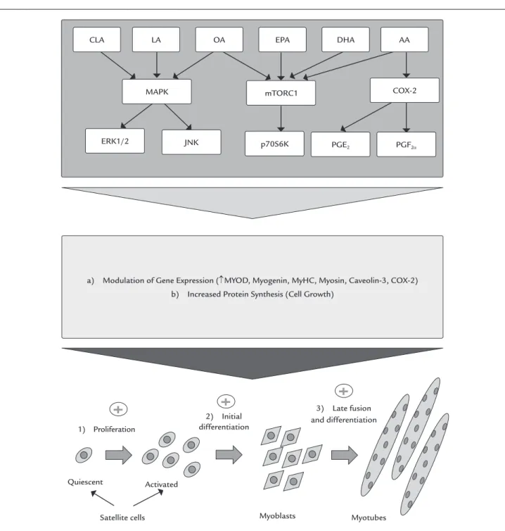
Briolay et al.10 showed that oleic (18:1 (n-9)),
arachi-donic (20:4 (n-6)), eicosapentaenoic (EPA) (20:5 (n-3)) and DHA (22:6 (n-3)) (20 µM) acids stimulate the myogenic differentiation of L6 myoblast. These fatty acids alter the lipid composition of the membrane and, during myogen-ic differentiation, promote phosphorylation of ribosomal protein S6 kinase beta-1 – 70 KDa (p70S6K1) and activation of mammalian target of rapamycin complex 1 (mTORC1),
an important cell cycle regulator and protein synthesis, without alteration of Akt phosphorylation. These results support the proposition that the fatty acid composition of the plasma membrane can control the activity of complex signaling pathways of myogenic differentiation. In this context, the treatment of isolated myoblasts with arachi-donic acid rapidly stimulates protein turnover(synthesis and degradation)48 (Figure 2).
Our group also assessedthe effect of oleic and linoleic fatty acids (100 μM) on myoblast differentiation, myotubes growth and ibroblast proliferation in primary culture (data still unpublished). Treatment of ibroblasts with linoleic acid decreases mRNA expression of proliferating cell nu-clear antigen (PCNA), collagen and ibronectin. Oleic acid, in turn, increases the content of MYOD mRNA in myoblasts,
increases desmin in previously differentiated myotubes, and inhibits mRNA expression of PCNA, collagen and ibronec-tin in ibroblasts. We conclude that oleic acid, in vitro, has a modulatory effect on the differentiation of satellite stem cellsand on the growth and maturation of myotubes.
F
INAL CONSIDERATIONSRecently, studies have revealed signiicant advances in the knowledge of mechanisms involved in the activation, proliferation and differentiation of muscle cells, as well as on fundamental processes of muscle trophism and plasticity. Dietary strategies have been investigated with a view to improving skeletal muscle regenerative capacity after injury. In this context, different fatty acids, such as oleic and linoleic acids, which are abundant in Western diets, have demonstrated in vitro modulatory effects on muscle cell differentiation, and in vivoeffectson muscle plasticity and trophism, with an emphasis on regeneration of skeletal muscle. Recent evidence on the regulatory ac-tion of fatty acids on muscle funcac-tion and muscle mass has been described and discussed in this review.
A
CKNOWLEDGMENTSThis study had inancial support from Fundação de Am-paro à Pesquisa do Estado de São Paulo (FAPESP), Coorde-nação de Aperfeiçoamento de Pessoal de Nível Superior (CAPES), Conselho Nacional de Desenvolvimento Cientí-ico e TecnológCientí-ico (CNPq), Instituto Nacional de Ciência e Tecnologia em Obesidade e Diabetes (INOD), Pró-reitoria de Pesquisa da Universidade de São Paulo (PRP/USP) and Pró-reitoria de Pós-graduação e Pesquisa da Universidade Cruzeiro do Sul (PRPGP/Universidade Cruzeiro do Sul).
C
ONFLICT OF INTEREST1) Proliferation
Satellite cells Myoblasts Myotubes
Quiescent Activated
2) Initial differentiation
a) Modulation of Gene Expression (↑MYOD, Myogenin, MyHC, Myosin, Caveolin-3, COX-2) b) Increased Protein Synthesis (Cell Growth)
3) Late fusion and differentiation
CLA LA OA EPA DHA AA
MAPK mTORC1 COX-2
PGF2α PGE2
p70S6K JNK
ERK1/2
FIGURE 2 Mechanisms possibly involved in the action of different fatty acids on the proliferation and differentiation of skeletal muscle cells. For details, see “Effects of fatty acids on muscle cell differentiation”.
R
ESUMORegulação da plasticidade e do troismo muscular pelos ácidos graxos: uma breve revisão
O tecido muscular esquelético possui a notável capacida-de plástica capacida-de alterar suas propriedacapacida-des estruturais e fun-cionais após um estímulo lesivo, regulando a expressão de proteínas durante eventos complexos como a regene-ração muscular. Nesse contexto, considerando que pos-síveis agentes terapêuticos vêm sendo amplamente estu-dados, estratégias nutricionais têm sido investigadas na perspectiva de melhorar a capacidade regenerativa do músculo esquelético. Há evidências da ação modulatória dos ácidos graxos, como os ácidos oleico e linoleico, que são abundantes nas dietas ocidentais, sobre a função muscular e o troismo. Nesse sentido, os ácidos graxos parecem ser potenciais candidatos para promover ou prejudicar a recuperação da massa e a função muscular durante a regeneração, uma vez que modulam vias intra-celulares reguladoras da miogênese. Este trabalho é o primeiro a descrever e discutir o efeito dos ácidos graxos sobre a plasticidade e o troismo muscular, com ênfase na regeneração do músculo esquelético e na diferenciação de células musculares in vitro.
Palavras-chave: diferenciação celular, reparo muscular, músculo esquelético, células satélites, ácidos graxos.
R
EFERENCES1. Chargé SB, Rudnicki MA. Cellular and molecular regulation of muscle regeneration. Physiol Rev. 2004; 84(1):209-38.
2. Serrano AL, Baeza-Raja B, Perdiguero E, Jardí M, Muñoz-Cánoves P. Interleukin-6 is an essential regulator of satellite cell-mediated skeletal muscle hypertrophy. Cell Metab. 2008; 7(1):33-44.
3. Järvinen TA, Järvinen M, Kalimo H. Regeneration of injured skeletal muscle after the injury. Muscles Ligaments Tendons J. 2014; 3(4):337-45. 4. Conte TC, Franco DV, Baptista IL, Bueno Jr CR, Selistre-de-Araújo HS, Brum
PC, et al. Radicicol improves regeneration of skeletal muscle previously damaged by crotoxin in mice. Toxicon. 2008; 52(1):146-55.
5. Sugita M, Sugita H, Kim M, Mao J, Yasuda Y, Habiro M, et al. Inducible nitric oxide synthase deiciency ameliorates skeletal muscle insulin resistance but does not alter unexpected lower blood glucose levels after burn injury in C57bl/6 mice. Metabolism. 2012; 61(1):127-36.
6. Chakravarthy MV, Abraha TW, Schwartz RJ, Fiorotto ML, Booth FW. Insulin-like growth factor-I extends in vitro replicative life span of skeletal muscle satellite cells by enhancing G1/S cell cycle progression via the activation of phosphatidylinositol 3’-kinase/Akt signaling pathway. J Biol Chem. 2000;275(46): 35942-52.
7. Lee JH, Tachibana H, Morinaga Y, Fujimura Y, Yamada K. Modulation of proliferation and differentiation of C2C12 skeletal muscle cells by fatty acids. Life Sci. 2009; 84(13-14):415-20.
8. Hurley MS, Flux C, Salter AM, Brameld JM. Effects of fatty acids on skeletal muscle cell differentiation in vitro. Br J Nutr. 2006; 95(3):623-30. 9. Markworth JF, Cameron-Smith D. Arachidonic acid supplementation
enhances in vitro skeletal muscle cell growth via a COX-2-dependent pathway. Am J Physiol Cell Physiol. 2013; 304(1):C56-67.
10. Briolay A, Jaafar R, Nemoz G, Bessueille L. Myogenic differentiation and lipid-raft composition of L6 skeletal muscle cells are modulated by PUFAs. Biochim Biophys Acta. 2013; 1828(2):602-13.
11. Zhang G, Chen X, Lin L, Wen C, Rao S. [Effects of fatty acids on proliferation and differentiation of myoblast]. Wei Sheng Yan Jiu. 2012; 41(6):883-8. 12. Sin DD, McAlister FA, Man SF, Anthonisen NR. Contemporary management
of chronic obstructive pulmonary disease: scientiic review. JAMA. 2003; 290(17):2301-12.
13. Mauro A. Satellite cell of skeletal muscle ibers. J Biophys Biochem Cytol. 1961; 9:493-5.
14. White RB, Biérinx AS, Gnocchi VF. Zammit PS. Dynamics of muscle ibre growth during postnatal mouse development. BMC Dev Biol. 2010; 10:21. 15. Lepper C, Conway SJ, Fan CM. Adult satellite cells and embryonic mus-cle progenitors have distinct genetic requirements. Nature. 2009; 460(7255): 627-31.
16. Bjornson CRR, Cheung TH, Liu L, Tripathi PV, Steeper KM. Rando TA. Notch signaling is necessary to maintain quiescence in adult muscle stem cells. Stem Cells. 2012; 30(2):232-42.
17. Shi X, Garry DJ. Muscle stem cells in development, regeneration, and disease. Genes Dev. 2006; 20:1692-708.
18. Lepper C, Partridge TA, Fan CM. An absolute requirement for Pax7-positive satellite cells in acute injury-induced skeletal muscle regeneration. Development. 2011; 138(17):3639-46.
19. von Maltzahn J, Jones AE, Parks RJ, Rudnicki MA. Pax7 is critical for the normal function of satellite cells in adult skeletal muscle. Proc Natl Acad Sci U S A. 2013; 110(41):16474-79.
20. Ono Y, Boldrin L, Knopp P, Morgan JE, Zammit PS. Muscle satellite cells are a functionally heterogeneous population in both somite-derived and branchiomeric muscles. Dev Biol. 2010; 337(1):29-41.
21. Davis RL, Weintraub H, Lassar AB. Expression of a single transfected cDNA converts ibroblasts to myoblasts. Cell. 1987; 51(6):987-1000.
22. Bentzinger CF, Wang YX, Rudnicki MA. Building muscle: molecular regulation of myogenesis. Cold Spring Harb Perspect Biol. 2012; 4(2):008342. 23. Wright WE, Sassoon DA, Lin VK. Myogenin, a factor regulating myogenesis,
has a domain homologous to MyoD. Cell. 1989; 56(4):607-17.
24. Rhodes SJ, Konieczny SF. Identiication of MRF4: a new member of the muscle regulatory factor gene family. Genes Dev. 1989; 3(12B):2050-61. 25. Wang YX, Rudnicki MA. Satellite cells, the engines of muscle repair. Nat
Rev Mol Cell Biol. 2011; 13(2):127-33.
26. Chan YS, Li Y, Foster W, Horaguchi T, Somogyi G, Fu FH, Huard J. Antiibrotic effects of suramin in injured skeletal muscle after laceration. J Appl Physiol (1985). 2003; 95(2):771-80.
27. Yaqoob P, Sherrington EJ, Jeffery NM, Sanderson P, Harvey DJ, Newsholme EA, et al. Comparison of the effects of a range of dietary lipids upon serum and tissue lipid composition in the rat. Int J Biochem Cell Biol. 1995; 27(3):297-310.
28. Vessby B. Dietary fat and insulin action in humans. Br J Nutr. 2000; 83(Suppl 1):S91-6.
29. Molee W, Bouillier-Oudot M, Auvergne A, Babilé R. Changes in lipid composition of hepatocyte plasma membrane induced by overfeeding in duck. Comp Biochem Physiol B Biochem Mol Biol. 2005; 141(4):437-44. 30. Lee AG. Some principles of membrane structure. Proc Nutr Soc. 1985;
44(2):147-56.
31. Meuillet EJ, Leray V, Hubert P, Leray C, Cremel G. Incorporation of exogenous lipids modulates insulin signaling in the hepatoma cell line, HepG2. Biochim Biophys Acta. 1999; 1454(1):38-48.
32. Pereira MG, Silva MT, Carlassara EO, Gonçalves DA, Abrahamsohn PA, Kettelhut IC, et al. Leucine supplementation accelerates connective tissue repair of injured tibialis anterior muscle. Nutrients. 2014; 6(10):3981-4001. 33. Baptista IL, Silva WJ, Artioli GG, Guilherme JP, Leal ML, Aoki MS, et al. Leucine and HMB differentially modulate proteasome system in skeletal muscle under different sarcopenic conditions. PLoS One. 2013; 8(10):752-7.
34. U.S. Department of Agriculture, A.R.S. Nutrient intakes from food: mean amounts consumed per individual, One Day, 2005–2006, 2008. Available from: http://www.ars.usda.gov/ba/bhnrc/fsrg.
35. Salvadó L, Coll T, Gómez-Foix AM, Salmerón E, Barroso E, Palomer X, et al. Oleate prevents saturated-fatty-acid-induced ER stress, inlammation and insulin resistance in skeletal muscle cells through an AMPK-dependent mechanism. Diabetologia. 2013; 56(6):1372-82.
37. Ye S, Tan L, Ma J, Shi Q, Li J. Polyunsaturated docosahexaenoic acid suppresses oxidative stress induced endothelial cell calcium inlux by altering lipid composition in membrane caveolar rafts. Prostaglandins Leukot Essent Fatty Acids. 2010; 83(1):37-43.
38. Tuazon MA, Henderson GC. Fatty acid proile of skeletal muscle phospholipid is altered in mdx mice and is predictive of disease markers. Metabolism. 2012; 61(6):801-11.
39. Høstmark AT, Haug A. The inverse association between relative abundances of oleic acid and arachidonic acid is related to alpha-linolenic acid. Lipids Health Dis. 2014; 3:76.
40. Kelley DS, Bartolini GL, Newman JW, Vemuri M, Mackey BE. Fatty acid composition of liver, adipose tissue, spleen, and heart of mice fed diets containing t10, c12-, and c9, t11-conjugated linoleic acid. Prostaglandins Leukot Essent Fatty Acids. 2006; 74(5):331-8.
41. Perdiconi MF, Politi LE, Bouzat CB, De Los Santos EB, Barrantes FJ. Myogenic differentiation of the muscle clonal cell line BC3H-1 is accompanied by changes in its lipid composition. Lipids. 1992; 27(9):669-75.
42. Sauro VS, Strick la nd K P. Cha nges in oleic acid ox idation a nd incorporation into lipids of differentiating L6 myoblasts cultured in normal or fatty acid-supplemented growth medium. Biochem J. 1987; 244(3):743-8.
43. Pinheiro, CHJ. Efeito da terapia com tronco musculares e células--tronco mesenguimais na regeneração do músculo esquelético: modulação por ácido oleico. [Tese]. São Paulo: Instituto de Ciências Biomédicas, Uni-versidade de São Paulo; 2012.
44. Prives J, Shinitzky M. Increased membrane luidity precedes fusion of muscle cells. Nature. 1977; 268(5622):761-3.
45. Abbott SK, Else PL, Hulbert AJ. Membrane fatty acid composition of rat skeletal muscle is most responsive to the balance of dietary n-3 and n-6 PUFA. Br J Nutr. 2010; 103(4):522-9.
46. Borkman M, Storlien LH, Pan DA, Jenkins AB, Chisholm DJ, Campbell LV. The relation between insulin sensitivity and the fatty-acid composition of skeletal-muscle phospholipids. N Engl J Med. 1993; 328(4):238-44. 47. Veliça PP, Khanim FL, Bunce CM. Prostaglandin D2 inhibits C2C12
myogenesis. Mol Cell Endocrinol. 2010; 319(1-2):71-8.
48. Rodeman HP, Goldberg AL. Arachidonic acid, PGE2 and F2α inluence rates of protein turnover in skeletal and cardiac muscle. J Biol Chem. 1982; 257(4):1632-8.
49. Hamai N, Nakamura M, Asano A. Inhibition of mitochondrial protein synthesis impaired C2C12 myoblast differentiation. Cell Struct Funct. 1997; 22(4):421-31.
50. Nguyen MH, Cheng M, Koh TJ. Impaired muscle regeneration in ob/ob and db/db mice. Scientiic World J. 2011; 11:1525-37.
51. Hu Z, Wang H, Lee IH, Modi S, Wang X, Du J, et al. PTEN inhibition improves muscle regeneration in mice fed a high-fat diet. Diabetes. 2010; 59(6):1312-20. 52. Pariza MW, Park Y, Cook ME. The biologically active isomers of conjugated
linoleic acid. Prog Lipid Res. 2001; 40(4):283-98.
53. Granados S, Quiles JL, Gil A, Ramírez-Tortosa AC. Dietary lipids and cancer. Nutr Hosp. 2006; 21(Suppl 2):42-52.
54. Magdalon J, Hatanaka E, Romanatto T, Rodrigues HG, Kuwabara WM, Scaife C, et al. Proteomic analysis of the functional effects of fatty acids in NIH 3T3 ibroblasts. Lipids Health Dis. 2011; 10:218.
55. Horwitz AF, Wight A, Ludwig P, Cornell R. Interrelated lipid alterations and their inluence on the proliferation and fusion of cultured myogenic cells. J Cell Biol. 1978; 77(2):334-57.
56. Allen RE, Luiten LS, Dodson MV. Effect of insulin and linoleic acid on satellite cell differentiation. J Anim Sci. 1985; 60(6):1571-79.