Instituto Dante Pazzanese de Cardiologia and Hospital do Coração – São Paulo Maling address: José Carlos Pachón Mateos – Av. Jamaris, 650/72 04078001 -São Paulo, SP - Brazil
Objective - To describe a new more efficient method
of endocardial cardiac stimulation, which produces a narrower QRS without using the coronary sinus or cardiac veins.
Methods - We studied 5 patients with severe dilated
cardiomyopathy, chronic atrial fibrillation and AV block, who underwent definitive endocardial pacemaker im-plantation, with 2 leads, in the RV, one in the apex and the other in the interventricular septum (sub pulmonary), connected, respectively, to ventricular and atrial bica-meral pacemaker outputs. Using Doppler echocar-diography, we compared, in the same patient, conven-tional (VVI), high septal (“AAI”) and bifocal (“DDT” with AV interval ≅ 0) stimulation.
Results - The RV bifocal stimulation had the best
resu-lts with an increase in ejection fraction and cardiac output and reduction in QRS duration, mitral regurgitation and in the left atrium area (p ≤ 0.01). The conventional method of stimulation showed the worst result.
Conclusion - These results suggest that, when left
ven-tricular stimulation is not possible, right venven-tricular bifo-cal stimulation should be used in patients with severe car-diomyopathy where a pacemaker is indicated.
Key words: pacemaker, multisite stimulation and heart failure
Arq Bras Cardiol, volume 73 (nº 6), 492-498, 1999
José Carlos Pachón Mateos, Remy N elson Albornoz, Enrique Indalécio Pachón Mateos, Vera Márcia Gimenez, Juán Carlos Pachón Mateos, Maria Zélia Cunha Pachón, Eusebio Ramos dos Santos Fº, Paulo de Tarso Jorge Medeiros, Marco Aurélio Dias da Silva, Paulo Paredes Paulista, José Eduardo
Moraes Rêgo Sousa, Adib Domingos Jatene
São Paulo, SP - Brazil
Right Ventricular Bifocal Stimulation in the Treatment of
Dilated Cardiomyopathy with Heart Failure
In dilated cardiomyopathy some degree of delay oc-curs in myocardial stimulus conduction, which causes QRS widening. Associated lesions in the conduction system also often cause QRS widening. In these cases, when a cardiac pacemaker is necessary, paced QRS is more enlar-ged, easily achieving 200 ms or even more. The delayed ventricular activation, by itself, provokes systolic and dias-tolic dysfunction, and increases mitral regurgitation 1.
Since the beginning of cardiac pacing, it has been known that the contraction caused by a paced QRS is less effective than the one resulting from a normal QRS. When the QRS is wide, the increased pressure caused by the first stimulated myocardium area is lessened by the natural complacence of other areas that will be activated later. On the other hand, in the normal contraction, the fast myocar-dial cell activation creates a mechanical synergism, extremely favorable for taking maximum advantage of the inotropic state. It causes a pressure wave with high dP/dt, which is a faster, highly efficient rise in pressure. In the dilated myocardium, the activation generated by a pacema-ker is distributed over a longer time, causing a pressure wave that is more attenuated proportionally to the paced QRS widening. To preserve systolic and diastolic func-tions, and reduce mitral insufficiency, it appears to be funda-mental to pace both ventricles with a normal QRS, or at least with the shortest PRS possible. This can be easily obtained by AAI pacing, when the patient has intra- and atrioventri-cular conduction systems preserved. In the case of AV block, the resulting ventricular paced QRS (almost always placed on the right ventricle) is very wide. It is possible to have a narrow QRS simultaneously pacing more than one point. Recent studies have shown narrow QRS and im-proved myocardial contractility, when both ventricles are si-multaneously paced 2. The problem is access to the left
ventricle. The first approach was epicardial, which requires a thoracotomy 3. The alternative is the use of cardiac veins;
thoracoto-my, because of the endocardial access. But technical diffi-culties related to the great anatomical variety of cardiac veins sometimes impede the implantation.
The main purposes of this work were: 1) To suggest an easy way for ventricular pacing, with narrower QRS, through the placement of two leads in the right ventricle, using an endocardial access, without a thoracotomy or coronary sinus access; 2) to propose a study method that allows the testing of hemodynamic efficiency of this kind of pacing compared with conventional heart stimulation, in the same patient, eliminating intervening variables that could occur among different individuals, and permit answers to the following questions: a) Is it possible to partially “resyn-chronize” the left ventricular myocardium, stimulating only the right ventricle? b) The resulting “resynchronization degree” has some beneficial effects in the heart stimulation in cases of severe heart failure of dilated cardiomyopathy? c) Is the implantation technique safe, reproducible and easy to perform with low risk in critical patients?
Methods
Five patients (1 female) with cardiac pacemaker indica-tion were included. Ages ranged from 37 to 66 years (mean of 52.2 ± 17.7 years). All patients had severe dilated cardio-myopathy (class III and IV NYHA), caused by Chagas’ di-sease in 4 cases and idiopathic in one case. All patients had atrial fibrillation with high degree or complete AV block and cardiac failure with radiological cardiac shapes 3 to 4+.
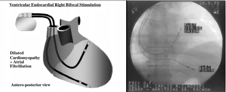
Implantation techniques - Two endocardial leads were implanted in the right ventricle. The first lead (“septal”) was implanted in the His bundle area, and the second lead was placed in the right ventricle apex according to the classical endocardial implantation technique (figure 1). The pacema-ker used in this series was the Biotronik Dromos (DDDR
pacemaker that allows AV delay of 15 ms, in the DDT mode). Ideally, the pacemaker should be able to use 0 ms of AV delay. The “septal” lead was connected to the atrial channel of the pacemaker and the conventional lead to the ven-tricular channel. To avoid the risk of pacemaker syndrome (due to ventricular stimulation) only patients with chronic atrial fibrillation and AV block were selected.
Programming and echocardiographic study - All pa-tients had cardiac insufficiency. Therefore, after the implant the pacemakers were programmed in “DDT” mode, with an AV delay of 15 ms (bifocal VVI pacing). After 2 weeks of evolution, with heart rate corrected by artificial pacing, all patients were evaluated by echocardiography in three pacing modes: 1) By programming the pacemaker to AAI mode (septal stimulation), the His bundle area, or the right ventricle outflow area, was stimulated; 2) By programming the pacemaker to VVI mode, conventional pacing was obtained and; 3) By programming the pacemaker to DDT mode (AV delay = 15 ms), it was possible to stimulate two points (high Septum and Apex), of right ventricle, almost simultaneously (figure 2). Our objective was to stimulate at the same time two distant points of the right ventricle.
All programming was performed in the echocardio-graphy laboratory by maintaining the same heart rate. The patient was under absolute rest, without positional chan-ges. After allowing 5 minutes for hemodynamic stabilization ehocardiographic parameters were measured. Subse-quently, mono and bidimensional Doppler modes were used to measure ejection fraction (EF), cardiac output (CO), functional mitral regurgitation area (MR) and length of QRS complex (in 3 simultaneous derivations DI, DII and DIII). Average values and standard deviation were calculated, using paired t test for equivalent variance series, comparing septal and bifocal right ventricular stimulation with conventional stimulation.
Ventricular Endocardial Right Bifocal Stimulation
Dilated
Cardiomyopathy + Atrial
Fibrillation
Antero-posterior view
Results
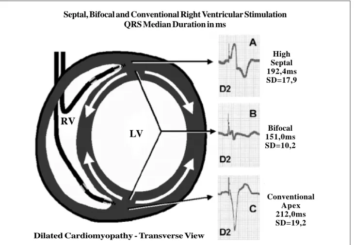
Narrowing the QRS – With bifocal stimulation, the mean duration of the QRS was 151 ms (SD=10.2), in other words, 61 ms narrower in relation to the mean QRS duration of the conventional stimulation (212 ms, SD=19.2). This narrowing was statistically significant p = 0.003. The septal pacing also showed a narrower QRS than conventional pacing, however without statistical significance (table I and figure 2).
Echocardiographic parameters – Comparing the conventional stimulation with the bifocal stimulation, it was verified that all the echocardiographic parameters had significant improvement. The average data show that the ejection fraction increased 6.8% (p=0.002), and the cardiac output increased 0.6 l/min (p=0.01). Similarly, there was a significant reduction of the left atrium, average 7.5 cm2
(p=0.004) due to an evident reduction in the degree of functional mitral regurgitation. The regurgitation area was reduced to an average of 7.4 cm2 (p=0.006).
Discussion
Because short AV delay was not clearly useful in the treatment of the heart failure, a greater interest exists in “ventricular resynchronization” 4.
Mechanically, wide QRS (common in the severe cardio-myopathy and in ventricular pacing by artificial pacemaker) (figure 2), is clearly less effective than narrow QRS 5,6. The
more enlarged the QRS, the greater the harm to contractility. The activation of all myocardial cells almost at the same time causes a synergic action with a contraction of great mecha-nical efficiency. However, in the presence of slow con-duction, other areas that will only be activated later lessen the contraction of the myocardial region. When the heart is very dilated, this phenomenon is more important, contri-buting to contractile dysfunction, in addition to the car-diomyopathy by itself (figure 3). Besides the systolic dys-function, the enlargement of QRS provokes an increase in functional mitral regurgitation and diastolic dysfunction 7,
reducing ventricular filling time.
Several studies 8 about process, test the viability and
clinical usefulness of “ventricular resynchronization”, stimulating the right and left ventricle at the same time. Left ventricular stimulation, however, faces some technical difficulties. The endocardial approach has to be performed in the coronary sinus, through cardiac veins. Otherwise, it must be performed by epicardial access with a thoracotomy. Chronic endocardial left ventricular stimulation through a transseptal puncture is not advisable due to the risk of
Septal, Bifocal and Conventional Right Ventricular Stimulation
QRS Median Duration in ms
High Septal 192,4ms SD=17,9
Bifocal 151,0ms SD=10,2
Conventional Apex 212,0ms SD=19,2 Dilated Cardiomyopathy - Transverse View
Fig. 2 - Scheme of the 3 stimulation modes used and tested in this study model with the stimulated QRS, the mean of durations and the standard deviation in the studied group A, B and C. The stimulated QRS resulting from the bifocal stimulation is clearly narrower (B).
systemic thromboembolism. Stimulation through cardiac veins, in addition to the access difficulty, causes these additional problems: the need of a special lead for cardiac veins, long-term stability of the lead, the tendency to higher stimulation thresholds, cardiac vein phlebitis, problems with removing chronicle leads, impossibility of access in cases where anatomical variations are present. On the other hand, left ventricular epicardial stimulation classically has higher acute and chronic thresholds, plus imposing the need of a thoracotomy, which is highly undesirable in patients with congestive heart failure, with severe and definitive cardiomyopathy, who are therefore at high surgical risk.
Due to these considerations, we decided to do a study
proposing a simple form of definitive right endocardial ventricular stimulation with narrower QRS in dilated cardiomyopathy and a protocol for answering the following questions:
Ventricular resynchronization - The results suggest that it is possible to partially resynchronize the ventricular myocardium with bifocal right ventricular pacing, resulting in a significant narrowing of QRS, and the improvement of cardiac contractility (figure 4).
Hemodynamics improvements - The comparison of three stimulation modes showed that the best hemodynamic efficiency was obtained with right bifocal stimulation. Comparison of conventional with bifocal stimulation reveals a significant increase in ejection fraction (+28.8%, p=0.0024), Table I - Echocardiographic parameters
P t EF/S EF/C EF/BF CO/S CO/C CO/BF LAS LA/C LA/BF MR/S MR/C MR/BF QRS/S QRS/C QRS/BF
1 0.09 0.13 0.18 2.26 2.35 2.42 32.3 49.4 36.4 24.1 31 20.2 185 220 140
2 0.27 0.32 0.35 3.7 3.58 3.99 29.2 34 27.1 12.1 14 7.1 200 190 155
3 0.17 0.18 0.27 2.39 1.99 2.97 18.1 22.1 18.3 8.5 7.5 6.3 175 240 160
4 0.34 0.27 0.35 3.65 3.19 3.93 7.11 12.8 3.93 5.6 10.7 3.48 220 200 160
5 0.32 0.28 0.37 3.2 2.9 3.9 18.2 24.1 19.1 7.6 12.5 5.5 182 210 140
M 0.24 0.24 0.30 3.0 2.8 3.4 21.0 28.5 21.0 11.6 15.1 8.5 192.4 212.0 151.0 D P 0.11 0.08 0.08 0.7 0.6 0.7 10.1 13.9 12.0 7.4 9.2 6.7 17.9 19.2 10.2
P 0.468 0.002 0.038 0.011 0.018 0.005 0.032 0.006 0.275 0.003
EF- ejection fraction; S- RV septal stimulation; C- RV conventional stimulation (right ventricular apex); CO- cardiac output (l/min), BF: RV bifocal stimulation between the high septal region and right ventricular apex; LAA- left atria area (cm2); MR- functional mitral regurgitation area (cm2); QRS- length of the QRS measured
in 3 simultaneous ECG leads (I, II, III); SD- standard deviation; p- p-value (paired t test comparing septal and bifocal right ventricular stimulation with the conventional stimulation).
A
B
Diastole
Diastole
Conventional Stimulation
QRS = 200 ms
Inotropic asynergism
Bifocal Stimulation
QRS = 150 ms
Inotropic synergism
Fig. 3 - Scheme of the contraction damage caused by the lack of inotropic synchronism due to very slow myocardial conduction. When the delayed region was activated, myofibril interposition was reduced by the contraction of the initial activated areas. The loss of optimal myofibril interposition and the activation when the intraventricular pressure is in-creasing are strong factors that appose the contractility.
Systole
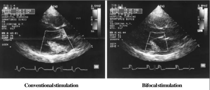
cardiac output (+21.4%, p=0.0114), a significant narrowing of QRS (-28.8%, p=0.003), mitral regurgitation (-43.7%, p=0.0063) and left atrium area (-26.3%, p=0.0049) (table I). A very interesting outcome was the significant reduction in mitral regurgitation. We believe that this highly desirable effect is due to the partial resynchronization of ventricular contraction, which favors mitral function (fig. 5). Except by the ejection fraction (p=0.468), the high septal stimulation also showed a better efficiency than conventional stimulation, but hemo-dynamic benefit was less evident when compared with that of bifocal stimulation (table I).
Implantation technique - The implantations were performed following the classical methodology of the endocardial bicameral pacemakers with both leads by the venous route, and easily reproducible. The first lead was positioned, preferably in the apex of the right ventricle (the most distant possible of the base). The second lead was positioned in the His bundle area, guided by the H potential and by anatomical elements. In four patients, due to severe cardiomyopathy, the H potential was difficult to locate and, in that case, the lead was positioned in the subpulmonary region (figure 6). This position was easily obtained by
Fig. 5 - Bidimensional echocardiogram showing significant reduction in functional mitral regurgitation with right ventricular bifocal stimulation. In addition to the reduction in regurgitation, evident change is also verified in the direction of the regurgitating flow, suggesting that the change in the ventricular activation modifies and favors synchronism among the mitral papillary muscles.
Conventional stimulation
Bifocal stimulation
Fig. 4 - Impact of the right ventricular bifocal stimulation in the QRS narrowing. In this example the QRS from 240 ms in the conventional pacing narrowed to 150 ms in the bifocal stimulation. After 8 days the hemodynamic atrial improvement provided conditions for chronic atrial fibrillation reversion to atrial tachycardia.
RV Apex >>> Bifocal
Conventional stimulation
(9 years)
Bifocal stimulation
(8 days)
Fig. 7 – Thoracic X-rays obtained in the same patient, the first with nine years of evolution with conventional ventricular pacing and the second, after 8 days of bifocal ventricular sti-mulation. There was an evident reduction in heart size. In this case the old VVI pacemaker was changed due to battery depletion, a bifocal system being implanted by adding a septal lead. entering in the pulmonary artery, slowly withdrawing the
lead that was previously connected to the stimulator until ventricular capture was obtained. The left anterior oblique position was used to advance the tip of the lead at its maximum, through the left ventricle. In all cases, this lead was bipolar, active fixation. The R wave, impedance and thresholds were obtained using conventional techniques. The first lead (RV apex) was connected to the ventricular channel, and the second lead (septal – His bundle – RV
outflow), was connected to the atrial channel of the pulse generator. Lead fixation and pocket closure were performed using conventional techniques. Finally, keeping in mind that AV synchronicity significantly contributes in the efficiency of an insufficient heart 9, we used the pacing configuration
shown in figure 6, in the dilated cardiomyopathy, functional mitral insufficiency and AV block, without atrial fibrillation. Clinical evaluation – In the short-term (mean follow-up of 5.6 ±1.1 months) a significant clinical improvement occurred in all cases that changed from NYHA class III and IV to class II (fig. 7). A long-term clinical follow-up will be very important to evaluate the constancy of the results.
Critical considerations - To obtain definitive informa-tion, the number of patients in this study is obviously low. However, each patient was his or her own control, which is a particular type of study that allows obtaining results with a smaller number of patients. In addition, our initial objec-tive was mainly to find trends and study models that can be useful in extensive new projects.
We believe that the positive point of this study is the pro-posed model. This model used ventricular stimulation (VVI) without harm because all patients had chronic atrial fibrillation. This way, without any technical barrier, we can use the bicameral pacemaker DDD to obtain unquestionable and safe long-term information, of the multisite ventricular stimulation. Even before the definitive results, a clear benefit to the patients is revealed in the measure that they receive safer stimulation (two independent stimulation points), which is certainly physiologically preferable (narrower QRS). In fact, based on Ventricular Endocardial Right Bifocal Stimulation
Dilated
Cardiomyopathy without Atrial Fibrillation
Antero-posterior view
1. Xiao HB, Brecker SJ, Gibson DG. Effects of abnormal activation on the time cour-se of the left ventricular pressure pulcour-se in dilated cardiomyopathy. Br Heart J 1992; 68: 403-7.
2. Gras D, Mabo P, Tang T, et al. Multisite pacing as a supplement treatment of con-gestive heart failure preliminary results of the Medtronic Inc. InsSync Study. PACE 1998; 21: 2249-55.
3. Cazeau S, Ritter P, Bakkdach S, et al. Four chamber pacing cardiomyopathy. PACE 1993; 17: 1974-9.
4. Buckingham TA, Candidas R, Fromer M, et al. Acute hemodynamic effects of atrio-ventricular pacing at differing sites in the reight ventricle individually and simultaneously. PACE 1995; 18: 1772.
5. Prinzen FW, Augusstijn CH, Allessie MA, et al. The time sequence of electrical
References
and mechanical activation during spontaneous beating and ectopic stimulati-on. Eur Heart J 1992; 13: 535-43.
6. Bakker PF, Meijburg H, de Jonge N, et al. Benefiicial effects of biventricular pa-cing in congestive heart failure. PACE 1994; 17: 820.
7. Xiao HB, Lee CH, Gibson DG. Effect of left bundle branch block on diastolic function in dilated cardiomyopathy. Br Heart J 1991; 66: 443-7.
8. Foster AH, Gold MR, Mc Laughlin JS. Acute hemodynamic effects of atriobiven-tricular pacing in humans. Ann Thorac Surg 1995; 59: 294-300.
9. Nishimura RA, Hayes DL, Holmes DR, et al. Mechanism of hemodynamic improvement by dual-chamber pacing for severe left ventricular dysfunction: An acute Doppler and catheterization hemodynamic study. J Am Coll Cardiol 1995; 25: 281-8.
these arguments we usually adopt this stimulation method for the cases of AV nodal ablation for refractory atrial tachyarrhy-thmia treatment. In case of exit block in the conventional lead, the other lead maintain ventricular stimulation.
The comparison of echocardiographic variables, chan-ging the ventricular stimulation points through noninvasive programming, without any change in patient position, heart frequency, respiratory frequency, physical activity, venous re-turn, sympathetic tone or psychological condition, comprises a valuable method capable of achieving minimum hemodyna-mic modifications.
Another critical feature is the ability to control by echocardiogram alone. This is justified by the fact that our
initial objective was to find clear and immediate evidence that justified the accomplishment of a larger number of implants without any harm to patients.


