http://doi.org/10.1590/0004-282X20160193
ARTICLE
Neuromyelitis optica: phenotypic
characteristics in a Brazilian case series
Neuromielite óptica: características fenotípicas em uma série de casos brasileiros
Maria Cristina Del Negro1, Patricia Beatriz Christino Marinho1, Regina Maria Papais-Alvarenga2
he association between optic neuritis (ON) and trans
-verse myelitis (TM) has been known since the 19th century.
he term neuromyelitis optica (NMO) was irst used in 1894 by Eugène Devic when describing the case of a female patient with bilateral ON and TM with severe functional deicits and subsequent death1. First considered a variant
of multiple sclerosis, NMO is currently regarded as a dis
-tinct disease, because it has clinical manifestations and radiological and pathological features that difer from mul
-tiple sclerosis2
.
he irst diagnostic criteria for NMO were formulated in 1999 and intended to distinguish NMO from multiple scle
-rosis. At that time, exclusive clinical dysfunction of the optic nerves and spinal cord was considered mandatory for diag
-nosis. Suggestive indings in diagnostic tests and clinical
characteristics emphasizing the severity of disease were con
-sidered major and minor criteria, respectively3
.
he discovery of a highly speciic antibody for NMO in 20044 and of its target antigen, aquaporin 4 (AQP4) in the fol
-lowing year5
, allowed the recognition of limited forms of the disease and the characterization of dysfunction at other sites in the central nervous system. hese developments led to a revision of the diagnostic criteria for NMO in 20066.
In 2007, the term NMO spectrum disorders (NMOSD) was introduced to extend to AQP4-IgG-seropositive patients with limited forms of NMO who were at high risk for future attacks7.
he reinement of the list of non-opticospinal disease characteristics and the presence of AQP4-IgG-seronegative patients or those with unknown serostatus have rendered the 2006 criteria inadequate for contemporary practice and
1Rede Sarah de Hospitais de Reabilitação, Serviço de Neurologia, Brasília DF, Brasil;
2Universidade Federal do Estado do Rio de Janeiro, Hospital Universitário Gaffrée & Guinle, Serviço de Neurologia, Rio de Janeiro RJ, Brasil. Correspondence: Maria Cristina Del Negro. SMHS Quadra 301/Bloco A; 70335-910 Brasília DF, Brasil; E-mail: [email protected]
Conflict of interest: There is no conlict of interest to declare.
Received 10 June 2015; Received in inal form 02 February 2016; Accepted 17 October 2016.
ABSTRACT
The deinition of neuromyelitis optica (NMO) is still evolving. In 2015, the International Panel for NMO Diagnosis was convened to develop revised diagnostic criteria. There have been few studies on NMO in the Brazilian population. Objective: To describe the characteristics of 34 Brazilian NMO patients. To evaluate the contribution of the 2015 criteria to the diagnosis of NMO spectrum disorders (NMOSD) in 40 patients with longitudinal extensive transverse myelitis (LEMT). Methods: This is a retrospective, descriptive and analytic study. Results:
Among NMO patients, there was a predominance of women, with onset in the fourth decade of life, and AQP4-IgG seropositivity in 73.5%. The diagnosis of NMOSD was established in 37.5% of LETM patients according to AQP4-IgG positivity and in 5% of LETM patients if the AQP4-IgG result was unknown. Conclusions: The characteristics of this series are similar to those of other Western populations. The AQP4-IgG testing assists in the diagnosis of NMOSD.
Keywords: neuromyelitis optica; epidemiology; cross-sectional studies. enzyme-linked immunosorbent assay.
RESUMO
Neuromielite óptica (NMO) é um conceito em evolução. Em 2015, o Painel Internacional para o diagnóstico de NMO apresentou novos critérios diagnósticos. Poucos são os estudos em NMO na população brasileira. Objetivos: Descrever as características de 34 casos brasileiros de NMO. Avaliar a contribuição dos critérios de 2015 para o diagnóstico de desordens do espectro NMO em 40 pacientes com mielite transversa longitudinal extensa (MTLE). Métodos: Estudo retrospectivo, descritivo e analítico. Resultados: Predomínio do sexo feminino, início na quarta década e anticorpo anti-AQP4 positivo em 73,5% dos casos de NMO. Diagnóstico de desordem do espectro NMO estabelecido em 37.5% dos casos de MTLE com positividade do anticorpo anti-AQP4 e em 5% se o resultado sorológico fosse desconhecido.
Conclusões: Esta série de casos de NMO tem características semelhantes às de outras séries ocidentais. A pesquisa do anticorpo anti-AQP4 é relevante para o diagnóstico das desordens do espectro da NMO.
research. he term NMOSD has also been used variably in the literature and requires clariication. In 2015, the International Panel for NMO Diagnosis was convened to develop revised diagnostic criteria and to deine the nomenclature. he panel recommends prospective validation of the criteria8.
here appear to be diferences among NMO patients according to serostatus. Some studies suggest that AQP4-IgG-seronegative patients are younger, less frequently female, and less likely to relapse9.
In this study, we describe the demographic, clinical, and paraclinical characteristics of a Brazilian series of patients with NMO, and search for diferences as a function of serosta
-tus. We also evaluate the contribution of the 2015 criteria to the diagnosis of NMOSD in a series of patients with longitu
-dinal extensive transverse myelitis (LETM).
METHODS
his is a retrospective, descriptive and analytic study. Patients of any age diagnosed with NMO, according to the 2006 Wingerchuk et al.6 criteria, or with LETM (three or more
contiguous spinal segments, without other known etiology), subjected to AQP4-IgG testing at the Brasilia unit of the Sarah Network of Rehabilitation Hospitals from November 2009 to July 2012, were included.
he AQP4-IgG testing was performed using a commercial ELISA-R kit (recombinant human AQP4, M1 isoform), with a cutof of 5 U/ml.
he variables were gender, age at initial event, skin color (white/non-white), initial clinical event (isolated TM, iso
-lated ON, simultaneous TM and ON, and brainstem syn
-drome), time from irst relapse to irst appointment, time interval between index events (ON and TM), duration of the disease, presence and total number of recurrences, score on the Expanded Disability Status Scale at last assessment, associated immune disease, AQP4-IgG serostatus, indings on spine magnetic resonance imaging (MRI) (the presence of extensive spinal cord injury, central lesions, and afected spinal cord segments), indings on brain MRI (normal, non
-speciic lesions, or typical NMO lesions according to Kim10),
the presence of other autoantibodies (antinuclear anti
-body, anti-Sjögren’s-syndrome-related antigen A, and anti
-cardiolipin), whether AQP4-IgG testing was performed during an attack, and whether testing occurred before or after immunosuppression.
For the statistical analyses, the variables are expressed as means ± standard deviations and were compared between groups using Student’s t-test or the Mann-Whitney test. For variables expressed as frequencies, the groups were compared using the chi-square test or Fisher’s exact test. P-values < 0.05 were considered signiicant.
he study was approved by the research ethics committee of the Sarah Network of Rehabilitation Hospitals.
RESULTS
A total of 34 patients diagnosed with NMO accord
-ing to the W-ingerchuk et al.6 2006 criteria were identified.
Most patients were female (7.5 females:1 male), and the average disease onset had occurred by the fourth decade of life (34.6 ± 17.2 years of age; range: 4–68 years of age). Six patients exhibited late disease onset, after 50 years of age, and five patients experienced onset before 16 years of age. Approximately 45% of the patients were non-white. The average time from the first attack to the first appointment was 23.7 ± 26,5 months (0–108 months). The initial clinical event was ON or TM in approximately 60% of the patients; simultaneous ON and TM (interval of up to one month between each event) occurred in 23.5% of the patients; and brainstem syndrome, mainly uncontrollable vomiting and persistent hiccups, was observed in 17.6% of the patients. Recovery after the initial clinical event was complete in slightly more than 25% of the patients and absent in almost 20%. The time between the index events was highly vari
-able (mean: 14.7 ± 29.5 months; range: 0–108 months). The mean duration of disease was 8.1 ± 5.5 years, and 73.5% of the patients were classified as recurrent NMO. In 73.5% of the patients, AQP4-IgG seropositivity was detected by ELISA. Spinal cord lesions were central in approximately 90% of the patients, and the cervicothoracic segment was the most often affected site. Brain MRI examinations exhibited typical NMO findings in 26.5% of the patients. An association with another autoimmune disease was found in approximately 30% of the patients (an association with thyroiditis was observed in 62.5% of these patients). Other autoantibodies were detected in more than 50% of patients and, in most instances, corresponded to ANA in low titers.
The average time from the last attack to the sero
-logic test was 28.4 ± 34,6 months (0–108 months). Only six patients (18%) had AQP4-IgG testing less than one month after relapse. Twenty-four patients (70%) were on immunosuppressive drug treatment (prednisone and azathioprine 12 patients, azathioprine 12 patients) when serological testing was performed. Neither factor signifi
-cantly affected the results of AQP4-IgG testing (Table 1). Thirty-three patients (97%) remained free of attacks dur
-ing immunosuppressive therapy.
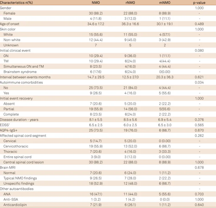
In a comparison of recurrent and monophasic NMO, the latter showed a greater prevalence of isolated TM and of simultaneous ON and TM as the initial clinical event, and of other autoimmune diseases (Table 2).
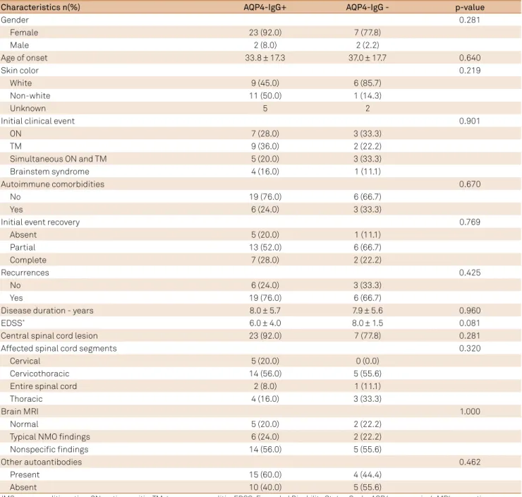
By comparing the patients according to their serosta
-tus, we found a greater proportion of women and non-white patients in the AQP4-IgG seropositive group and greater func
Forty patients were diagnosed with LETM (25 monophasic LETM, 15 recurrent LETM). hirteen (32.5%) were AQP4-IgG sero
-positive and, as a result, deined as NMOSD. Only two recurrent
NMO patients had brainstem syndrome and neuroimaging sug
-gestive of NMOSD, matching the diagnosis of NMOSD according to the 2015 criteria, but both were AQP4-IgG seropositive.
Table 2. Demographic, clinical and paraclinical characteristics of the patients with NMO. n(%)
Characteristics n(%) NMO rNMO mNMO p-value
Gender 1.000
Female 30 (88.2) 22 (88.0) 8 (88.9) -
Male 4 (11.8) 3 (12.0) 1 (11.1)
-Age of onset 34.6 ± 17.2 36.3 ± 16.6 30.1 ± 19.1 0.489
Skin color 1.000
White 15 (55.6) 11 (55.0) 4 (57.1) -
Non-white 12 (44.4) 9 (45.0) 3 (42.9)
-Unknown 7 5 2 -
Initial clinical event 0.080
ON 10 (29.4) 9 (36.0) 1 (11.1)
-TM 10 (29.4) 6(24.0) 4(44.4)
-Simultaneous ON and TM 8 (23.5) 4(16.0) 4 (44.4)
-Brainstem syndrome 6 (17.6) 6(24.0) 0(0.00)
-Interval between events months 14.7 ± 29.5 12.5 ± 27.0 20.3 ± 36.3 0.621
Autoimmune comorbidities 0.034
No 25 (73.5) 21 (84.0) 4 (44.4) -
Yes 9 (26.5) 4 (16.0) 5 (55.6)
-Initial event recovery 1.000
Absent 7 (20.6) 5 (20.0) 2 (22.2)
-Partial 19 (55.9) 14 (56.0) 5(55.6) -
Complete 8 (23.5) 6(24.0) 2 (22.2)
-Disease duration - years 8.1 ± 5.5 8.5 ± 5.6 6.9 ± 5.4 0.376
EDSS* 6.5 ± 2.5 6.0 ± 2.5 6.5 ± 3.0 0.565
AQP4-IgG+ 25 (73.5) 19 (76.0) 6 (66.7) 0.670
Affected spinal cord segment 0.262
Cervical 5 (14.7) 5 (20.0) 0 (0.00)
-Cervicothoracic 19 (55.9) 13 (52.0) 6 (66.7)
-Thoracic 7 (20.6) 4 (16.0) 3 (33.3)
-Entire spinal cord 3 (9.0) 3 (12.0) 0 (0.00)
-Central spinal cord lesion 30 (88.2) 22 (88.0) 8 (88.9) 1.000
Brain MRI 0.678
Normal 7 (20.6) 6 (24.0) 1 (11.2)
-Typical NMO indings 9 (26.5) 7 (28.0) 2 (22.2)
-Unspeciic indings 18 (52.9) 12 (48.0) 6 (66.7)
-Other autoantibodies
-ANA 16 (47.1) 11 (44.0) 5 (55.6) 0.703
Anti-SSA 1 (3.2) 1 (4.2) 0 (0.0) 1.000
Anticardiolipin 7 (21.9) 6 (26.1) 1 (11.2) 0.640
NMO: neuromyelitis optica; r: recurrent; m: monophasic; ON: optic neuritis; TM: transverse myelitis; EDSS: expanded disability status scale; AQP4: aquaporin 4; MRI: magnetic resonance imaging; ANA: antinuclear antibody; SSA: anti-Sjögren’s-syndrome-related antigen A. *Values expressed as the median; ± interquartile range.
Table 1. Characteristics of the patients when AQP4-IgG was performed.
Characteristics n(%) Total AQP4-IgG+ AQP4-IgG- p-value
Time from attack 1.0000
> 1 month 28(82.4) 20 (80.0) 8 (88.9)
-≤ 1 month 6 (17.6) 5 (20.0) 1 (11.1)
-Immunosuppressive drugs 0.6921
No 10 (29.4) 8 (32.0) 2 (22.2) -
Yes 24 (70.6) 17 (68.0) 7 (77.8) -
DISCUSSION
he deinition of NMO is still evolving. he International Panel for NMO Diagnosis airmed the decision to unify the terms NMO and NMOSD8. he index events required
for diagnosis, now termed core clinical characteristics, include ON, TM, area postrema syndrome (episode of oth
-erwise unexplained hiccups or nausea and vomiting), acute brainstem syndrome, symptomatic narcolepsy or acute diencephalic clinical syndrome with NMOSD-typical dien
-cephalic MRI lesions, and symptomatic cerebral syndrome with NMOSD-typical brain lesions8. Bilateral and/or recur
-rent ON, poor recovery of visual acuity, lesions extending over half the optic nerve length or involving the optic chiasm,
and the absence of visual evoked potentials are indicative of NMOSD11. Painful tonic spasms, sensory level, bilateral
motor impairment, persistent sphincter dysfunction, exten
-sive and central spinal cord lesions on MRI performed within the irst weeks after the onset of symptoms, spinal cord cavi
-tation and late atrophy are common indings in NMOSD3,6.
he demographic characteristics of NMO patients are reinforced by each successive published study. Neuromyelitis optica patients are predominantly female, with a ratio higher than that of multiple sclerosis; the average age at onset is older, usually by the end of the fourth decade of life; and the prevalence of non-white patients is higher than that of other demyelinating diseases12,13,14,15,16,17,18. hese demographic char
-acteristics were also present in the current Brazilian NMO
Table 3. Demographic, clinical and paraclinical characteristics by serological status.
Characteristics n(%) AQP4-IgG+ AQP4-IgG - p-value
Gender 0.281
Female 23 (92.0) 7 (77.8)
Male 2 (8.0) 2 (2.2)
Age of onset 33.8 ± 17.3 37.0 ± 17.7 0.640
Skin color 0.219
White 9 (45.0) 6 (85.7)
Non-white 11 (50.0) 1 (14.3)
Unknown 5 2
Initial clinical event 0.901
ON 7 (28.0) 3 (33.3)
TM 9 (36.0) 2 (22.2)
Simultaneous ON and TM 5 (20.0) 3 (33.3)
Brainstem syndrome 4 (16.0) 1 (11.1)
Autoimmune comorbidities 0.670
No 19 (76.0) 6 (66.7)
Yes 6 (24.0) 3 (33.3)
Initial event recovery 0.769
Absent 5 (20.0) 1 (11.1)
Partial 13 (52.0) 6 (66.7)
Complete 7 (28.0) 2 (22.2)
Recurrences 0.425
No 6 (24.0) 3 (33.3)
Yes 19 (76.0) 6 (66.7)
Disease duration - years 8.0 ± 5.7 7.9 ± 5.6 0.960
EDSS* 6.0 ± 4.0 8.0 ± 1.5 0.081
Central spinal cord lesion 23 (92.0) 7 (77.8) 0.281
Affected spinal cord segments 0.320
Cervical 5 (20.0) 0 (0.0)
Cervicothoracic 14 (56.0) 5 (55.6)
Entire spinal cord 2 (8.0) 1 (11.1)
Thoracic 4 (16.0) 3 (33.3)
Brain MRI 1.000
Normal 5 (20.0) 2 (22.2)
Typical NMO indings 6 (24.0) 2 (22.2)
Nonspeciic indings 14 (56.0) 5 (55.6)
Other autoantibodies 0.462
Present 15 (60.0) 4 (44.4)
Absent 10 (40.0) 5 (55.6)
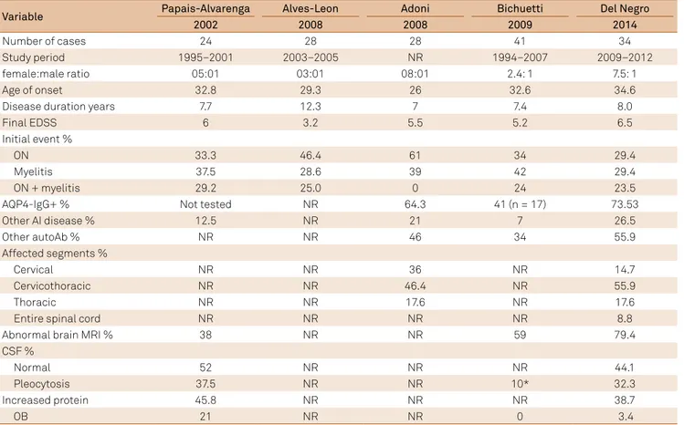
case series and were similar to those reported for other case series in the country19,20,21,22. he exceptions include
a younger age at onset in the Adoni et al.21 series, in which
only patients with recurrent NMO were analyzed, and lower female-to-male ratios in the Alves-Leon et al.20 and Bichuetti
et al.22 series(Table 4).
All NMO patients in our series exhibited extensive lon
-gitudinal spinal cord lesions in at least one spinal MRI, and none had imaging features suggestive of multiple sclerosis. herefore, AQP4-IgG seropositivity was not mandatory to establish the diagnosis of NMO6,8.
he most common initial events were isolated ON or iso
-lated TM, recovery was generally partial or absent, the aver
-age time to the second core event was longer than one year, and immunosuppressive therapy appeared highly efective at reducing relapses. hese indings reinforce the relevance of early diagnosis and early onset of immunosuppressive ther
-apy. he AQP4-IgG testing may help the diagnosis of NMOSD. Diagnostic requirements are more stringent for patients in whom AQP4-IgG is not detected or for whom testing is unavailable8. In this series of patients with LETM, the diagno
-sis of NMOSD was established in 37.5%, based on AQP4-IgG positivity. If the AQP4-IgG result was unknown, only 5% would meet the diagnosis, according to 2015 criteria.
In the present case series, AQP4-IgG seropositivity was similar to that reported by Lennon et al.4 (73%) and was
higher than that found by Adoni et al.23
in Brazilian patients with recurrent NMO; both studies used the IF method.
An AQP4-IgG seropositivity by the ELISA method reported for other Western populations varies from 60 to 76%9,24,25. he
use of the isotype AQP4 M23 increases the sensitivity of this method26,27. Currently, cell-based assays are strongly recom
-mended8,25, but the expertise and resources required to per
-form those assays preclude their use in small-scale clinical diagnostic laboratories. A previous study airms that com
-mercially available kit assays (ELISA-R and CBA-E) are both sensitive and speciic for AQP4-IgG detection, and their rela
-tive simplicity to perform allows small-scale laboratories to ofer sensitive and speciic AQP4-IgG testing25
.
We did not ind any signiicant diference among NMO patients according to their serological status. he propor
-tions of women and of people of African descent were higher in the AQP4-IgG seropositive group. he small numbers of seropositive and seronegative patients may account for the lack of signiicant diferences between the groups.
In our series, an association with other autoimmune dis
-eases was more frequent among the monophasic patients. Isolated TM or simultaneous ON and TM were more preva
-lent as initial events in that group, although this diference was non-signiicant. Once again, the small number of recur
-rent and monophasic patients reduced the statistical power of the sample.
Table 4. Characteristic of Brazilian NMO case series.
Variable Papais-Alvarenga Alves-Leon Adoni Bichuetti Del Negro
2002 2008 2008 2009 2014
Number of cases 24 28 28 41 34
Study period 1995–2001 2003–2005 NR 1994–2007 2009–2012
female:male ratio 05:01 03:01 08:01 2.4: 1 7.5: 1
Age of onset 32.8 29.3 26 32.6 34.6
Disease duration years 7.7 12.3 7 7.4 8.0
Final EDSS 6 3.2 5.5 5.2 6.5
Initial event %
ON 33.3 46.4 61 34 29.4
Myelitis 37.5 28.6 39 42 29.4
ON + myelitis 29.2 25.0 0 24 23.5
AQP4-IgG+ % Not tested NR 64.3 41 (n = 17) 73.53
Other AI disease % 12.5 NR 21 7 26.5
Other autoAb % NR NR 46 34 55.9
Affected segments %
Cervical NR NR 36 NR 14.7
Cervicothoracic NR NR 46.4 NR 55.9
Thoracic NR NR 17.6 NR 17.6
Entire spinal cord NR NR NR NR 8.8
Abnormal brain MRI % 38 NR NR 59 79.4
CSF %
Normal 52 NR NR NR 44.1
Pleocytosis 37.5 NR NR 10* 32.3
Increased protein 45.8 NR NR NR 38.7
OB 21 NR NR 0 3.4
Because it is a rare disease, multicenter studies and meta-analyses are needed to achieve better epidemiological characterization of NMO and NMOSD. he establishment of databases, such as NEMOS17 and NMO-DBr28, is fundamental.
In conclusion, the characteristics exhibited by the pres
-ent series of Brazilian pati-ents with NMO reinforce those reported for other Western populations. he ELISA-R
method exhibited satisfactory sensitivity for the detection of AQP4-IgG. he most common initial events included lim
-ited forms of the disease, which emphasizes the relevance of AQP4-IgG testing in such patients. No statistically-signif
-icant diference was found between patients as a function of their serological status, but the small sample size may have led to this result.
References
1. Jarius S, Wildemann B. The history of neuromyelitis optica. J
Neuroinlammation. 2013;10(1):8. http://doi.org/10.1186/1742-2094-10-8
2. O’Riordan JI, Gallagher HL, Thompson AJ, Howard RS, Kingsley DP, Thompson EJ et al. Clinical, CSF, and MRI indings in Devic’s neuromyelitis optica. J Neurol Neurosurg Psychiatry. 1996;60(4):382-7. http://doi.org/10.1136/jnnp.60.4.382
3. Wingerchuk DM, Hogancamp WF, O’Brien PC, Weinshenker BG. The clinical course of neuromyelitis optica (Devic’s syndrome). Neurology. 1999;53(5):1107-14. http://doi.org/10.1212/WNL.53.5.1107
4. Lennon VA, Wingerchuk DM, Kryzer TJ, Pittock SJ, Lucchinetti CF, Fujihara K et al. A serum autoantibody marker of neuromyelitis optica: distinction from multiple sclerosis. Lancet. 2004;364(9451):2106-12. http://doi.org/10.1016/S0140-6736(04)17551-X
5. Lennon VA, Kryzer TJ, Pittock SJ, Verkman AS, Hinson SR. IgG marker of optic-spinal multiple sclerosis binds to the aquaporin-4 water channel. J Exp Med. 2005;202(4):473-7. http://doi.org/10.1084/jem.20050304
6. Wingerchuk DM, Lennon VA, Pittock SJ, Lucchinetti CF, Weinshenker BG et al. Revised diagnostic criteria for neuromyelitis optica. Neurology. 2006;66(10):1485-9. http://doi.org/10.1212/01.wnl.0000216139.44259.74
7. Wingerchuk DM, Lennon VA, Lucchinetti CF, Pittock SJ, Weinshenker BG. The spectrum of neuromyelitis optica. Lancet Neurol. 2007;6(9):805-15. http://doi.org/10.1016/S1474-4422(07)70216-8
8. Wingerchuk DM, Banwell B, Bennett JL, Cabre P, Carroll W, Chitnis T et al. International consensus diagnostic criteria for neuromyelitis optica spectrum disorders. Neurology. 2015;85(2):177-89. http://doi.org/10.1212/WNL.0000000000001729
9. Jiao Y, Fryer JP, Lennon VA, Jenkins SM, Quek AM, Smith CY et al. Updated estimate of AQP4-IgG serostatus and disability outcome in neuromyelitis optica. Neurology. 2013;81(14):1197-204. http://doi.org/10.1212/WNL.0b013e3182a6cb5c
10. Kim W, Park MS, Lee SH, Kim SH, Jung IJ, Takahashi T et al. Characteristic brain magnetic resonance imaging abnormalities in central nervous system aquaporin-4 autoimmunity. Mult Scler. 2010;16(10):1229-36. http://doi.org/10.1177/1352458510376640
11. Lim YM, Pyun SY, Lim HT, Jeong IH, Kim KK. First-ever optic neuritis: distinguishing subsequent neuromyelitis optica from multiple sclerosis. Neurol Sci. 2014;35(5):781-3. http://doi.org/10.1007/s10072-014-1635-6
12. Cabre P, González-Quevedo A, Bonnan M, Saiz A, Olindo S, Graus F et al. Relapsing neuromyelitis optica: long term history and clinical predictors of death. J Neurol Neurosurg Psychiatry. 2009;80(10):1162-4. http://doi.org/10.1136/jnnp.2007.143529
13. Bizzoco E, Lolli F, Repice AM, Hakiki B, Falcini M, Barilaro A et al. Prevalence of neuromyelitis optica spectrum disorder and phenotype distribution. J Neurol. 2009;256(11):1891-8. http://doi.org/10.1007/s00415-009-5171-x
14. Collongues N, Marignier R, Zéphir H, Papeix C, Blanc F, Ritleng C et al. Neuromyelitis optica in France: a multicenter study of 125 patients. Neurol. 2010;74(9):736-42. http://doi.org/10.1212/WNL.0b013e3181d31e35
15. Cossburn M, Tackley G, Baker K, Ingram G, Burtonwood M, Malik G et al. The prevalence of neuromyelitis optica in south east Wales. Eur J Neur. 2012;19(4):655-9. http://doi.org/10.1111/j.1468-1331.2011.03529.x
16. Asgari N, Lillevang ST, Skejoe HP, Falah M, Stenager E, Kyvik KO. A population-based study of neuromyelitis optica in Caucasians. Neurology. 2011;76(18):1589-95. http://doi.org/10.1212/WNL.0b013e3182190f74
17. Jarius S, Ruprecht K, Wildemann B, Kuempfel T, Ringelstein M, Geis C et al. Contrasting disease patterns in seropositive and seronegative neuromyelitis optica: a multicentre study of 175 patients. J
Neuroinlammation. 2012;9(1):14. http://doi.org/10.1186/1742-2094-9-14
18. Mealy MA, Wingerchuk DM, Greenberg BM, Levy M. Epidemiology of neuromyelitis optica in the United States: a multicenter analysis. Arch Neurol. 2012;69(9):1176-80. http://doi.org/10.1001/archneurol.2012.314
19. Papais-Alvarenga RM, Miranda-Santos CM, Puccioni-Sohler M, Almeida AM, Oliveira S, Oliveira CAB et al. Optic neuromyelitis syndrome in Brazilian patients. J Neurol Neurosurg Psychiatry. 2002;73(4):429-35. http://doi.org/10.1136/jnnp.73.4.429
20. Alves-Leon SV, Pimentel ML, Sant’anna G, Malfetano FR, Estrada CD, Quirico-Santos T. Immune system markers of neuroinlammation in patients with clinical diagnose of neuromyelitis optica. Arq Neuropsiquiatr. 2008;66(3B):678-84. http://doi.org/10.1590/S0004-282X2008000500013
21. Adoni T, Lino AM, Gama PD, Apóstolos-Pereira SL, Marchiori PE, Kok F et al. Recurrent neuromyelitis optica in Brazilian patients: clinical, immunological, and neuroimaging characteristics. Mult Scler. 2010;16(1):81-6. http://doi.org/10.1177/1352458509353651
22. Bichuetti DB, Oliveira EM, Souza NA, Rivero RL, Gabbai AA.
Neuromyelitis optica in Brazil: a study on clinical and prognostic factors. Mult Scler. 2009;15(5):61-9. http://doi.org/10.1177/1352458508101935
23. Adoni T, Lino AM, Marchiori PE, Kok F, Callegaro D.
Seroprevalence of NMO-IgG antibody in Brazilian patients with neuromyelitis optica. Arq Neuropsiquiatr. 2008;66(2B):295-7. http://doi.org/10.1590/S0004-282X2008000300001
24. Jarius S, Franciotta D, Paul F, Bergamaschi R, Rommer PS, Ruprecht K et al. Testing for antibodies to human aquaporin-4 by ELISA: sensitivity, speciicity, and direct comparison with immunohistochemistry. J Neurol Sci. 2012;320(1-2):32-7. http://doi.org/10.1016/j.jns.2012.06.002
25. Waters PJ, McKeon A, Leite MI, Rajasekharan S, Lennon VA, Villalobos A et al. Serologic diagnosis of NMO: a multicenter comparison of aquaporin-4-IgG assays. Neurology.
2012;78(9):665-71. http://doi.org/10.1212/WNL.0b013e318248dec1
26. Isobe N, Yonekawa T, Matsushita T, Kawano Y, Masaki K, Yoshimura S et al. Quantitative assays for anti-aquaporin-4 antibody with subclass analysis in neuromyelitis optica. Mult Scler. 2012;18(11):1541-51. http://doi.org/10.1177/1352458512443917
27. Fryer JP, Lennon VA, Pittock SJ, Jenkins SM, Fallier-Becker P, Clardy SL et al. AQP4 autoantibody assay performance in clinical laboratory service. Neurol Neuroimmunol Neuroinlammation. 2014;11(1): e11. http://doi.org/10.1212/NXI.0000000000000011