CLINICAL SCIENCE
I Plastic Surgery Service, Brigadeiro Hospital - São Paulo/SP, Brazil. II Division of Plastic Surgery, Hospital das Clinicas da Faculdade de Medicina da Universidade de São Paulo - São Paulo/SP, Brazil.
III Pathologic Anatomy Department, A. C. Camargo Hospital, São Paulo/ SP, Brazil.
Email: [email protected] Tel.: 55 11 3873.2366
Received for publication August 26, 2009 Accepted for publication on November 3, 2009
PRIMARY CUTANEOUS MELANOMA: AN 18-YEAR
STUDY
Moris Anger,I Henri Friedhofer,II Marina Fussae Fukutaki,I Marcus Castro
Ferreira,II Gilles LandmanIII
doi: 10.1590/S1807-59322010000300004
Anger M, Friedhofer H, Fukutaki MF, Ferreira MC, Landman G. Primary cutaneous melanoma: an 18-year study. Clinics. 2010;65(3):257-63.
BACKGROUND: Primary cutaneous melanoma still constitutes the main cause of skin cancer death in developed countries, and its incidence in recent years has been increasing in a steady, worrisome manner.
OBJECTIVES: This study evaluated the clinical, epidemiological and demographic aspects of this disease, and correlated them with patient prognosis.
METHODS: Using epidemiologic and clinical data, we analyzed 84 patients with mild to severe primary cutaneous melanoma treated between 1990 and 2007. Slides containing surgical specimens were analyzed, and new slides were made from archived parafin sections when necessary.
RESULTS: The melanoma incidence was higher in areas of sun exposure, with lesions commonly observed in the trunk for males, and lower limbs for females. In addition to Breslow’s thickness and ulceration (p = 0.043 and p < 0.001, respectively), the mitotic rate per mm2 also correlated with worse patient outcome (p = 0.0007). The sum of ulceration (0 when absent or 1 when present), the
Breslow index (1 when <1 mm, 2 when >1 mm and <4 mm, 3 when >4 mm) and the mitotic index (0 when absent or 1 when >1 per mm2)
allowed the establishment of a prognostic score: if the sum was equal to or over three, nearly all (91.7%) patients had systemic disease. The 5-year survival was approximately seventy percent.
CONCLUSION: Because American Join Committee of Cancer Staging will update the classiication of malignant tumors (TNM) staging in the near future, and introduce mitosis as a prognostic factor, our results show the importance of such a feature. Additional studies are necessary to conirm the importance of a prognostic score as proposed herein.
KEYWORDS: Melanoma; Skin neoplasm; Epidemiology; Risk factors; Prognosis.
INTRODUCTION
Melanoma is a tumor of neuroectodermal origin that is formed from melanocytes - cells that are found along the basal layer of the epidermis with the main function of producing the pigment melanin.1 It has been showing a
steady, worrisome increase in incidence that is likely due to the intermittent habit of sun exposure, which is considered
to be the major etiologic factor for skin cancer.2
Melanoma is a neoplasm with good prognosis when diagnosed early. Nevertheless for intermediate thickness (1 to 4 mm in depth), it is not possible to predict the disease progression, varying from 63 to 89% for 5-year survival. However, when deeper than 4 mm, the 5-year survival rates are low (33 to 55%).3-5 The transition from radial growth to
vertical growth grants melanoma with higher aggressiveness, the larger and deeper the lesion is.1
Melanoma shows great potential for dissemination, which is the reason it constitutes one of the most severe tumors among skin lesions, with high mortality rates when diagnosed late.6-8
However, even with large tumor resections,9 the 5-year
1950s and 1960s sought to establish melanoma-related predictive factors.
In 1969, Clark et al.10 proposed staging criteria for
lesions on the basis of skin invasion levels. Subsequently, Breslow and Macht 11 evidenced the importance of the
primary melanoma thickness in millimeters, and this index became the most importantprognostic indicator,12, 13 in association with data on ulceration, mitosis and
regression.5,14,15
Preventive campaigns, dermatoscopy, therapeutic approach protocols, determination of anatomic-pathologic prognostic factors, and more accurate staging through the study of sentinel lymph nodes have allowed for the early diagnosis, and treatment of melanomas in the last decade.16
Although variables such as clinical presentation, histopathological data and staging are considered to be appropriate instruments for evaluating the prognosis of melanoma cases, these variables are not always suficient to accurately predict the evolution of a specific case.14
Patients with similar clinical and anatomic-pathologic prognostic factors may progress in different manners, sometimes progressing favorably, sometimes with disease dissemination, probably because of the intrinsic properties of these tumor cells. A number of studies have examined molecular factors that may explain such differences, particularly focusing on genetic changes involved in tumorigenesis and melanoma progression.2, 17- 19
OBJECTIVES
• To study the clinical and epidemiologic aspects in pri-mary skin melanomas.
• To survey histopathological data (Breslow index, Clark level, ulceration, mitotic index and regression) revised according to the Brazilian Melanoma Group’s protocol. • To correlate these data with primary cutaneous
melano-ma-treated patient evolution.
METHODS
This retrospective study comprises 84 primary cutaneous melanoma-diagnosed patients who underwent surgical treatment between 1990 and 2007, in private practice (50 cases) and at the Sistema Unico de Saude (SUS, Uniied Health System) unit (34 cases).
Clinical, epidemiologic, histological and disease evolution data were collected at the pre- and post-operative follow-up of study patients. The histological preparations were reviewed, and new slides were obtained from parafin blocks when necessary, and stained with hematoxylin and eosin (HE), with the prognostic parameters being evaluated
in compliance with the protocol set by the Brazilian Melanoma Group.20
Demographic data (age, sex and ethnicity), topographic location and histopathological data (Breslow index, Clark level, ulceration, mitotic index, regression and peri- and intra-tumor iniltration) were surveyed for the treated cases.
Variables were described in frequencies or means and standard deviations, and they were compared with the Chi-square test, Fisher exact test and Student’s t test.
The evaluation of disease-free survival and cancer-speciic survival was performed through actuarial analysis, and conirmed using the Kaplan-Meier Product.21 In this
analysis, only melanomas > 1 mm were included.
For result analysis, the rejection probability of the 95% hypothesis (p < 0.05) was adopted.
The study was approved by the Research Ethics Committees of the University of Sao Paulo Medical School (16/02/2005), A C Camargo Hospital (15/10/2007), and Brigadeiro Hospital (05/09/2007).
RESULTS
There were no significant differences of frequency between genders (Table 1), (p = 0.1266). Only 1 African-American patient participated in the study (1%); the remaining patients were Caucasian.
This patient group’s mean age was 54.8+18.1 years, which was similar for both genders: 56.0+17.9 for males and 53.9+18.4 for females (p = 0.6190), and ranged from 13-91 years old. In addition, there were no signiicant age differences between genders when they were compared by decade of life (p = 0.1942).
Melanomas are predominant in the trunk in males (42.8%), and the lower limbs in females (36.7%) (p = 0.0113).
There was no evidence of differences between genders when the mean ages and standard deviations were assessed for the location of lesions.
Comparing location of the lesions in relation of the patient gender there was a significant difference (p = 0.0113), being lesions in the head and neck and in the trunk particularly more frequent in males, while lesions in the upper and lower limbs are more frequent in females (Figure 1).
distribution regarding Clark levels (p = 0.461).
There was no evidence of differences in the tumor Breslow index, in terms of mean age (and standard deviations); however, there was a wide age range in these groups. In addition, no signiicant differences were observed when Clark levels were analyzed with respect to patient age. Breslow index frequencies, as distributed according to lesion sites, were widely varied. In Figure 2, we observed that there was a higher frequency of tumors thicker than 1 mm (65.2%) in the lower limb lesions, while tumors less than 1 mm thick were signiicantly predominant in trunk lesions (p < 0.001).
The Clark level distribution according to tumor location demonstrated that level III tumors were predominant in the lower and upper limbs and the trunk (p < 0.001), while there was no difference in the distribution in the head and neck.
To ensure a minimum 24 months follow-up in patient disease-free and overall survival evaluation, melanoma diagnosed and treated as of October 2006 were excluded, as well as in situ cutaneous melanoma, and predisposing disease (xeroderma pigmentosum).
Of the 54 patients (27 with melanoma > 1 mm and 27 with melanoma < 1 mm) included in this evaluation, disease dissemination was observed in 12 patients (22.2%), of which 10 (18.5%) progressed to death, while 2 were still on oncological follow-up (3.7%). Disease dissemination was not observed in any of the patients with < 1 mm-thick melanoma.
There was no difference of disease dissemination between genders (p = 0.577), and was observed in 50% of females and 53.3% of males. The mean age of patients with disseminated disease (60.7+21.1) was not different (p = 0.850) from that of patients without relapse (59.3+15.2). In addition, there was no difference in the distribution of frequencies relative to the melanoma site (p = 0.950).
The differences found in the patient evolution analysis in relation to the Breslow indexes (Table 2) were
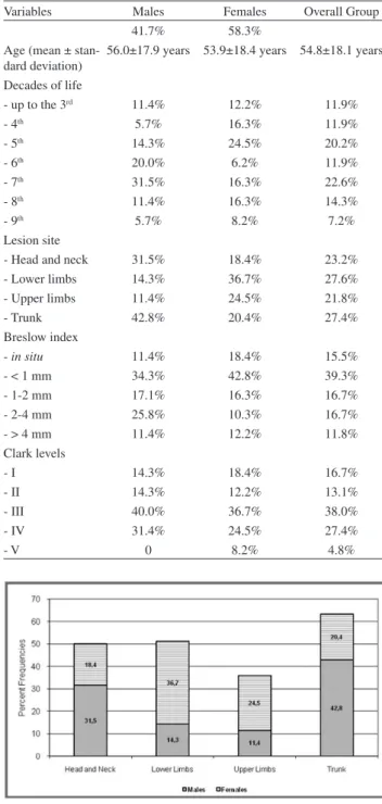
Table 1- Clinical and epidemiologic indings in 84 patients
with primary cutaneous melanoma
Variables Males Females Overall Group
41.7% 58.3%
Age (mean ± stan-dard deviation)
56.0±17.9 years 53.9±18.4 years 54.8±18.1 years Decades of life
- up to the 3rd 11.4% 12.2% 11.9%
- 4th 5.7% 16.3% 11.9%
- 5th 14.3% 24.5% 20.2%
- 6th 20.0% 6.2% 11.9%
- 7th 31.5% 16.3% 22.6%
- 8th 11.4% 16.3% 14.3%
- 9th 5.7% 8.2% 7.2%
Lesion site
- Head and neck 31.5% 18.4% 23.2%
- Lower limbs 14.3% 36.7% 27.6%
- Upper limbs 11.4% 24.5% 21.8%
- Trunk 42.8% 20.4% 27.4%
Breslow index
- in situ 11.4% 18.4% 15.5%
- < 1 mm 34.3% 42.8% 39.3%
- 1-2 mm 17.1% 16.3% 16.7%
- 2-4 mm 25.8% 10.3% 16.7%
- > 4 mm 11.4% 12.2% 11.8%
Clark levels
- I 14.3% 18.4% 16.7%
- II 14.3% 12.2% 13.1%
- III 40.0% 36.7% 38.0%
- IV 31.4% 24.5% 27.4%
- V 0 8.2% 4.8%
Figure 1 - Distribution of frequencies relative to primary cutaneous
mela-noma site
In the female group, head and neck lesions affected older patients signiicantly more than lower limb (p = 0.020) and trunk (p = 0.004) lesions; in the remaining locations, there were no signiicant differences.
Among the morphological parameters, there was homogeneous distribution of patients regarding depth (Breslow) of lesions, although there was a higher frequency of lesions smaller than 1 mm (54.8%). There was uniform
Figure 2 - Distribution of frequencies relative to Breslow index in accordance
statistically significant (p = 0.042), revealing that 50% of patients with disseminated disease had > 4 mm-thick melanomas as compared to 13.3% of patients with no disease dissemination. This difference was signiicant (p = 0.043) when Breslow index means were compared between disseminated (4.6+2.8 mm) and non-disseminated (2.6+2.3 mm) melanomas.
Ulceration was also significantly associated with unfavorable patient evolution, affecting nine out of twelve
cases with melanoma dissemination (p < 0.001), as seen in Table 3.
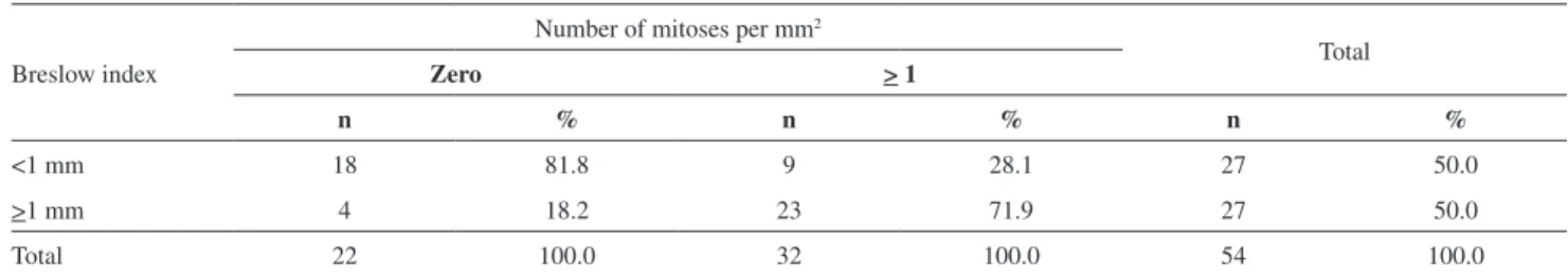
The mitotic index was correlated to Breslow index (p = 0.0002), as seen in Table 4. The number of mitoses was signiicantly higher in patients with disease progression (p = 0.0171), as shown in Table 5.
A sentinel lymph node biopsy was performed in 19/54 patients, of which 2 had lymph nodes metastasis. One case evolved to dissemination and death, while the other is still on clinical follow-up.
Among patients with melanoma dissemination, the mean disease-free survival time was 17.8+18.3 months (range, 4-65 months). The actuarial analysis, as conirmed using the Kaplan-Meier Product, revealed an 82% disease-free rate for 1 year, 70% for 2 years and 62% for 5 years.
Cancer-speciic mean survival time was 24.5+20.7 months (range, 5-67 months). Survival rates, as calculated by the actuarial analysis and Kaplan-Meier Product, revealed an 85% survival rate for 1 year, 82% for 2 years and 70% for 5 years.
Table 2 - Percent distribution of frequencies relative to the
evolution of 27 patients with Breslow index > 1 mm
Breslow index Disseminated disease P value
Absent Present
1-2 mm 9 2
2-4 mm 4 4
> 4 mm 2 6 0.042
Table 3 - Distribution of frequencies relative to the evolution of 54 patients with melanoma according to the presence or
absence of ulceration
Ulceration
Disseminated disease
Total
Absent Present
N % N % n %
Yes 3 7.1 9 75.0 12 22.2
No 39 92.9 3 25.0 42 77.8
Total 42 100.0 12 100.0 54 100.0
Freedom degree: 1. Calculated chi-square: 28.94. Calculated p value: < 0.001
Table 4 - Distribution of frequencies relative to the number of mitoses per mm2 according to Breslow’s index
Breslow index
Number of mitoses per mm2
Total
Zero > 1
n % n % n %
<1 mm 18 81.8 9 28.1 27 50.0
>1 mm 4 18.2 23 71.9 27 50.0
Total 22 100.0 32 100.0 54 100.0
Fisher exact test. Calculated p value: 0.0002.
Table 5 - Distribution of frequencies relative to the evolution of 54 patients with melanoma according to the number of mitoses per mm2
Number of
mitoses per
mm2
Disseminated disease Total
Absent Present
n % n % n %
Zero 21 50.0 1 8.3 22 40.7
>1 21 50.0 11 91.7 32 59.3
Total 42 100.0 12 100.0 54 100.0
DISCUSSION
Malignant cutaneous melanoma is still a major clinical challenge, especially when we take into consideration the increasing incidence of smaller lesions that have been recorded in the last decades in developed countries,22 and
more notably, in their Caucasian populations.3 Melanoma is
still the main cause of death due to skin cancer.14
In the south region of Brazil, the relative increase of melanoma incidence between 1979 and 1987 was 38% among men and 11% among women.3 Such an increase,
although in lower proportions, was subsequently conirmed in surveys carried out in Sao Paulo between 1963 and 1997,
23 and between 1993 and 2006,24 and in Goiania from 1988
to 2000.25
These Brazilian data are not different from the results of epidemiologic studies conducted in other countries, like Scotland (a rise of 303% for men and 187% for women over eleven years),13 the United States (increasing incidence of
cutaneous melanoma),26,27 Queensland, Australia (increasing
by more than 50% in women and more than doubling in men), 28 Sweden (5% annual increase),29 New South Wales,
Australia (incidence rates are much higher than a few decades before),30 Japan (annual increase ratio for deaths
of 6.3%)31 and British Columbia, Canada (3-7% a year
frequency increase for new cases, even doubling at each 10-15 year period, depending on the population studied)32.
The available diagnostic methods for early detection of the disease, based on microscopic indings of the tissue morphology, still lack accuracy.1 Likewise, the clinical and
histological variables determining the disease prognosis include the Breslow index, tumor size, presence of ulceration, mitotic index and level of vascular invasion, but gaps in the deinition of risk groups persist, as this neoplasm’s clinical manifestation is quite variable from case to case.14, 33
From an epidemiological perspective, we noticed that this pathology affected men and women equally often and at similar mean ages, corroborating the data found in the international and national literature.4,22,23,34 Reports of
differences between genders in the incidence of cutaneous melanoma appear to be related to each country’s culture, in terms of which part of the body is exposed to the sun more often and by whom (Jordan).35
Among males trunk lesions were more frequent (42.8%), while among females the lower limb lesions prevaled (36.6%). Such results conirm the relation of cutaneous melanoma with solar radiation, as the higher melanoma incidence in areas that were more exposed to the sun resulted in a disease incidence in different locations in males and females, according to their habits and vestments in Brazil.
These indings are consistent with reports identifying the most frequent tumor sites as the backs of males (49.5%), and the lower limbs of females (33.1%) in the southern region of Brazil.36 Criado et al.23 had even observed a likelihood
2.26-times as high for males to develop cutaneous melanoma in the posterior region of the trunk, while the likelihood for the disease to occur on the lower limbs was 2.4-times as high in females.
There was no evidence of differences between genders when assessing mean age, Clark level and Breslow index, except for lesions having a thickness of 2 to 4 mm, which were signiicantly more frequent in males (p = 0.049).
The increased incidence currently being reported for this neoplasm generally refers to thinner or in situ lesions.13,29
However in males, an increase in the number of patients with thicker lesions has also been demonstrated.16,27
This study identified an 18.5% rate of disease dissemination resulting in death, and a 3.7% rate of disease dissemination without death. Therefore, 22.2% of patients had disease dissemination, with no differences between males and females and no inluence by patient age or lesion site.
Disease dissemination was observed an average of 17.8 months from lesion diagnosis, and deaths occurred an average of 24.5 months from the initial diagnosis. There was no correlation between these mean times and lesion thickness, suggesting an association of > 1 mm-thick lesion dissemination risk with other characteristics.
The Breslow index constituted a predictive variable of disease evolution, as no patient with a < 1 mm-thick tumor progressed to disease dissemination, while patients with > 1 mm-thick tumors had dissemination, particularly those with > 4 mm-thick tumors, conirming the reports of other publications.4,9,15
Likewise, the number of detected mitoses in the primary tumor may follow the progression of the disease (r = 0.43), and it may constitute, along with ulceration and Breslow index, the group of associated factors that can best predict patient tumor evolution.
Of the 27 > 1 mm melanoma cases used for disease-free and survival time analysis, 12 had ulcerated lesions, and 9 of those 12 lesions had disease dissemination. Of the patients with a Breslow index greater than 4 mm, 7 had ulceration, and 6 of those 7 ulcerations progressed to death or dissemination.
Breslow index association with the presence of ulceration and mitotic index showed a clear correlation with patient evolution when converted into a numeric score: sum of
ulceration (0 when absent or 1 when present), Breslow index
(1 when < 1 mm, 2 when > 1 and < 4 mm, 3 when > 4 mm)
The study also identiied a 63% disease-free survival time rate, and a 70% cancer-speciic 5-year survival rate. In São Paulo, Brazil, Criado et al.22 reported a similar
5-year survival (67.4%) to that found in Great Britain by Carmichael et al.32 (55.5% for males and 70.3% for females).
CONCLUSIONS
• Melanoma is a low-incidence skin tumor that is showing an increasing incidence in the last decades.
• A higher incidence of melanoma was found in sun-exposed areas, with trunk lesions being more frequent
in males, and lower limb lesions being predominant in females.
• The mean time elapsed from treatment to melanoma dis-semination diagnosis was 17.8 months.
• The survival rates calculated through actuarial analysis and the Kaplan-Meier Product revealed a 5-year survival rate of seventy percent.
• Deeper tumors correlated with more unfavorable patient disease evolution, which correlated with the Breslow index.
• The presence of ulcer and mitotic index had also impact in patient disease evolution.
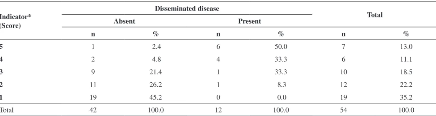
Table 6 - Distribution of frequencies relative to the disease evolution of 54 patients with melanoma in relation to the sum of
the Breslow index, ulceration and the mitotic index
Indicator* (Score)
Disseminated disease
Total
Absent Present
n % n % n %
5 1 2.4 6 50.0 7 13.0
4 2 4.8 4 33.3 6 11.1
3 9 21.4 1 33.3 10 18.5
2 11 26.2 1 8.3 12 22.2
1 19 45.2 0 0.0 19 35.2
Total 42 100.0 12 100.0 54 100.0
* Sum of ulceration (0 when absent or 1 when present), Breslow index (1 when < 1 mm, 2 when > 1 and < 4 mm, 3 when > 4 mm) and mitotic index (0 when absent or 1 when >1 per mm2)
REFERENCES
1. Wainstein AJA, Belfort FA. Conduta para o melanoma cutâneo. Ver Col Bras Cir. 2004; 31:204-14.
2. Sekulik A, Haluska P Jr, Miller AJ, De Lamo JG, Ejadi S, Pulido JS, et al. Malignant melanoma in the 21st century: The emerging molecular
landscape. Mayo Clin Proc. 2008; 83:825-46.
3. Tomatis L. Cancer: Causes, occurrence and control. IARC Scientiic Publication. 1990; 368:100.
4. Azevedo G, Mendonça S. Risco crescente de melanoma de pele no Brasil. Rev Saude Publica. 1992;26:290-4.
5. Balch CM, Buzaid AC, Soong SJ, Atkins MB, Cascinelli N, Coit DG, et al. Final version of the American Joint Committee on Cancer staging system for cutaneous melanoma. J Clin Oncol. 2001;19:3635-48. 6. Benett DC. Human melanocyte senescence and melanoma susceptibility
genes. Oncogene 2003;22:3063-9.
7. Howe HL, Wingo PA, Thun MJ, Ries LAG, Rosenberg HM, Feigal EG, et al. Annual report to the nation on the status of cancer (1973 through 198), featuring cancers with recent increasing trends. J Natl Cancer Inst. 2001;93:824-42.
8. INCA – Instituto Nacional de Câncer. Câncer de pele: Melanoma. Disponível em: www.inca.gov.br. Acesso em: 12 de agosto de 2008.
9. Essner R. Surgical treatment of malignant melanoma. Surg Clin N Am. 2003;83:109-56.
10. Clark Jr. WH, From L, Bernardino EA, Mihm MC. The histogenesis and biologic behavior of primary human malignant melanomas of the skin. Cancer Res. 1969;29:705-27.
11. Breslow A, Macht SD. Optimal size of resection margin for thin cutaneous melanoma. Surg Gynecol Obstet 1977;145:691-2. 12. Elder DE, Guerry D 4th, Heiberger RM, LaRossa D, Goldman LI,
Clark WH Jr, et al. Optimal resection margin for cutaneous malignant melanoma. Plast Reconstr Surg. 1983; 71:66-72.
13. MacKie RM, Bray CA, Hole DJ, Morris A, Nicolson M, Evans A, et al. Incidence of and survival from malignant melanoma in Scotland: An epidemiological study. Lancet. 2002; 360:587-91.
14. Alonso SR, Otiz P, Pollán M, Pérez-Gomez B, Sánchez L, Acuña MJ, et al. Progression in cutaneous malignant melanoma is associated with distinct expression proiles: A tissue microarray-based study. Am J Pathol. 2004;164:193-203.
16. Kuo TC, Hoon DSB, Takeuchi H, Turner R, Wang He-Jing, Morton DL, et al. Prediction of disease outcome in melanoma patients by molecular analysis of parafin-embedded sentinel lymph nodes. J Clin Oncol. 2003;19:3566-72.
17. Baldi A, Santini D, De Luca A, Paggi MG. cDNA array technology in melanoma: An overview. J Cell Physiol. 2003;196:219-23.
18. Bar-Eli M. Molecular mechanisms of melanoma metastasis. J Cell Physiol. 1997; 173:275-8.
19. Welch DR, Goldberg SF. Molecular mechanisms controlling human melanoma progression and metastasis. Pathobiology. 1997;65:311-30. 20. Landman G, Muller H, Neto JF, Maceira JMP, Costa M, Costa MB, et al.
Consenso para o laudo anatomopatológico do melanoma cutâneo. Acta Oncol Brasil. 2003;23:504-10.
21. Dawson-Saunders B, Trapp RG. Basic and clinical biostatistics. Norwalk: Appleton & Lange; 1994.
22. Tamir G, Milo Y, Rothem A, Sulkes J, Hauben DJ. Cutaneous malignant melanoma in young adults under age 30. Isr J Med Sci. 1996;32:1290-6. 23. Criado PR, Vasconcelos C, Sittart JAS, Valente NYS, Moura BPS, Barbosa
GL, et al. Melanoma maligno cutâneo primário: Estudo retrospectivo de 1963 a 1997 no Hospital do Servidor Público Estadual de São Paulo. Rev Ass Méd Brasil. 1999;45:157-62.
24. Ferrari Júnior NM, Muller H, Ribeiro M, Maia M, Sanches Júnior JA. Cutaneous melanoma: descriptive epidemiological study. Sao Paulo Med J. 2008;126:41-7.
25. Sortino-Rachou AM, Curado MP, Latorre MRDO. Melanoma cutâneo: estudo de base populacional em Goiânia, Brasil, 1988 - 2000. An Bras Dermatol. 2006;81:449-55.
26. Dennis LK. Analysis of the melanoma epidemic, both apparent and real: Data from the 1973 through 1994 surveillance, epidemiology, and end results program registry. Arch Dermatol. 1999;135:275-80.
27. Jemal A, Devesa SS, Hartge P, Tucker MA. Recent trends in melanoma incidence among whites in the United States. J Natl Cancer Inst. 2001;93:678-83.
28. MacLennan R, Green AC, McLeod GR, Martin NG. Increasing incidence of cutaneous melanoma in Queensland, Australia. J Natl Cancer Inst. 1992; 84:1427-32.
29. Mansson-Brahme E, Johansson H, Larsson O, Rutqvist LE, Ringborg U. Trends in incidence of cutaneous malignant melanoma in a Swedish population 1976-1994. Acta Oncol 2002;41:138-46.
30. Marrett LD, Nguyen HL, Armstrong BK. Recent trends in the incidence of malignant melanoma in New South Wales 1983-1996. Int J Cancer. 2001;92:457-62.
31. Ohtsuka H, Nagamatsu S. Changing trends in numbers of deaths from Malignant Melanoma in Japan, 1955-2000. Dermatology. 2003;207:162-5. 32. Carmichael VE, Wilson KS. Primary cutaneous malignant melanoma: Experience of British Columbia Cancer Agency from 1972 to 1981. CJS. 1992;35:589-97.
33. Tovo LFR, Belfort FA, Sanches Jr JA. Melanoma cutâneo primário. Rev Assoc Med Bras. 2005;51:1-10.
34. Holme SA, Malinovsky K, Roberts DL. Malignant melanoma in South Wales: Changing trends in presentation (1986-1998). Clin Exp Dermatol. 2001;26:484-9.
35. Oumeish OU. Epidemiology of primary cutaneous malignant melanoma in Jordan. Intern J Dermatol. 1995;36:113-5.