INTRODUCTION
Address to: Dra Marina Loyola Dantas. CPqGM/FIOCRUZ. R. Waldemar Falcão 121,
Candeal, 40296-710 Salvador, Bahia, Brasil.
Phone: 55 71 3176-2232; Fax: 55 71 3176-2327
e-mail: [email protected]
Received 3 September 2013
Accepted 22 November 2013
CD8+ T cells
in situ
in different clinical forms of human
cutaneous leishmaniasis
Marina Loyola Dantas
[1], Juliana Cabral de Oliveira
[1], Lucas Carvalho
[2],[3],
Sara Timóteo Passos
[2],[3],
Adriano Queiroz
[2],[3],
Paulo Machado
[2],[3],
Edgar Carvalho
[2],[3]and Sérgio Arruda
[1][1]. Laboratório Avançado de Saúde Pública, Centro de Pesquisas Gonçalo Moniz, Fundação Oswaldo Cruz, Salvador, BA. [2]. Serviço de Imunologia, Hospital Universitário Professor Edgard Santos, Universidade Federal da Bahia, Salvador, BA. [3]. Instituto Nacional de Ciência e Tecnologia de Doenças Tropicais,
Conselho Nacional de Desenvolvimento Científi co e Tecnológico, Salvador, BA.
ABSTRACT
Introduction: Leishmania braziliensis infection induces a large spectrum of lesions that clinically manifest as nodules or papules that progress to ulcers. Although it is already known that T helper cells predominate in the lesions, cytotoxic T cells have also been reported to be present, and their role in leishmaniasis immunopathogenesis is not well known. This study investigated the amounts of CD8+ and granzyme B+ cells in different clinical forms of human cutaneous leishmaniasis (CL). Methods:
Forty tissue fragments from early (E-CL) and late CL (L-CL) lesions and from disseminated leishmaniasis (DL) - papules and ulcers - were characterized. The infl amed area per fragment was calculated, and the CD8 and granzyme B expression levels in the infi ltrates were quantifi ed by counting positive cells in 15 fi elds. The localization of CD8 and granzyme B was graded subjectively. Results: Infl ammation was higher in L-CL and DL ulcers. CD8 expression was increased in late ulcerated lesions
compared to recent lesions. The increase in CD8+ cells also correlated with the duration of the lesion. Papules had a higher frequency of granzyme B+ cells than E-CL lesions, although the frequency was similar to those for late and DL ulcers. CD8+ cells were mostly found in the papillary dermis. Conclusions: CD8+ T and granzyme B+ cells are present in the infl ammatory
infi ltrates of CL and DL and may participate in the immunopathogenesis of Leishmania infection.
Keywords: Leishmaniasis. Cytotoxicity. Infl ammation. Cutaneous. CD8. Human.
Leishmaniasis, a disease caused by a protozoan of the genus Leishmania, affects millions of individuals in many regions of the world. Infections by Leishmania braziliensis, the most prevalent species in Brazil, lead to lesions in the skin or mucosa that become cutaneous leishmaniasis (CL) or mucosal leishmaniasis (ML). Cutaneous leishmaniasis lesions start as small papules in the presence or absence of lymphadenopathy, and they progress into one or more skin ulcers. This localized skin ulceration is characterized by a chronic infl ammatory response involving lymphocytes, plasma cells and macrophages, with and an eventual granulomatous reaction and necrosis1,2.
The tissue response in the initial disease manifestation, including early ulcers (≤ 20 days of lesion appearance) and papules from disseminated leishmaniasis (DL), has not been well studied. DL is an emerging form of leishmaniasis that corresponds to 1.9% of all CL cases; it is characterized by the
appearance of multiple pleomorphic lesions in more than two noncontiguous areas of the body, starting from a primary ulcer2-5.
The mechanisms involved in DL have not been elucidated. It has been suggested that parasite, host and environmental factors may contribute to the dissemination of the parasite throughout the body5.
Cluster of differentiation 8 (CD8)+ T cells are essential in the response against viruses and malignant cells, but few studies report the role of these cells in the defense against Leishmania
parasites6-8. Although the presence of cluster of differentiation
4 (CD4)+ T cells within the inflammatory infiltrate in leishmaniasis lesions has already been shown, cytotoxic T cells in the lesions of CL patients6 have also been reported.
The role of CD8+ T cells remains unclear, and contradictory results have been obtained for Leishmania infection; these cells may contribute to the killing of Leishmania or to the immunopathology of this parasite. A signifi cant proportion of CD8+ cells in human leishmaniasis tissue fragments has been reported9. The CD8+ lymphocytes could participate in parasite
growth control through different mechanisms. These cells have been associated with cure after treatment through interferon
gamma (IFN-γproduction, and they can also interact directly
with Leishmania-infected macrophages by killing them through cytotoxic activity via granzyme granules10-12. Thus, CD8+ T
RESULTS METHODS
composition and the localization of granzyme B in all types of cutaneous leishmaniasis lesions, including early events in the immune response, may help to elucidate the mechanism of tissue damage.
In the present study, we investigated the proportions of CD8+ T cells and granzyme B+ cells in primary and secondary lesions from DL and in early and late CL lesions to better understand the roles of these cells in the pathogenesis of the disease.
Patients
Thirty patients from Corte de Pedra, an area of Leishmania braziliensis transmission in the State of Bahia, Brazil, were enrolled in this study. A diagnosis of cutaneous leishmaniasis was initially made based on the presence of a typical lesion and a positive Leishmania skin test, and the diagnosis was confi rmed
by parasite isolation or identifi cation of amastigote forms by histopathological analysis. Disseminated leishmaniasis was clinically diagnosed based on the presence of multiple (more than 10 lesions in at least two different areas of the body) acneiform, papular or ulcerated lesions. Confi rmation of the diagnosis was performed as established above for CL. Patients were not under treatment at the time of tissue fragment collection. Cases with durations of more than 20 days were classifi ed as late cutaneous lesions (L-CL), and cases with durations of 20 days or less were grouped as early cutaneous lesions (E-CL). For each patient with disseminated leishmaniasis, one tissue fragment was collected from primary ulcers of DL (U-DL), and one was collected from secondary papular lesions of DL (P-DL). Each group of patients consisted of 10 subjects, with a total of 40 tissue fragments. None of the patients had been previously diagnosed with or treated for leishmaniasis.
Tissue fragment collection
Tissue fragments were collected under local anesthesia using a 4mm punch. The specimens were collected from the border of the ulcer and, in case of a secondary lesion in DL, from papules.
The fragments were fi xed in 10% formaldehyde, and the slides were prepared at the histotechnology laboratory at
Fundação Oswaldo Cruz (FIOCRUZ). The tissue samples were dehydrated and embedded into paraffi n blocks, and cryosections (4 to 6µm) were placed on silane-pre-coated slides.
Hematoxylin-eosin and immunohistochemistry staining
Sections obtained from paraffi n were routinely deparaffi nized and rehydrated. Slides were stained with hematoxylin-eosin
(HE) for histological diagnosis, for infl ammatory infi ltrate
analysis and to perform immunohistochemistry (IHC) reactions. Immunohistochemistry reactions were followed by blocking peroxidase activity with 3% hydrogen peroxide for 5min. The slides were incubated for 30min with anti-CD8antibody (M3164, Spring Bioscience Corp., Pleasanton, CA, USA) at a dilution of 1:100 at 25°C or overnight with anti-granzyme B (262A-16, Cell Marque Corp., Rocklin, CA, USA) at a dilution
of 1:100 at 4°C. Immunostaining was performed using the Peroxidase Mouse & Rabbit Kit - HRP (DBS KP500, US) according to the manufacturer’s recommendations. All slides were counter-stained with Harris hematoxylin, dehydrated and mounted with Canadian balsam and glass cover slips.
Morphometric analysis
Images from all HE-stained sections were acquired using an optical microscope (NIKON E-600) coupled to a digital color video camera at 400x magnifi cation. The infl amed area was calculated through measurements of the total fragment and infl ammatory infi ltrate areas. The CD8 and granzyme B expression levels in the infi ltrate were quantifi ed by counting the number of CD8+ T and granzyme B+ cells present in 15 fi elds per fragment. The frequencies of CD8+ and granzyme B+ cells in the papillary and reticular dermis were graded subjectively, and the values indicate the frequency of positive immunostained cells (0, absent; 1, low frequency; 2, medium frequency; 3, high frequency). ImageJ software (National Institute of Mental Health, USA) was used to measure areas and count cells.
Statistical analysis
Statistical analysis was performed using GraphPad Prism 5.0 software. The Spearman test was used to analyze correlations of infl ammation between both groups of cells. The Kruskal-Wallis non-parametric test was performed to compare L-CL, E-CL, U-DL and P-DL groups according to the number of CD8+ T and granzyme B+ cells. Differences in the frequencies of immunostained cells in the papillary dermis and the reticular dermis between patient groups were analyzed using the Chi-square test. Results were considered statistically signifi cant when p < 0.05.
Ethical considerations
Informed consent was obtained before the collection of tissue fragments, and all patients received treatment despite their enrollment in this study. The study was approved by the Human Ethics Committee of the Gonçalo Moniz Research Center, FIOCRUZ protocol number 321-2009.
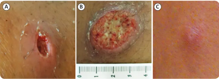
The clinical profi le of the 30 patients is shown in Table 1,
and the clinical characteristics of different lesions are shown in
Figure 1. Twenty-one men and 9 women were enrolled, with a mean age ± standard deviation (SD) of 31.52 ± 16.4 years (ages ranged from 9 to 76 years). The duration of lesions ranged from 25 to 60 days for L-CL (34 ± 10 days), 15 to 20 days for E-CL (17 ± 2 days), 20 to 90 days for U-DL (43 ± 20 days) and 8 to 90 days for P-DL (30 ± 25 days). Lesion size varied from 56 to 750mm2 for L-CL, 9 to 200mm2 for E-CL and 56 to
460mm2 for U-DL. P-DL lesions were not measured, and the
TABLE 1 - Clinical characteristics of cutaneous leishmaniasis patients.
Clinical Duration of Leishmania infection Location of Ulcer area DTH
Patient Age/gender form lesions (days) detection in HE ulcerated lesion (mm x mm) (mm x mm)
1 22/M L-CL 60 0 IM 30 x 20 15 x 15
2 19/M L-CL 30 0 IM 30 x 20 8 x 7
3 42/F L-CL 30 + IM 30 x 15 14 x 14
4 18/M L-CL 45 + IM 30 x 25 20 x 22
5 19/M L-CL 30 0 IM 10 x 8 19 x 16
6 39/F L-CL 25 + IM 20 x 15 15 x 16
7 9/M L-CL 30 + IM 10 x 10 14 x 10
8 27/M L-CL 30 + trunk 20 x 15 16 x 11
9 27/M L-CL 30 0 IL 20 x 20 12 x 10
10 37/M L-CL 30 0 IL ND negative
11 15/M E-CL 20 0 SL 6 x 9 30 x 30
12 18/M E-CL 15 0 SL 4 x 4 10 x 12
13 9/M E-CL 15 + SL 3 x 3 negative
14 36/F E-CL 15 + IL 8 x 5 20 x 14
15 26/F E-CL 15 + SL 3 x 3 14 x 13
16 25/M E-CL 15 + trunk 6 x 5 9 x 9
17 16/F E-CL 20 + IL 20 x 10 19 x 16
18 37/M E-CL 20 + IL 16 x 9 21 x 19
19 16/F E-CL 20 + IL 4 x 8 negative
20 23/M E-CL 20 0 IL 8 x 6 21 x 15
21 50/F DL (U/P) 60/15 +/0 IL 7 x 8 16 x 16
22 53/M DL (U/P) 60/30 +/+ face 20 x 15 negative
23 37/M DL (U/P) 30/15 +/+ IM ND 15 x 12
24 37/M DL (U/P) 30/10 0/+ SM 10 x 9 10 x 9
25 59/F DL (U/P) 30/30 0/0 SM 20 x 23 5 x 5
26 60/M DL (U/P) 30/60 +/0 IM 10 x 10 ND
27 17/M DL (U/P) 90/15 0/0 SM 10 x 15 22 x 18
28 37/F DL (U/P) 30/30 0/0 SM 10 x 11 18 x 15
29 30/M DL (U/P) 30/90 0/+ IM 10 x 10 ND
30 76/M DL (U/P) 38/8 +/0 IM 10 x 30 10 x 10
DTH: delayed type hypersensitivity; HE: hematoxylin-eosin; M: male; F: female; L-CL: late cutaneous leishmaniasis; E-CL: early cutaneous leishmaniasis; DL: disseminated leishmaniasis; U: ulcer; P: papule; +: presence; 0: absence; IM: inferior limb; SM: superior limb; ND: not determined. Papules were not measured.
Histopathological examination of tissue fragments from ulcerated or papular lesions revealed chronic infl ammatory reactions in the tissue fragment sections from all of the clinical forms. In all stages of CL, infl ammation was characterized by the presence of lymphocytes, plasmocytes and macrophages. Other general histopathological patterns were also found in
tissue fragments, such as perivascular and papillary edema (45%), necrosis (43%), granuloma (43%), giant cells (30%), rare neutrophils (18%) and pigmentary incontinence (28%).
A
B
C
FIGURE 1 - Typical lesions caused by different clinical forms of cutaneous leishmaniasis. A: Early cutaneous leishmaniasis lesion. B: Late cutaneous leishmaniasis lesion. C: Papule lesion, typical non-ulcerated lesion from disseminated leishmaniasis.
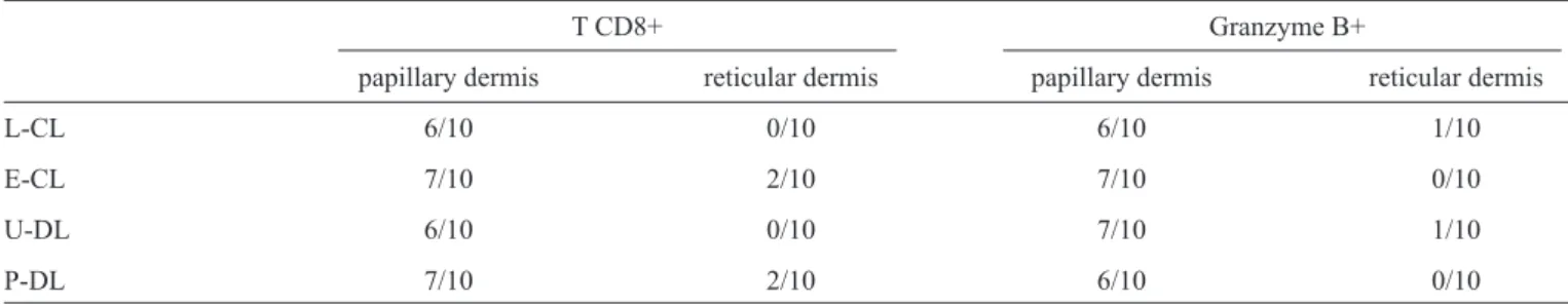
B+ cells was lower (Figure 2); the co-localization of the two types of cells was not analyzed. The majority of L-CL lesions (6/10) and U-DL lesions (6/10) were grade 3 (high frequency) for CD8+ frequency in the papillary dermis. Among the recent lesions (E-CL and P-DL), the intensity of stained cells had a greater variation, but a majority of the tissue fragments presented higher numbers of cells in the papillary dermis (Table 2). The granzyme B+ cell frequency was classifi ed as grade 2 in the majority of the groups. Only 4 tissue fragments in the CD8+ T cell analysis and 2 in the granzyme B+ analysis had a greater grade in the reticular dermis than the papillary dermis. Cells also appeared in a homogenous distribution (10 to 40% of cases per group).
Comparing the infl ammatory infi ltrate percentage between groups, P-DL lesions had the lowest percentage (median=21%; range 6-38%). A higher percentage of inflammation was present in L-CL (median=37%; range 21-52%) and U-DL (median=34%; range 20-56%) patients (Figure 3A).
Analyses of the frequencies of CD8+ and granzyme B+ cells were performed using ImageJ software, which allows manual counting of cells.
CD8 expression, similar to the infi ltrate extension, was increased in late ulcerated lesions compared to recent lesions (papules and lesions present < 20 days), which had low absolute numbers of CD8+ lymphocytes (Figure 3B).
To determine the contribution of CD8+ cells in the infl ammatory infi ltrate, the correlation of these cells and the infl ammation percentage was analyzed for each group. Only E-CL (R=0.7; p=0.02) and P-DL (R=0.79; p=0.005) lesions exhibited a correlation between the number of CD8+ T cells and greater infl ammation (not shown).
The papular lesions from DL had a higher frequency of granzyme B+ cells than E-CL lesions (p=0.0302) and a frequency similar to those of late and DL ulcers (Figure 3C). Interestingly, only E-CL lesions had a positive correlation
between granzyme B+ cells and the infl ammatory infi ltrate (R=0.7; p=0.0216). The analysis of late (R=-0.29; p<0.05) and DL ulcers (R=-0.22; p<0.05) revealed that the higher the infl ammation, the lower the amount of granzyme B+ cells (data not shown).
DISCUSSION
Cutaneous leishmaniasis is the most frequent form of leishmaniasis due to L. braziliensis infection. It is clinically characterized by the presence of single or multiple ulcers with a well-delimited and raised border that can persist for months or heal spontaneously. DL, similar to CL, presents initially with an ulcer and then disseminates throughout the body with multiple acneiform and papular secondary lesions5.
CD8+ and granzyme B+ lymphocytes were more frequent in the papillary dermis. Histopathological analysis of tissue fragments showed the presence of spongiosis. The aggregation of both cells in the papillary dermis, sometimes also in the epidermis, seems to lead to injury in the basal layer and may contribute to ulcer formation. Recently, it has been suggested that the movement of T cells through the dermis and epidermis and their permanence in one layer or another correlates with the effector or memory phase of the immune response13. This
fi nding suggests another mechanism for these cells, which together with cytotoxic activity, may contribute to leishmaniasis immunopathology.
TABLE 2 - Number of cases with a high frequency of immunostained cells in the papillary dermis or reticular dermis.
T CD8+ Granzyme B+
papillary dermis reticular dermis papillary dermis reticular dermis
L-CL 6/10 0/10 6/10 1/10
E-CL 7/10 2/10 7/10 0/10
U-DL 6/10 0/10 7/10 1/10
P-DL 7/10 2/10 6/10 0/10
There were 10 patients per group. Ten tissue fragments stained for CD8+ T cells and twelve for granzyme B+ cells were found to have a homogenous distribution of cells. p=0.0105 for CD8+ T cells and granzyme B+ cells (Chi-square test). CD8+: cluster of differentiation 8; L-CL: late cutaneous lesions; E-CL: early cutaneous lesions; U-DL: primary ulcers of DL; P-DL: secondary papular lesions of DL.
FIGURE 2 - Frequency of CD8+ cells in the papillary dermis: A: Tissue fragment graded as low frequency. B: Tissue fragment graded as medium frequency. C: Tissue fragment graded as high frequency. Frequency of granzyme B+ cells in the papillary dermis. D: Tissue fragment graded as low frequency. E: Tissue
fragment graded as medium frequency. F: Tissue fragment graded as high frequency. All images have the same magnifi cation (400x).
A
B
C
D
E
F
x400
x400
x400
x400
x400
Recent studies have demonstrated an increased frequency of CD8+ T cells over CD4+ T cells in ulcerated lesions as well as correlations between the frequencies of CD8+ T cells and CD8+ T cells that express granzyme and the intensity of
the infl ammation, which indicate a role for these cells in the
pathogenesis of CL14-16. Santos et al8. have also shown the
participation of CD8+ granzyme B+ T cells in tissue damage in CL caused by L. braziliensis, data that reinforce the association of the intensity of necrosis and the percentage of these cells8.
CD8+ lymphocytes with cytotoxic function seem to execute a cytolytic activity that may be important for tissue destruction with ulcer formation, as is seen in ML, where the same CD8+ T cells seem to be involved in disease exacerbation17.
Vieira et al18.showed that both localized CL and papular DL
lesions presented a higher frequency of CD8+ T cells than CD4+
T cells in infl ammatory infi ltrates, suggesting that CD8+ cells
participate directly in the immune response against parasites, principally by IFN-γ production, which induces macrophage activation18. However, CD4+ T cells appear to be the main
source of IFN-γ and CD8+ T cells have a principal function of cytotoxicity, contributing to lesion formation8. In experimental
models, CD8+ T cells have already been shown to be necessary for a protective response and also for the immunopathological development of CL17.
The evaluation of the presence of cytotoxic activity in
infl ammatory infi ltrates revealed that lesions of less than 20 days
had lesser quantities of cells expressing granzyme B than late ulcers and ulcers from DL. This result agrees with a previous study that showed granzyme expression was associated with lesion progression14. Interestingly, papular lesions from DL
showed higher numbers of granzyme B+ cells than early ulcers. The association of DL with mucosal disease due to the rapid dissemination of lesions throughout the body and mucosa and the association of cytotoxic CD8+ T cells to tissue damage in ML have previously been reported5,19. Furthermore, granzyme
B does not seem to participate in parasite growth control8. The
presence of granzyme B+ cells in DL papular lesions is indicative of their role in pathology. Granzyme B seems to participate in
0 5 10 15 20 25 30 35 40 45
L-CL E-CL U-DL P-DL
Inflam m a tion (% ) 0 50 100 150 200 250 300 350 400 T CD8+ c e ll s U-DL P-DL 0 50 100 150 200
L-CL E-CL U-DL P-DL
G ra n z y m e B + c e ll s 0 5 10 15 20 25 30 35 40 45
L-CL E-CL U-DL P-DL
Inflam m a tion (% ) 0 50 100 150 200 250 300 350 400 L-CL E-CL T C D8+ c e ll s
FIGURE 3 - A: Comparison of the infl ammation percentage in different lesions from cutaneous leishmaniasis. B: Comparison of the numbers of CD8+ T cells
in different lesions from cutaneous leishmaniasis. C: Comparison of the numbers of granzyme B+ cells in different lesions from cutaneous leishmaniasis. Median ± standard error is represented with bars. The Kruskal-Wallis test was performed. L-CL: late cutaneous lesions; E-CL; early cutaneous lesions; U-DL: primary ulcers of DL; P-DL: secondary papular lesions of DL.
lesion progression in E-CL, as the expression of granzyme B and the infl ammation percentage show simultaneous increases in E-CL.
In summary, CD8+ T cells and granzyme B+ cells are present in the infl ammatory infi ltrates of CL and DL. Further studies are necessary to elucidate the contribution of these cells to the killing or dissemination of the parasite in Leishmania
infections. Their cytotoxic function and specifi c localization may be responsible for tissue destruction and may explain the mechanism of ulceration. Due to the scarcity of patients with the DL form who simultaneously present papules and ulcers, a low number of patients was recruited. Further characterization
of the infl ammatory reaction in situ in the two clinical forms
and the four-lesion spectrum of human leishmaniasis may help elucidate the role of CD8+ cells and their cytotoxic activity in the immunopathogenesis of this disease.
ACKNOWLEDGMENTS
We thank Mr. Lago, Ms. Neuza and Ms. Renda from Corte de Pedra Health Service and the technicians of the histotechnology service of Centro de Pesquisas Gonçalo Moniz, Fundação Oswaldo Cruz.
REFERENCES
The authors declare that there is no confl ict of interest. CONFLICT OF INTEREST
1. Magalhães AV, Moraes MAP, Raick AN, Cuentas AL, Costa JML, Cuba CAC, et al. Histopathology of tegumentary leishmaniasis caused by
Leishmania braziliensis braziliensis: 2. Tissue Humoral Response. Rev Inst Med Trop 1986; 28:293-299.
2. Jones TC, Johnson WD, Barretto AC, Lago E, Badaro R, Cerf B, et al. Epidemiology of american cutaneous leishmaniasis due to Leishmania braziliensis braziliensis. J Infect Dis 1987; 156:73-83.
3. Costa JM, Marsden PD, Llanos-Cuentas EA, Netto EM, Carvalho EM,
Barral A, et al. Disseminated cutaneous leishmaniasis in a fi eld clinic in
Bahia, Brazil: a report of eight cases. J Trop Med Hyg 1986; 89:319-323.
4. Carvalho EM, Barral A, Costa JM, Bittencourt A, Marsden P. Clinical and immunopathological aspects of disseminated cutaneous leishmaniasis. Acta Trop 1994; 56:315-325.
5. Turetz ML, Machado PR, Ko AI, Alves F, Bittencourt A, Almeida RP, et al. Disseminated leishmaniasis: a new and emerging form of leishmaniasis observed in northeastern Brazil. J Infect Dis 2002; 186:1829-1834.
6. Machado P, Kanitakis J, Almeida R, Chalon A, Araújo C, Carvalho EM. Evidence of in situ cytotoxicity in american cutaneous leishmaniasis. Eur J Dermatol 2002; 12:449-451.
7. Ruiz JH, Becker I. CD8 cytotoxic t cells in cutaneous leishmaniasis. Parasite Immunol 2007; 29:671-678.
8. Santos CS, Boaventura V, Ribeiro Cardoso C, Tavares N, Lordelo MJ, Noronha A, et al. CD8 (+) Granzyme B (+)-mediated tissue injury vs. CD4
(+) IFNγ (+)-mediated parasite killing in human cutaneous leishmaniasis.
J Invest Dermatol 2013; 133:1533-1540.
9. Campanelli AP, Roselino AM, Cavassani K, Pereira MSF, Mortara R, Brodskyn CI, et al. CD4+ CD25+ T cells in skin lesions of patients with cutaneous leishmaniasis exhibit phenotypic and functional characteristics of natural regulatory T cells. J Infect Dis2006; 193:1313-1322.
10. Smith LE, Rodrigues M, Russell DG. The interaction between CD8+ cytotoxic t cells and Leishmania-infected macrophages. J Exp Med 1991; 174:499-505.
11. Da-Cruz AM, Conceição-Silva F, Bertho AL, Coutinho SG. Leishmania -reactive CD4+ and CD8+ T cells associated with cure of human cutaneous leishmaniasis. Infect Immun 1994; 62:2614-2618.
12. Coutinho SG, Oliveira MP, Da-Cruz AM, De Luca PM, Mendonça SC, Bertho L, et al. T-cell responsiveness of american cutaneous
leishmaniasis patients to purifi ed Leishmania pifanoi amastigote antigens and Leishmania braziliensis promastigote antigens: immunologic patterns associated with cure. Exp Parasitol 1996; 84:144-155.
13. Gebhardt T, Whitney PG, Zaid A, Mackay LK, Brooks AG, Heath WR, et al. Different patterns of peripheral migration by memory CD4+ and CD8+ T cells. Nature 2011; 477:216-219.
14. Faria DR, Souza PE, Durães FV, Carvalho EM, Gollob KJ, Machado PR, et al. Recruitment of CD8(+) T cells expressing granzyme A is associated with lesion progression in human cutaneous leishmaniasis. Parasite Immunol 2009; 31:432-439.
15. Faria DR, Gollob KJ, Barbosa Jr J. Decreased in situ expression of
interleukin-10 receptor is correlated with the exacerbated infl ammatory
and cytotoxic responses observed in mucosal leishmaniasis. Infect Immun 2005; 73:7853-7859.
16. Da-Cruz AM, Bertho AL, Oliveira-Neto MP, Coutinho SG. Flow cytometric
analysis of cellular infi ltrate from american tegumentary leishmaniasis
lesions. Br J Dermatol2005; 153:537-543.
17. Stäger S, Rafati S. CD8+ T cells in Leishmania infections: friends or foes? Front Immunol 2012; 3:1-8
18. Vieira MGS, Oliveira F, Arruda S, Bittencourt AL, Barbosa AA,
Barral-Netto M, et al. B-cell infi ltration and frequency of cytokine producing
cells differ between localized and disseminated human cutaneous leishmaniasis. Mem Inst Oswaldo Cruz 2002; 97:979-983.