Tumor Necrosis Factor-
αα
αα
α
in Human American Tegumentary
Leishmaniasis
Alda Maria Da-Cruz, Márcia Pereira de Oliveira, Paula Mello De Luca,
Sergio CF Mendonça, Sergio G Coutinho
+Laboratório Imunidade Celular e Humoral, Departamento de Protozoologia, Instituto Oswaldo Cruz, Av. Brasil 4365, 21045-900 Rio de Janeiro, RJ, Brasil
Tumor necrosis factor-alpha (TNF-α) is a cytokine produced by activated macrophages and other cells. In order to verify whether the serum levels of TNF-α in American tegumentary leishmaniasis patients are associated with the process of cure or aggravation of the disease, 41 patients were studied: 26 cases of cutaneous leishmaniasis (CL) and 15 of mucocutaneous leishmaniasis (MCL). During active disease the serum levels of TNF-α of MCL patients were significantly higher than those of CL patients and control subjects (healthy individuals and cutaneous lesions from other etiologies). The MCL pa-tients had serum titers of TNF-α significantly lower at the end of antimonial therapy than before therapy. After a six-month follow-up, the MCL patients had serum levels of TNF-α similar to those observed at the end of the therapy as well as to those of CL patients and control subjects. No significant variation in the serum levels of TNF-α was observed in CL patients throughout the study period (before, at the end of therapy and after a six-month follow-up). The possible relationship between the high TNF-α serum levels and severity of the disease is discussed.
Key words: TNF-α - cytokine - tegumentary leishmaniasis - Leishmania braziliensis
Supported by grants from the UNDP/World Bank/WHO Special Program for Research and Training in Tropical Diseases, The Europeean Community, Conselho Nacional de Desenvolvimento Científico e Tecnológico (CNPq) and Coordenadoria de Apoio à Pesquisa e Ensino Superior (CAPES).
+Corresponding author. Fax: 55-21-280.1589
Received 4 September 1995 Accepted 19 October 1995
In Brazil, American tegumentary leishmaniasis (ATL) is mainly caused by the protozoan parasite Leishmania braziliensis. Infection occurs by para-sitization of phagocytic cells at the site where the infected Phlebotominae bites the host when taking bloodmeal. The most frequent clinical form of the disease, cutaneous leishmaniasis (CL), is charac-terized by single or multiple skin ulcers that may heal spontaneously or after therapy. However, a minority of patients develop a severe chronic form of the disease named mucocutaneous leishmaniasis (MCL) with secondary metastatic lesions in mu-cosae of the mouth and nose, that may result in severe destruction of the face. Moreover, MCL is frequently resistant to therapy.
Tumor necrosis factor alpha (TNF-α) is a cytokine produced mainly by activated macroph-ages but also by T lymphocytes, natural killer cells, and other cell types (Vassalli et al. 1992). It has a broad spectrum of biological functions on many different target cells, including cytotoxicity,
inflam-matory mediation, tissue remodeling and host de-fense against microbes (Beutler & Cerami 1988, Camussi et al. 1991). The two biological active forms of TNF-α, a soluble molecule (sTNF-α) and a membrane-associated protein (mTNF-α), seem to play different roles in the pathogenesis of sev-eral diseases, including experimental leishmaniasis (Kriegler et al. 1988, Birkland et al. 1992).
In experimental leishmaniasis TNF-α appears to play an important role in host defense (Titus et al. 1989, Liew et al. 1990a, b, 1991). On the other hand, in humans, higher titers of serum TNF-α do not appear to be associated with immune protec-tion against Leishmania infecprotec-tion (Pisa et al. 1990) and also have been implicated in aggravation of many infectious diseases (Grau et al. 1989, Sarno et al. 1991, Barnes et al. 1992). In sera from pa-tients with visceral leishmaniasis and diffuse cuta-neous leishmaniasis relatively high titers of
TNF-α have been observed (Pisa et al. 1990, Barral-Neto et al. 1991). The purpose of this study was to com-pare the serum levels of TNF-α in ATL patients with different clinical forms, namely CL and MCL, throughout their clinical evolution, from active disease to cure.
MATERIALS AND METHODS
TNF-α in Human Leishmaniasis Alda M Da-Cruz et al.
to leishmanial antigens. The test was considered positive when a enduration higher than 5 mm in diameter was observed after 48 hr; (c) IgG and/or IgM Leishmania-reactive antibodies, detected by indirect immunofluorescence in the serum. Titres were considered positive when fluorescence was observed at a 1:45 serum dilution or higher; (d) Detection of Leishmania in lesions either by mi-croscopic examination of histological sections from biopsy samples or by culture in McNeal, Novy and Nicolle (NNN) medium (Nicolle 1908).
All patients were treated with antimonial (Glucantime®, Rhodia, São Paulo, SP, Brazil; three 10-day courses of N-methylglucamine at a dose of 15-20 mg Sb5+ daily i.m.). After a six-month fol-low-up the patients were considered cured if their lesions had completely healed.
Patients were evaluated before therapy (twenty CL patients and eleven MCL patients), at the end of therapy (fourteen CL patients and nine MCL patients) and after a six-month follow-up (thirteen CL patients and seven MCL patients). Some pa-tients were evaluated in all periods of the study. As controls, five patients with cutaneous lesions from other etiologies and ten healthy individuals were also studied.
Serum samples - The sera were collected in ster-ile vacutainer blood collection tubes (Becton, Dickinson and Company, Rutherford, NJ, USA) before therapy, at the end of therapy and after a six-month follow-up period. Serum samples were immediately frozen at - 70°C and stored for a maxi-mum of twelve months. No serum sample was fro-zen and thawed more than once.
TNF-α assay - Levels of TNF-α were deter-mined by testing the sera using a solid phase en-zyme-immunoassay employing the multiple anti-body sandwich principle, as described in the kit specifications (Factor-TestTMh TNF-α ELISA Test
Kit, Genzyme, Cambridge, MA, USA). Briefly, hu-man recombinant TNF-α, used as a standard in this assay, was serially diluted from 800 to 12 pg/ml. A 96-well microtiter plate was coated with mouse monoclonal antibody specific for human TNF-α and incubated overnight. Standard amounts of hu-man recombinant TNF-α and serum samples were added in duplicate. Afterwards, the second anti-body (rabbit anti-human TNF-α polyclonal anti-body) and the third antibody (biotin-conjugated goat anti-rabbit IgG) were sequentially applied. Streptavidin-conjugated peroxidase was distributed into each well and a substrate reagent was added in order to obtain color reaction. The absorbance was measured at 492 nm in Multiskan Plus MK II spectrophotometer (Titertek®, Flow Laboratories, McLean, Virginia, USA). The results were ex-pressed in pg/ml.
Statistical analysis -The Mann-Whitney two tail U test and Spearman correlation test were utilized.
RESULTS
Clinical findings - All patients had active
leish-manial lesions at the beginning of the study. The mean age was 36.5±13.7 years for CL patients and 49.8±8.7 years for MCL patients. The mean pe-riod of illness was 2.4±1.8 months and 15.4±30.3 months, respectively for CL and MCL patients. With regard to the number of lesions in CL pa-tients, 54% had a single lesion, 38% had two to ten lesions and 8% had more than ten lesions. All the MCL patients displayed lesions on the mucous membranes of the mouth and nose. All patients came from endemic areas of Rio de Janeiro, Bra-zil, where the only species of Leishmania that has been found infecting humans and dogs is L. braziliensis (Grimaldi Jr et al. 1991). The MST was positive in 88.4% of CL patients (mean = 10.3±3.5 mm in diameter) and 93.3% of MCL pa-tients (mean = 16.5±8.1 mm in diameter). The di-agnosis of leishmaniasis was established based in at least two of the criteria mentioned in the mate-rial and methods. At the end of therapy the lesions of all CL patients had healed while the lesions of MCL patients still displayed mild signs of activ-ity. After the six-month follow-up all patients were considered clinically healed.
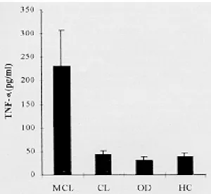
Serum levels of TNF-α - Sera from the patients with CL and MCL, as well as individuals with cu-taneous lesions from other etiologies and from the healthy control subjects were tested for TNF-α. Before therapy, MCL patients displayed higher levels of TNF-α (mean = 231.4±76.3 pg/ml) than CL patients (mean = 43.5±8.2 pg/ml) (p = 0.0004). The latter had levels of TNF-α similar to those from patients with other cutaneous diseases (mean = 30.6±9.3 pg/ml) and healthy individuals (mean = 39.2±7.7 pg/ml) (Fig. 1). No significant correla-tion was observed between time evolucorrela-tion of the lesions and TNF-α serum levels in the leishmaniasis patients.
The mean levels of TNF-α in the sera from CL patients studied before therapy (twenty cases), at the end of therapy (fourteen cases) and after a six-month follow-up (thirteen cases) were respec-tively 43.5±8.2 pg/ml, 32.4±10.1 pg/ml and 37.4± 8.3 pg/ml. No significant difference among the lev-els of TNF-α of these three groups of CL patients (Fig. 2) was observed.
DISCUSSION
The active form of TNF-α is a trimer molecule which can be preserved at -70°C (Camussi et al. 1991, Thavasu et al. 1992). Under other conditions it is cleaved in monomer molecules that can also be measured, resulting in false results that are much higher than the actual levels. There is also a possi-bility of ex vivo induction of cytokines, including TNF-α, during the processing of blood samples (Leroux-Roels et al. 1988). For these reasons it is clear that a critical point for the measurement of the serum levels of circulating cytokines has been the establishment of optimal conditions for blood collection, processing and storage of the sample (Thavasu et al. 1992). In this study the samples were collected following these criteria, which sug-gests that the values found represent the actual lev-els of serum TNF-α.
The present results show that patients with ac-tive MCL, a severe clinical form of ATL, had lev-els of serum TNF-α significantly higher than CL patients (either active or healed). Clinically cured MCL patients had TNF-α levels significantly lower than those observed during active disease and simi-lar to those of patients with CL and control sub-jects. The levels of TNF-α among CL patients did not show significant variations throughout the study period, which is in accordance with results reported by other authors (Pisa et al. 1990, Barral-Neto et al. 1991), although different parasite spe-cies and different human populations had been studied.
In human leishmaniasis high levels of TNF-α have also been associated with severe clinical forms such as active visceral leishmaniasis (Barral-Neto et al. 1991) and diffuse cutaneous leishmaniasis (Pisa et al. 1990), both characterized by a failure of the Leishmania-specific T cell-mediated immune responses (Bryceson 1970, Carvalho et al. 1988). In those cases, the observed high titers of TNF-α in the absence of an efficient T cell-mediated im-mune response was apparently not beneficial for the patient (Pisa et al. 1990). On the other hand, in the majority of MCL patients there is an exacerba-tion of the immune responses (Coutinho et al. 1987) with relatively higher frequencies of L. braziliensis-reactive T cells in the lesions (Conceição-Silva et al. 1990). In such cases the T cell-mediated immune responses appear to be as-sociated with the process of aggravation of the le-sions. It is possible that the higher levels of soluble TNF-α in the sera of MCL patients could be in-volved with pathogenic mechanisms. However, patients with CL, a clinical form characterized by the development of a well modulated T cell-medi-ated immune response, have TNF-α levels in their
Fig 1: TNF-α levels (mean ± SE) in sera from twenty cutane-ous leishmaniasis patients (CL) and eleven mucocutanecutane-ous leishmaniasis patients (MCL) before therapy, as well as pa-tients with cutaneous diseases from other etiologies (OD) and healthy controls (HC).
MCL patients after a six-month follow-up (mean = 45±15.5 pg/ml) (Fig. 2). The results of the latter two groups were not different from those of the CL patients (at any time of study) and control sub-jects (patients with cutaneous diseases from other etiologies and healthy individuals).
Fig 2: TNF-α levels (mean ± SE) in sera from patients with
cutaneous leishmaniasis (CL) ( n ) and mucocutaneous
TNF-α in Human Leishmaniasis Alda M Da-Cruz et al.
sera similar to healthy subjects. It has also been demonstrated that the production of TNF-α by hu-man T lymphocytes can be induced by the activa-tion of molecules such as CD3 (Sung et al. 1988). Studies of in situ production of TNF-α in hu-man American tegumentary leishhu-maniasis. (Cáceres-Dittmar et al. 1993, Pirmez et al. 1993) have not shown significant differences in the
TNF-α mRNA expression between MCL and CL lesions. However, the detection of mRNA may not neces-sarily correspond to the protein production in situ and/or seric levels, and also it did not discriminate between the two biological forms of TNF-α (soluble and membrane-associated) that appear to play different roles in the immunopatho-genesis of experimental leishmaniasis (Birkland et al. 1992). The soluble form having deleterious effects and the membrane-associated form with beneficial effects.
Studies on experimental murine leishmaniasis have indicated that TNF plays an important role in the immune protection against the disease. Local treatment of the leishmanial lesions with TNF sig-nificantly reduced their development (Liew et al. 1990b) not only by reducing the pathological dam-age but also inhibiting parasite replication. The last effect may be due to its ability to induce macroph-age leishmanicidal activity mediated by nitric ox-ide (Liew et al. 1990a). Lymph node cells (LNC) from genetically resistant mice infected with L. major are able to produce higher levels of TNF than those from susceptible mice (Titus et al. 1989), although it was not clear which cells (macroph-ages or T lymphocytes) were the main producers of the cytokine. The production of TNF-α can be associated with unspecific tissue reactions or re-lated to the presence of Leishmania-specific CD4+ T cells able to produce and express mTNF on their cell surfaces leading to a increased ability to acti-vate antileishmanial mechanisms of macrophages from resistant mouse strains (Birkland et al. 1992). In leishmaniasis the host-protective effect of TNF-α that prevents lesion development could be associated with the presence of the mTNF-α ex-pressed on macrophages or lymphocytes in the le-sions. In CL patients an optimal production of mTNF-α in association with other cytokines could induce mechanisms involved in the healing of the lesions. On the other hand, an inappropriate release of soluble TNF-α could have deleterious effects, as suggested by the present results concerning MCL patients. High levels of TNF-α have also been as-sociated with the pathogenesis of several damage conditions such as in Plasmodium falciparum ma-laria (Grau et al. 1989), streptococcal toxic shock syndrome (Hackett & Stevens 1992), meningococcemia (Waage et al. 1987), septicemic
melioidosis (Suputtamongkol et al. 1992), leprosy reactions (Sarno et al. 1991), bacterial meningitis (Glimaker et al. 1993) and AIDS (Ayehunie et al. 1993).
The present results indicate an association be-tween elevated concentrations of TNF-α in the sera of leishmaniasis patients and the development of MCL, a severe form of the disease characterized by the presence of destructive mucosal lesions, suggesting that overproduction of this cytokine could be one of the factors which have deleterious effects leading to aggravation of the disease.
ACKNOWLEDGMENTS
To Dr F Conceição-Silva for valuable discussions, to Mr R Nogueira and Mr PRZ Antas for assistance in the laboratory and also to Mrs R Pellegrino for excel-lent secretarial assistance.
REFERENCES
Ayehunie S, Sonnerborg A, Yemane-Berhan T, Zewdie DW, Britton S, Strannegard O 1993. Raised levels of tumour necrosis factor-alpha and neopterin, but not interferon-alpha, in serum of HIV-1-infected patients from Ethiopia. Clin Exp Immunol 91: 37-42.
Barnes PF, Chatterjee D, Brennan PJ, Rea TH, Modlin RL 1992. Tumor necrosis factor production in pa-tients with leprosy. Infect Immun 60: 1441-1446. Barral-Neto M, Badaró R, Barral A, Almeida RP, Santos
SB, Badaró F, Pedral-Sampaio D, Carvalho EM, Falcoff E, Falcoff R 1991. Tumor necrosis factor in human visceral leishmaniasis. J Infect Dis 163: 853-857.
Beutler B, Cerami A 1988. Tumor necrosis, cachexia, shock, and inflammation: a common mediator. Ann Rev Biochem 57: 505-518.
Birkland TP, Sypek JP, Wyler DJ 1992. Soluble TNF and membrane TNF expressed on CD4+T lympho-cytes differ in their ability to activate macrophage antileishmanial defense. J Leu Biol 51: 296-299. Bryceson ADM 1970. Diffuse cutaneous leishmaniasis
in Ethiopia. III. Immunological studies. Trans R Soc Trop Med Hyg 64: 380-393.
Cáceres-Dittmar G, Tapia FJ, Sánchez MA, Yamamura M, Uyemura K, Modlin RL, Bloom BR, Convit J 1993. Determination of the cytokine profile in American cutaneous leishmaniasis using polymerase chain reaction. Clin Exp Immunol 91: 500-505. Camussi G, Albano E, Tetta C, Bussolino F 1991. The
molecular action of tumor necrosis factor-α. Eur J Biochem 202: 3-14.
Carvalho EM, Bacellar O, Reed SG, Barral A, Rocha H 1988. Visceral leishmaniasis: a disease associated with inability of lymphocyte to activate macroph-age to kill Leishmania. Braz J Med Biol Res 21: 85-92.
American mucocutaneous leishmaniasis. Clin Exp Immunol 79: 221-226.
Coutinho SG, Pirmez C, Mendonça SCF, Conceição-Silva F, Dórea RCC 1987. Pathogenesis and immu-nopathology of leishmaniasis. Mem Inst Oswaldo Cruz 82: 214-228.
Glimaker M, Kragsbjerg P, Forsgren M, Olcén P 1993. Tumor necrosis factor-α (TNFα) in cerebrospinal fluid from patients with meningitis of different eti-ologies: high levels of TNFα indicate bacterial men-ingitis. J Infect Dis 167: 882-889.
Grau GE, Taylor TE, Molyneux ME, Wirima JJ, Vassalli P, Hommel M, Lambert PH 1989. Tumor necrosis factor and disease severity in children with falciparum malaria. N Engl J Med 320: 1586-1591. Grimaldi Jr G, Momen H, Naiff RD, McMahon-Pratt D, Barrett TV 1991. Characterization and classifica-tion of leishmanial parasites from humans, wild mammals and sand flies in the Amazon region of Brazil. Am J Trop Med Hyg 44: 645-661.
Hackett SP, Stevens DL 1992. Streptococcal toxic shock syndrome synthesis of tumor necrosis factor and interleukin-1 by monocytes stimulated with pyro-genic exotoxin A and streptolysin O. J Infect Dis 165: 879-885.
Kriegler M, Perez C, DeFay K, Albert I, Lu SD 1988. A novel form of TNF/cachectin is a cell surface cyto-toxic transmembrane protein: ramifications for com-plex physiology of TNF. Cell 53: 45-53.
Leroux-Roels G, Offner F, Philippé J, Vermeulen A 1988. Influence of blood-collecting systems on con-centrations of tumor necrosis factor in serum and plasma. Clin Chem 34: 2373-2374.
Liew FY, Li Y, Millott S 1990a. Tumor necrosis fac-tor-α synergizes with IFN-γ in mediating killing of Leishmania major through the induction of ni-tric oxide. J Immunol 145: 4306-4310.
Liew FY, Li Y, Yang DM, Severn A, Cox FEG 1991. TNF-α reverses the disease-exacerbating effect of subcutaneous immunization against murine cutane-ous leishmaniasis. Immunol 74: 304-309.
Liew FY, Parkinson C, Millott S, Severn A, Carrier M
1990b. Tumor necrosis factor (TNFα) in leishmaniasis. I. TNFα mediates host protection against cutaneous leishmaniasis. Immunol 69: 570-573.
Nicolle MC 1908. Culture du parasite du bouton d’Orient. C R Acad Sci 146: 842-843.
Pirmez C, Yamamura M, Uyemura K, Paes-Oliveira M, Conceição-Silva F, Modlin RL 1993. Cytokine pat-terns in the pathogenesis of human leishmaniasis. J Clin Invest 91: 1390-1395.
Pisa P, Gennene M, Söder O, Ottenhoff T, Hansson M, Kiessling R 1990. Serum tumor necrosis factor lev-els and disease dissemination in leprosy and leishmaniasis. J Infect Dis 161: 988-991.
Sarno EN, Grau GE, Vieira LMM, Nery JA 1991. Se-rum levels of tumour necrosis factor-alpha and interleukin-1β during leprosy reactional states. Clin Exp Immunol 84: 103-108.
Sung S-SJ, Bjorndahl JM, Wang CY, Kao HT, Fu SM 1988. Production of tumor necrosis factor/cachectin by human T cell lines and peripheral blood T lym-phocytes stimulated by phorbol myristate acetate and anti-CD3 antibody. J Exp Med 167: 937-953. Suputtamongkol Y, Kwiatkowski D, Dance DAB,
Chaowagul W, White NJ 1992. Tumor necrosis fac-tor in septicemic melioidosis. J Infect Dis 165: 561-564.
Thavasu PW, Longhurst S, Joel SP, Slevin ML, Balkwill FR 1992. Measuring cytokine levels in blood. Im-portance of anticoagulants, processing and storage conditions. J Immunol Methods 153: 115-124. Titus RG, Sherry B, Cerami A 1989. Tumor necrosis
factor plays a protective role in experimental mu-rine cutaneous leishmaniasis. J Exp Med 170: 2097-2104.
Vassalli P 1992. The pathophysiology of tumor necro-sis factors. Annu Rev Immunol 10: 411-452. Waage A, Halstensen A, Espevik T 1987. Association