INTRODUCTION
Article/Artigo
1. Serviço de Reabilitação Cardíaca, Instituto Nacional de Cardiologia, Rio de Janeiro, RJ. 2.Instituto de Pesquisa Clínica Evandro Chagas, Fundação Oswaldo Cruz, Rio de Janeiro RJ.
Address to: Dra. Paloma Hargreaves Fialho. Rua das Laranjeiras 174, Laranjeiras, 22240-006 Rio de Janeiro, RJ, Brasil.
Phone: 55 21 2285-3344 e-mail: [email protected]
Received in 17/02/2011
Accepted in 30/09/2011
Efects of an exercise program on the functional capacity of patients
with chronic Chagas’ heart disease, evaluated by cardiopulmonary
testing
Efeitos de um programa de exercícios sobre a capacidade funcional de pacientes com cardiopatia
chagásica crônica, avaliados por teste cardiopulmonar
Paloma Hargreaves Fialho
1, Bernardo Rangel Tura
1, Andréa Silvestre de Sousa
1.2, Claudia Rosa de Oliveira
1,
Carla Cristiane Santos Soares
1, Juliana Rega de Oliveira
1, Marcus Vinícius Souza
1, Marina Pereira Coelho
1,
Fernando César de Castro e Souza
1, Ademir Batista da Cunha
1and Daniel Arkader Kopiler
1ABSTACT
Introduction: Despite all eforts to restrict its transmission, Chagas’ disease remains a severe public health problem in Latin America, afecting 8-12 million individuals. Chronic Chagas’ heart disease, the chief factor in the high mortality rate associated with the illness, afects more than half a million Brazilians. Its evolution may result in severe heart failure associated with loss of functional capacity and quality of life, with important social and medical/labor consequences. Many studies have shown the beneicial efect of regular exercise on cardiac patients, but few of them have focused on chronic Chagas’ heart disease. Methods: his study evaluated the efects of an exercise program on the functional capacity of patients with chronic Chagas’ disease who were treated in outpatient clinics at the Evandro Chagas Institute of Clinical Research and the National Institute of Cardiology, Rio de Janeiro, Brazil. he exercises were performed 3 times a week for 1 h (30 min of aerobic activity and 30 min of resistance exercises and extension) over 6 months in 2010. Functional capacity was evaluated by comparing the direct measurement of the O2 uptake volume (VO2) obtained by a cardiopulmonary exercise test before and ater the program (p < 0.05). Results: Eighteen patients (13 females) were followed, with minimum and maximum ages of 30 and 72 years, respectively. We observed an average increase of VO2peak > 10% (p = 0.01949). Conclusions:he results suggest a statistically signiicant improvement in functional capacity with regular exercise of the right intensity.
Keywords: Chagas’ heart disease. Exercises. Functional capacity. Cardiopulmonary exercise test. VO2.
RESUMO
Introdução: Mesmo com todos os esforços para interrupção de sua transmissão, a doença de Chagas permanece como grave problema de saúde pública na América Latina, onde atinge entre 8 e 12 milhões de indivíduos. A cardiopatia chagásica crônica, principal responsável pela elevada morbimortalidade da doença, chega a acometer mais de meio milhão de brasileiros. Sua evolução atinge estágios graves de insuficiência cardíaca com perda de capacidade funcional e qualidade de vida, com grande impacto social e médico-trabalhista. Muitos estudos demonstram o resultado benéico da prática regular de exercícios em cardiopatas, porém, há escassez de investigações em cardiopatia chagásica. Métodos:O presente estudo avaliou efeitos de um programa de exercícios sobre a capacidade funcional de dezoito pacientes (13 mulheres) com cardiopatia chagásica crônica, com idade entre 30 e 72 anos, atendidos nos ambulatórios do Instituto de Pesquisa Clínica Evandro Chagas e do Instituto Nacional de Cardiologia, na cidade do Rio de Janeiro. Os exercícios foram executados 3 vezes por semana, durante 1 hora (30 minutos de atividade aeróbica e 30 minutos de exercícios contra-resistência e alongamentos), ao longo de 6 meses, no ano de 2010. A avaliação da capacidade funcional foi realizada pela comparação da medida direta do VO2 obtido pelo Teste de Exercício Cardiopulmonar, antes e depois do programa. Para análise estatística foram utilizados testes T de Student pareado e de Wilcoxon. Resultados: Os resultados mostram aumento médio do VO2pico acima de 10%(p=0,01949). Conclusões:Os resultados sugerem melhora signiicativa da capacidade funcional com prática regular de exercícios na população amostral.
Palavras-chaves: Cardiopatia chagásica. Exercícios. Capacidade funcional. Teste de exercício cardiopulmonar. VO2.
Since the discovery of Chagas’ disease by Carlos Chagas in 1909, there have been signiicant advances in the efort to interrupt the cycle of transmission of this disease in Brazil. However, it still remains an important public health problem in Latin America, and is present from Chile and Argentina to the south of the United States1. In Brazil, it is the fourth highest
cause of death by parasitic infection2. It is estimated
that there are 2 million infected people in Brazil3 and
between 8-12 million in Latin America4, with 60
million people exposed to the danger of infection5.
Chagas’ disease is an infectious parasitic illness caused by the flagellate protozoan Trypanosoma cruzi, whose main transmission route is by an insect vector, through infection via the fecal material of bloodsucking insects where the bite occurs6. Among
the different forms of the evolution of Chagas’ disease, chronic Chagas’ heart disease (CHD) is the chief reason for the elevated rate of mortality associated with this illness and afects as many as half a million Brazilians7. Its evolution may result
in severe stages of heart failure associated with a loss of functional capacity and quality of life, with enormous social and medical/labor consequences8.
Many studies have shown the beneicial efect of regular exercise on cardiac patients. Such an efect is a consequence, among other factors, of the increase in functional capacity by central and peripheral responses9,10.
here is litle speciic scientiic evidence relating to the beneit of physical exercise for patients with Chagas’ disease11. he objective of this study was to
METHODS
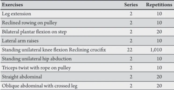
TABLE 1 - Resistance exercises.
Exercises Series Repetitions
Leg extension 2 10
Reclined rowing on pulley 2 10 Bilateral plantar lexion on step 2 20
Lateral arm raises 2 10
Standing unilateral knee lexion Reclining cruciix 22 1,010 Standing unilateral hip abduction 2 10 Triceps twist with rope on pulley 2 10
Straight abdominal 2 20
Oblique abdominal with crossed leg 2 20
Eighteen patients with chronic CHD were followed in this prospective intervention study. The patients participated in a program of exercises in the Cardiac Rehabilitation Service of the National Institute of Cardiology (INC in Portuguese). he exercises were performed 3 times a week for 1h (30 min of aerobic activity and 30 min of resistance exercises and extension) over 6 months in 2010. Functional capacity was evaluated by comparing the direct measurement of the O2 uptake volume (VO2) obtained by the cardiopulmonary exercise test (CPET) before and ater the program.
he exercise program was structured as given below.
I) thirty minutes of aerobic exercise on an Inbrasport2000®
treadmill, which was divided into the following 3 phases: a. Five minutes of warm-up with progressive speed acceleration. b. Twenty minutes of exertion aiming for the target cardiac frequency zone (established for each patient by the CPET − 5% above the anaerobic threshold and 10% under the maximum heartbeat or the respiratory compensation point)12, associated
with perceived exertion according to the modiied Borg scale13,
and maintaining the intensity of the efort between moderate and moderate/intense. To ensure the achievement of the target cardiac frequency zone, heartbeat was measured using a Polar®
cardiac monitor during the aerobic training.
c. Five minutes of cool-down until the treadmill reached a complete stop.
II) Twenty minutes of empirically programmed resistance exercise for the main muscle groups, with 2 series of 10 repetitions for each of the main muscle groups, applying a load that provided the patient with a sensation of moderate efort according to the modiied Borg scale13.
III) Ten minutes of stretching for all of the exercised muscle groups, with each position held for 20 s14 (Table 1).
outpatient clinic of the Evandro Chagas Institute of Clinical Research (IPEC in Portuguese) or the INC, Rio de Janeiro, Brazil.
Patients were excluded because of the following reasons: associated angina pectoris, suspension of stress tests due to clinical or electrocardiographic evidence of myocardial ischemia;clinically evident thyroidal dysfunction;orthopedic involvement that limited treadmill use;cancer; hepatopathy; serious alcoholism; and chronic nephropathies.
he admission protocol of the Cardiac Rehabilitation Sector of the INC required the following exams prior to participation: general clinical examination, cardiopulmonary exercise test,conventional electrocardiogram, and Doppler echocardiogram.
he analysis of the potential beneits of regular exercise on functional capacity was carried out using direct measurement of the gases exhaled during the CPET. he tests were carried out in the exercise sector of the INC, utilizing the Bruce protocol15,and applied by a single
examiner. An Inbrasport® treadmill linked to a computer with the
Elite® Micromed® sotware was used. In preparation, the patients were
depilated in the thoracic region, where necessary, and rubbed with gauze and alcohol to remove any grease. hirteen electrodes were used, corresponding to the following shunts: DI, DII, DIII, aVR, aVL, aVF, V1, V2, V3, V4, V5, V6, and MC5. he temperature of the test room was maintained at 18-22°C. Exhaled gases were analyzed by VO2000®
Aerosport® system, acquired breath-by breath, averaged over 20 seconds.
he traditional methods of Wasserman et al.16 were used for obtaining
results for the ventilatory variables. During the test, the patients were given instructions and encouraged to reach their exhaustion point.
he primary result of the study used the comparison of the maximum values of O2 consumption at the peak of exertion (VO2peak), pre- and post-training. Other variables of the CPET were studied as secondary results. he irst of them was VO2 in the irst ventilatory threshold, also known as the lactate threshold or anaerobic threshold (VO2AT). Another variable evaluated as a secondary result was the O2 pulse, which analyzes the relationship of VO2 with the heartbeat (VO2/beat) during exercise and permits an estimate of systolic volume (SV). he third variable evaluated as a secondary result was the ventilatory equivalent of CO2 or the VE/VCO2 slope, which represents the quantity of air that needs to be ventilated for 1 min to eliminate 1 L of CO27.
he data were entered into an Excel® spreadsheet, always by the
same typist. For the database, a standard comma separated value ile was used. For statistical analysis, the program R 2.10 was used with Students’ paired t-test and the Wilcoxon test. he level of statistical signiicance was set at a value of p < 0.05. According to the sample calculation, 12 patients were the minimum necessary to guarantee a power of 80% and conidence of 95%, considering an improvement of 10% in the primary results.
Exploratory data analysis utilized the descriptions of the relative and absolute frequencies of the categorical variables and the description of the summary measurements of the quantitative variables (i.e., VO2peak, VO2AT, and VO2/beat), such as average, median, standard deviation, and interquartile range (IQR). Where statistically signiicant diference occurred, these were compared to the values of the median and IQR.
Ethical considerations
For this study, the recommendations of the World Health Organization, the Helsinki Statement of Rights, and the National The subjects consisted of men and women aged between
30-72 years and having 2 diferent positive results for serological tests for Chagas’ disease enzyme-linked immunosorbent assay hemagglutination and indirect fluorescence and for electrocardiographic or echocardiographic characteristic alterations compatible with chronic CHD7; who did not engage in regular
RESULTS
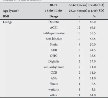
TABLE 2 - General features.
30-72 56.67 (mean) ± 9.46 (SD)
Age (years) 15.60-37.60 26.24 (mean) ± 4.46 (SD)
BMI Drugs n %
Using: Diuretic 15 83.0
ACEI 12 66.5 antihypertensive 10 55.5 beta-blocker 10 55.5 Statin 9 50.0
ARB 8 44.5
OHG 6 33.5
Digitalis 3 17.0 anti-arrhythmic 2 11.0
CCB 2 11.0
ASA 2 11.0
ibrate 1 5.5 warfarin 1 5.5 other 11 61.0
BMI: body mass index; SD: standard-deviation; ACEI: angiotensin-converter enzyme inhibitor; ARB: angiotensin-receptor blocker; OHG: oral hypoglycemic;
CCB: calcium channel blocker; ASA: acetylsalicylic acid; other: drugs not used for cardiovascular control.
Commission on Research Ethics (CONEP in Portuguese) Resolution 196/96 were respected. he entire study process was explained to the patients, and their authorizationwas registered in an informed consent form. his study was approved by the ethics commitees of the INC and IPEC under numbers 0237/26.05.2009 and P 065/2010, respectively.
We selected 54 patients. Of this total, 18 concluded the program, exceeding the value of the sample calculation. Before the study began, 30 patients were excluded due to their inability to join the exercise program (lack of time/availability and/or distance of residence) or associated comorbidities. he study was initiated with 24 patients; however, 6 did not inish the program: 1 had severe pneumonia, 1 had a transitory ischemic accident, 1 had lumbago related to work activities, 1 had acute peripheral vascular disease (none of them were related to exercise), and 2 dropped out.
Of the 18 patients that inished the study, 13 (72.2%) were women. heir functional class was I/II according to the New York Heart Association, and their mean ejection fraction was 54%.
Table 2 shows the general characteristics of the subjects with regard
to age (years), body mass index in kg/m2, and medications in use
during the training period.
Regarding the primary result, the pre-conditioning VO2peak (mL·kg-1·min-1) varied between 9.32 and 33.43, with an average of
21.81, median of 21.11, and interquartile range of 18.69-26.94. he post-conditioning VO2peak (mL·kg-1·min-1) varied between 12.45 and
37.93, with an average of 24.24, median of 24.48, and interquartile range of 18.36-29.15. he average increase in VO2peak was equivalent to 11.14% (p = 0.019). he variation can be seen in Figure 1.
FIGURE 1 - O2 uptake at exertion peak - VO2peak (mL·kg-1·min-1).
Regarding the secondary results, the pre-conditioning O2 pulse (mL.beat -1) varied between 4.80 and 19.80, with an average of 10.76,
median of 10.65, and interquartile range of 6.90-13.30. he post-conditioning O2 pulse (mL.beat -1) varied between 6.00 and 18.30,
with an average of 11.85, median of 11.30, and interquartile range of 9.07-1.67. he average increase of O2 pulse was equivalent to 10.18% (p = 0.044). he variation can be seen in Figure 2.
20
15
10
5 O2
p
ul
se
(m
L.
be
at
-1)
Distribu�on of O2 pulse pre and post-condi�oning
FIGURE 2 - O2 pulse - VO2/beat (mL.beat-1).
he pre-conditioning VO2AT (mL.kg-1.min-1) varied between
5.24 and 19.04, with an average of 14.73, median of 15.74, and interquartile range of 11.21-18.52. he post-conditioning VO2AT (mL.kg-1.min-1) varied between 11.32 and 25.00, with an average of
17.49, median of 17.75, and interquartile range of 13.31-21.33. he average increase of VO2AT was equivalent to 18.74% (p = 0.016). he variation can be seen in Figure 3.
40
30
20
10
0
Distribu�on of O2 consump�on at anaerobic
threshold pre and post-condi�oning
VO
2
AT (mL.kg
-1.min -1)
FIGURE 3 -Anaerobic threshold - VO2AT (mL.kg-1.min-1).
V
O2peak
(mL.kg
-1.min -1)
ACKNOWLEDGMENTS DISCUSSION
he pre-conditioning VE/VCO2 slopevaried between 17.40 and 30.60, with an average of 24.15 and median of 24.27. he post-conditioning VE/VCO2 slope varied between 19.40 and 32.40, with an average of 24.95 and median of 24.69. he average increase of VE/ VCO2 was equivalent to 1.73% (p = 0.582), a value not considered signiicant.
he objective of this study was to evaluate the potential efect of an exercise program on the functional capacity of patients with chronic CHD,thus creating a basis for the practice of regular exercise as an additional medical therapy for this disease. In the literature reviewed, only 1 study correlated the efects of regular physical training with CHD18.
As previously mentioned, many studies have shown the beneicial effect of regular exercise on cardiac patients. Regular exercise generates cardiovascular, metabolic, and ventilatory modiications, both acute and chronic, in response to increased physiological demands19. Such modiications provoke an increase in functional
capacity with central and peripheral responses10.
Myers et al. considered functional capacity a strong predictor of mortality in cardiac patients and normal individuals, more so than other pre-established risk factors20. In heart failure, functional
capacity is considered an important predictive marker. In this context, functional capacity can be represented by the consumption of O2 during exercise17.
he maximum consumption of O2 (VO2max) has been considered the best indicator of human capacity to sustain prolonged exertion. However, faced with the technical diiculties of measurement in cardiac patients or individuals with poor conditioning, it must be said that the highest measurement of O2 consumption attained during exercise (VO2peak) would be an objective indicator of functional capacity, especially when associated with the measurement of anaerobic metabolism through records of ventilatory variables obtained in the CPET21. In addition, VO
2peak
is an important predictor, as much for deaths by cardiac events as for deaths due to other diseases. In this way, even a small gain in aerobic conditioning can improve not only functional capacity but also life expectancy20.
he value of the primary post-conditioning measure (VO2peak) had a comparative average increase that was statistically signiicant22
(11.14%, p = 0.01949). As this measurement is extremely dependent on the collaboration of the patient in really making the maximum efort, other variables were studied as secondary measures so that important information could be obtained, even from sub-maximum efort, because during the CPET, the patients were stimulated to achieve their maximum efort, but not all of them felt comfortable doing so.
Considering the secondary measures, the study of VO2AT is relevant because it corresponds to the moment at which the accumulation of plasma lactate occurs with subsequent bufering by bicarbonate, resulting in the elevation of CO2 output17.
Concerning O2 pulse (VO2/beat), VO2 is well known to be directly proportional to sistolic volume (SV) versus the arteriovenous O2 content diference (C(A-V)O2). In the absence of illness such as anemia, hemoglobinopathy, hypoxic pulmonary disease and cardiomyopathy
due to shunting, we can consider that C(A-V)O2 rises exaggeratedly without signiicant deviation and that VO2 becomes dependent on the cardiac output (i.e., VO2 = SV × HB). As such, we can infer that the O2 pulse (VO2/HB) = SV13.
In heart disease patients with alterations in pulmonary difusion and perfusion, e.g., pulmonary hypertension, edema or interstitial pulmonary ibrosis, elevated anaerobic metabolism, and central hyperventilation, the value of the VE/VCO2 slopemay be increased due to alterations in chemoreceptors and ergoreceptors. It is a value that varies with the moment at which it is measured, and for this reason, the analysis continues throughout the entire exertion period by using linear regression or slope17. A VE/VCO
2 slope value of up
to 30 is considered normal, while a value more than 36 is related to a worse prognosis13.
In the present sample, the value of the VE/VCO2 slope was found to be within the normal range, and therefore did not sufer alterations that could be considered significant. The remaining secondary results evaluated had statistically signiicant alterations in terms of the improvement of functional capacity and physical conditioning22.
he elevated number of women (72.2%) found in this sample may be related to the fact that they may have more available time to commit. Considering that the wide age range used in this study could inluence its results, if we exclude the youngest (30 years) and oldest (72 years) patients, the remaining patients are aged between 44-62 years, with a mean of 56.67 years and a median of 58.50 years, which minimizes this possible inluence.
In a review of the literature, just 1 similar study was found. he study by Lima et al. correlated the efect of regular physical training with CHD18. he article was published in the European Journal
of Heart Failure in June 2010 and reported the improvement of functional capacity in patients with Chagas’ cardiomyopathy undergoing a 12-week exercise program. This single-blind, randomized study compared 21 cases and 19 control subjects.
In the present study, there was no control group, which is a limitation; nevertheless, the results are in accordance with the indings of the group from Minas Gerais18. In the study performed
by Lima et al., the exercise program was executed over a 3-month period, which is half the duration of the present study. Besides, in the study of Lima et al., VO2 was measured indirectly (inferred or approximate value) through a standard exercise test. In the present study, a more precise measurement system was used, i.e., the cardiopulmonary exercise test, in which VO2 was measured directly via the exhaled gases.
In conclusion, during the course of the exercise program there was neither an improvement nor worsening of cardiac symptoms. he results suggest that regular exercise was beneicial to the sample studied, in terms of the improvement of physical conditioning and functional capacity. his study may provide a basis for the prescription of exercise in the treatment of chronic CHD in association with medical therapy.
he authors declare that there is no conlict of interest. CONFLICT OF INTEREST
FINANCIAL SUPPORT
REFERENCES
Fundação Carlos Chagas Filho de Amparo à Pesquisa do Estado do Rio de Janeiro (FAPERJ).
1. Hagar JM, Rahimtoola SH. Chagas’ heart disease in the United States. New Engl J Med 1991; 325:763-768.
2. National School of Public Health-Fiocruz [Internet]. Epidemiology: current situation. [cited 2010 Jan 27]. Available from: htp: www.ioruz.br/chagas. 3. Anonymous. Chagas’ disease. An epidemic that can no longer be ignored. Lancet
2006; 368:619.
4. Rassi Jr A, Rassi A, Marim-Neto JA. Chagas’ disease. Lancet 2010; 375:1388-402. 5. Dias JC. Globalization, iniquity and Chagas disease. Cad Saúde Pública booklet
2007; 23 (supl 1):13-22.
6. Marin-Neto JA, Simões MV, Sarabanda AVL. Chagas’ Heart Disease. Arq Bras Cardiol 1999; 72:247-263.
7. Sociedade Brasileira de Medicina Tropical. Consenso Brasileiro em Doença de Chagas. Rev Soc Bras Med Trop 2005; 38 (supl III):1-29.
8. Pereira-Barreto AC. Polymorphic aspects of heart disease in the indeterminate form of Chagas disease: studies based on results of non-invasive methods. [Habilitation thesis]. [São Paulo]: University of São Paulo; 1985. 185 p. 9. Casillas JM, Gremeaux V, Damak S, Feki A, Pérennou D. Exercise training for
patients with cardiovascular disease. Ann Réadapt Med Phys 2007; 50:403-418. 10. Crimi E, Ignarro LJ, Cacciatore F, Napoli C. Mechanisms by which exercise training beneits patients with heart failure. Nat Mag Cardiol 2009; 6:292-300. 11. Bocchi EA. Exercise training in Chagas’ cardiomyopathy: trails are welcome for
this neglected heart disease. Eur Jour Heart Fail 2010; 12:782-784.
12. Negrão CE, Barreto ACP. Cardiology of Exercise. 3rd ed. São Paulo: Manole;
2010.
13. Costa RVC, Carreira MAMQ. Ergometry: ergospirometry, scintillography and exertion echocardiography. São Paulo: Atheneu; 2007.
14. Anderson B. Stretching, 20th ed. California: Shelter Publications: 2000;
15. Hespanha R. Ergometry: physiological bases and methodology for the prescription of exercise. Rio de Janeiro: Rubio; 2004.
16. Wasserman K, Hansen J, Sue D, Stringer W, Whipp B. Principles of Exercise Testing and Interpretation, 3rd ed. Philadelphia, USA: Lippincot Williams &
Wilkins; 1999.
17. Yazbek Jr P, Carvalho RT, Sabbag LMS, Battistella LR. Ergospirometry. Cardiopulmonary Exercise Test, methodology and interpretation. Arq Bras Cardiol 1998; 71:719-724.
18. Lima MMO, Rocha MOC, Nunes MCP, Sousa L, Costa HS, Alencar MCN, et al. A randomized trail of the efects of exercise training in Chagas cardiomyopathy. Eur J Heart Fail 2010; 12:866-873.
19. Brum PC, Forjaz CLM, Tinucci T, Negrão CE. Acute and chronic adaptations of physical exercise on the cardiovascular system. Paul Mag Ed Fis I Act 2004;18:21-31.
20. Myers J, Prakash M, Froelicher V, Dat Do, Partington S, Atwood, JE. Exercise capacity and mortality among referred for exercise testing. N Engl J Med 2002; 346:793-800.
21. Berry JRS. Evaluation of the efects of cardiac rehabilitation in post-myocardial infarction patients. Niterói. Dissertation [Master Dissertation]. [Rio de Janeiro]: Federal University of Rio de Janeiro; 2008. 186 p.
22. Froelicher VF, Myers JN. Exercise and the Heart, 3rd ed. Philadelphia: