DOI: 10.5935/2359-4802.20180073
ORIGINAL ARTICLE
Mailing Address: Pablo Marino Corrêa Nascimento
Rua das Laranjeiras, 374. Postal Code: 22240-006, Laranjeiras, Rio de Janeiro, RJ - Brazil. E-mail: marino_pablo@yahoo.com.br, pablomarino@cardiol,br
Hemodynamic, Metabolic and Ventilatory Responses to Exercise in Adults with
Congenital Heart Disease
Pablo Marino Corrêa Nascimento,1 Daniel Arkader Kopiler,1 Fernando Cesar de Castro e Souza,1 Maria Carolina Terra Cola,1 Marina Pereira Coelho,1 Gabriella de Oliveira Lopes,1 Eduardo Tibiriçá2
Instituto Nacional de Cardiologia, INC,1 Rio de Janeiro, RJ - Brazil Instituto Oswaldo Cruz, FIOCRUZ,2 Rio de Janeiro, RJ - Brazil
Manuscript received November 16, 2017, revised manuscript May 08, 2018, accepted May 14, 2018.
Abstract
Background: Congenital heart disease in adults shares some features with heart failure (HF), including exercise intolerance, ventilatory inefficiency, inflammatory and neurohormonal activation, cardiac arrhythmias and myocardial fibrosis. Over the last years, cardiopulmonary exercise test has gained importance in the diagnostic and prognostic evaluation of congenital heart diseases, as has already occurred in HF.
Objective: To describe the behavior of hemodynamic, metabolic and ventilatory parameters in response to exercise in adults with congenital heart disease.
Methods: Observational cross-sectional study evaluating 31 adults with congenital acyanotic or cyanotic heart disease, treated clinically, surgically or percutaneously, referred for cardiopulmonary exercise test. A descriptive analysis of the data was performed.
Results: Patients aged 35.7 ± 14.2 years were included. Oxygen consumption (VO2) was 44.86 ± 18.01% of predicted
at peak exercise and 36.92 ± 12.93% of predicted maximal VO2 at anaerobic threshold. We found an oxygen uptake
efficiency slope (OUES) of 1.49 ± 0.89 (61.43 ± 26.63% of predicted), oxygen pulse of 58.90 ± 22.24% and increment
in systolic arterial pressure during exercise was 31.42 ± 21.60 mmHg.
Conclusion: Adults with congenital heart disease had similar responses to heart failure patients during exercise – reduced aerobic capacity, ventilatory inefficiency for oxygen consumption and limited inotropic response to exercise, characterized by reduced oxygen pulse and small increase in systolic arterial pressure. (Int J Cardiovasc Sci. 2019;32(1)41-47)
Keywords: Heart Defects, Congenital; Exercise; Adults; Exercise Test; Cyanosis; Hemodynamics; Metabolism.
Introduction
Congenital heart diseases are abnormalities in the structure of the heart at birth that may involve the interior surface of the walls of the heart, cardiac valves or blood vessels that carry blood to and from the heart to the body.1
The incidence of congenital heart disease is eight cases per 1,000 live births and the estimated prevalence is more than 1,000,000 of adults in the USA.1,2 The prevalence of
congenital heart disease in adults has increased in the last five decades due to significant progression in the
treatment of these conditions during childhood.3 These
patients, however, have decreased functional capacity4-7
and higher morbidity and mortality rates as compared with healthy individuals.8-14
Congenital heart diseases in adults have characteristics similar to heart failure (HF) caused by other etiologies, such as exercise intolerance,8-11 ventricular dysfunction
(right or left),15 cardiac arrhythmias,16 myocardial fibrosis,17
ventilatory inefficiency,3,11,18 increased inflammatory
The most precise method to quantify aerobic capacity is the direct measure of peak exercise oxygen consumption (VO2 max) obtained exclusively by cardiopulmonary exercise testing (CPET). Reduced values of VO2 max not only indicate more accentuated functional impairment, but also imply an adverse prognosis especially in HF,21
and also in other clinical conditions including congenital heart diseases in adults.8-11
The aim of the present study was to describe the level of functional limitation and hemodynamic, ventilatory and metabolic responses to exercise in adults with congenital heart disease referred for CPET in a tertiary cardiology hospital.
Methods
This was an observational, cross-sectional study that evaluated adults with congenital heart disease (cyanotic or acyanotic), under medical, surgical or percutaneous treatment. The study was approved by the local ethics committee (approval number 47563315.2.0000.5272). All patients were informed about the aim of the study and signed an informed consent form. The study was conducted in accordance with the World Health Organization recommendations and the Helsinki Declaration (October 2013) and the Brazilian National Health Council resolution number 466/2012.
Patients referred for CPET between April 2016 and August 2017 were included in this study. Exclusion criteria were: age younger than 18 years, unwillingness to sign the informed consent form, contraindications for CPET according to the Guidelines on Exercise Tests of the Brazilian Society of Cardiology.22
Cardiopulmonary exercise testing
A symptom-limited treadmill (Inbramed® - Porto
Alegre - Brazil) exercise test was performed, using ramp protocol, with duration of approximately eight to 12 minutes. Patients were encouraged to continue exercise until exhaustion. Participants had a minimum of six minute-resting phase, with speed of 1.5 mph and slope of 2.5% in the first minute. For measurements of the gases, a clip was placed on patients’ nose, a mouthpiece with saliva trap was connected to a pneumotachograph which, in turn, was connected to a VO2000® gas
analyzer (MedGraphics® - St Paul - USA) coupled to a
computer. Analysis was performed using the Ergo PC Elite® software (Micromed® - Brasília - Brazil). Every
20 seconds, the following parameters were analyzed in a breath-by-breath format: peak oxygen consumption (VO2 max), expressed as percentage of the predicted value and related to body mass;23 oxygen consumption
at anaerobic threshold (AT), expressed as percentage of the predicted value and related to body mass; slope of the ratio of ventilation (VE) to CO2 production (VCO2) (VE/VCO2 slope); oxygen uptake efficiency slope (OUES), expressed as absolute value and percentage of the predicted value;24 respiratory exchange ratio (RER);
changes in systolic arterial pressure (SAP) from resting
to maximal exercise (∆SAP); heart rate reserve (HRR)
calculated as heart rate (HR) changes from resting to maximal exercise; chronotropic index (proportion between measured and predicted HRR); HR decrease during the first minute of recovery (HRrec); peak exercise oxygen pulse (O2P); circulatory power (CP); ventilatory power (VP). Peak VO2 was defined as the maximal value detected during the last 20 seconds of exercise or at the first measurement performed during the resting phase. AT was identified by the ventilatory equivalent method, and 40% of the predicted VO2 max defined as the lower normal limit.25 VE/VCO
2 slope was calculated during
the whole test period.
Statistical analysis
A descriptive analysis of the data was performed. Categorical variables were described as frequency and percentage, whereas continuous variables as mean ± standard deviation (SD). Data were analyzed using Prism statistics software, version 5.0 (GraphPad Software Inc. La Jolla, CA, USA). Patients were selected by convenience.
Results
Table 1 - Clinical characteristics of the patients
Characteristics n %
Female/male sex 17/14 54.8/45.2
Age, mean ± SD 35.7 ± 14.2
-Acyanotic /cyanotic 27/4 87.1/12.9
Primary diagnosis
Tetralogy of Fallot 9 29.1
Single ventricle 4 12.9
Interventricular communication 3 9.7
Ebstein 2 6.4
Interatrial communication 2 6.4
TGA 2 6.4
Non-compacted myocardium 1 3.2
Tricuspid insufficiency 1 3.2
Tricuspid stenosis 1 3.2
Pulmonary stenosis 1 3.2
Aorta coarctation 1 3.2
Truncus arteriosus 1 3.2
TGA cc 1 3.2
cAVSD 1 3.2
ALCAPA syndrome 1 3.2
TGA: transposition of the great arteries; TGAcc: transposition of the great arteries congenitally corrected; cAVSD: complete atrioventricular septal defect, ALCAPA: Anomalous origin of the left coronary artery from the pulmonary artery.
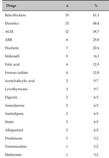
Table 2 - Medications used by the study population
Drugs n %
Beta-blockers 19 61.3
Diuretics 15 48.4
ACEI 12 38.7
ARB 8 25.8
Warfarin 7 22.6
Sildenafil 5 16.1
Folic acid 4 12.9
Ferrous sulfate 4 12.9
Acetylsalicylic acid 3 9.7
Levothyroxine 3 9.7
Digoxin 2 6.5
Amiodarone 2 6.5
Amlodipine 2 6.5
Statin 2 6.5
Allopurinol 2 6.5
Prednisone 1 3.2
Trimetazidine 1 3.2
Metformin 1 3.2
ACEI: angiotensin converting enzyme inhibitors; ARB: angiotensin II receptor blockers.
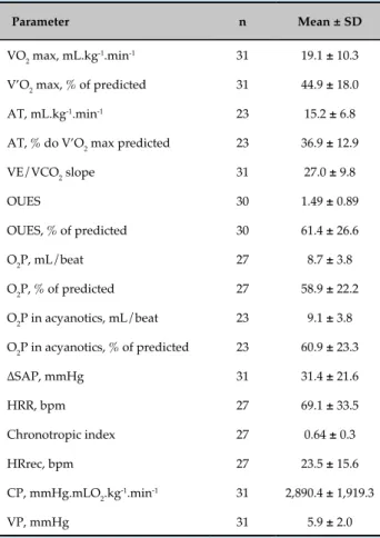
CPET results are described in Table 3. Patients with congenital heart disease evaluated in the present study had reduced aerobic capacity, with a VO2 max of 44.9% of predicted. AT could not be identified in 31 (25.8%) patients.
In addition, our study population showed ventilatory inefficiency for oxygen consumption, with OUES of 1.49 (61.4% of predicted) and limited SAP increment
in response to exercise (∆SAP: 31.4 mmHg). Also,
patients showed reduced O2P (8.7 mL/beat; 58.9% of the predicted value), even when only acyanotic subgroup was considered in the analysis (9.1 mL/beat; 60.9% of the predicted value).
Duration of the ramp exercise test was 9.2 ± 3.6 minutes, which was considered adequate. R value was 1.21 ± 0.26, indicating maximum efficiency of the test.
According to the Brazilian Society of Cardiology criteria,22 the prevalence of parasympathetic autonomic
dysfunction was 22.2%, and the small increase in SAP during incremental exercise indicated depressed response of this parameter. The prevalence of chronotropic incompetence was 44.4% based on different cut-off points for beta-blocker users26 and
non-users22 (0.62 and 0.80, respectively).
Given the impossibility of HR measurement, patients with a permanent pacemaker (n = 3) and patients with atrial fibrillation (n = 1) were excluded from O2P analysis, chronotropic response (HRR and chronotropic index) and parasympathetic autonomous modulation (HRrec).
Discussion
Table 3 - Results of the cardiopulmonary exercise test
Parameter n Mean ± SD
VO2 max, mL.kg-1.min-1 31 19.1 ± 10.3
V’O2 max, % of predicted 31 44.9 ± 18.0
AT, mL.kg-1.min-1 23 15.2 ± 6.8
AT, % do V’O2 max predicted 23 36.9 ± 12.9
VE/VCO2 slope 31 27.0 ± 9.8
OUES 30 1.49 ± 0.89
OUES, % of predicted 30 61.4 ± 26.6
O2P, mL/beat 27 8.7 ± 3.8
O2P, % of predicted 27 58.9 ± 22.2
O2P in acyanotics, mL/beat 23 9.1 ± 3.8
O2P in acyanotics, % of predicted 23 60.9 ± 23.3
∆SAP, mmHg 31 31.4 ± 21.6
HRR, bpm 27 69.1 ± 33.5
Chronotropic index 27 0.64 ± 0.3
HRrec, bpm 27 23.5 ± 15.6
CP, mmHg.mLO2.kg-1.min-1 31 2,890.4 ± 1,919.3
VP, mmHg 31 5.9 ± 2.0
VO2 max: peak exercise oxygen consumption; AT: anaerobic threshold; VE/VCO2 slope: ventilatory equivalent for carbon dioxide; OUES: oxygen uptake efficiency plateau; O2P: peak exercise oxygen pulse;
∆SAP: changes in systolic arterial pressure, from resting to peak exercise; HRR: heart rate reserve; HRrec: heart rate recovery, from peak exercise to the first minute of recovery phase; CP: circulatory power; VP: ventilatory power.
capacity, with a decrease not only in maximum aerobic power (indicated by VO2 max), but also in aerobic capacity for submaximal activities (indicated by the AT). In addition, results of the CPET revealed ventilatory inefficiency for oxygen consumption and limited inotropic response to exercise.
Previous studies have reported lower values of VO2 max4-6,9,27 and AT5 in adults with congenital heart disease
as compared with healthy adults.5 Reduced aerobic
capacity is a common condition in this population, and 80% of these patients have a VO2 max lower than predicted for age and sex.4 It is worth mentioning that
many adults with congenital heart disease overestimate their own clinical conditions in light of the long period of exercise restriction. The level of exercise intolerance is more accurately assessed by measurement of the
VO2max, and even asymptomatic patients considered as New York Heart Association class 1 have lower VO2max when compared with healthy individuals of the same age,9
suggesting a discrepancy between a subjective and an objective approach of exercise capacity in this population.
The decrease in maximal aerobic capacity in congenital heart disease adults is so important that VO2 max in these patients is comparable to that in patients with HF caused by other conditions.9 VO
2 max is a traditional marker of
an unfavorable prognosis of HF and has a central role in the evaluation of eligibility for heart transplantation in this group.28 Similarly, a reduced VO
2 max is associated
with higher morbidity and mortality in adults with congenital heart disease.8,9
We found a mean VO2 max of 19.1 mL.kg-1.min-1.
Diller et al.,9 evaluating a large group of patients with
congenital heart disease, suggested a VO2 max of 15.5 mL.kg-1.min-1 as a cut-off for predicting cardiac events.
Patients with a VO2 max lower than this had a three-time higher risk of death and hospitalization. Based on the studies by Diller et al.,9,10 our patients had a good
prognosis. However, this was a young population (< 35.7 years of age) and, in fact, VO2 max found in these patients was only 44.9% of predicted, i.e., considerably lower than that expected for healthy individuals of the same sex and age. Inuzuka et al.,8 also evaluated adults with
congenital heart disease and suggested a cut-off value of 64% of predicted VO2 max to identify patients with a low or high 5-year survival mortality risk.8 Therefore,
according to these authors, our study group could be, in fact, at risk of a poor prognosis.
Modern concepts of HF classify this syndrome in progressive stages. Patients with cardiac structural abnormalities but no signs of HF would be designated as stage B.29,30 Although this classification did not
include congenital heart disease, in a scientific statement published in 2016, the American Heart Association recommended that patients with congenital structural heart disease should be classified as being at least stage B of HF.31 In agreement with this position, our results demonstrated a depressed response of SAP (∆SAP: 31.4
patients in our sample (87.1%) allow us to infer that the decreased O2P was probably a consequence of a limited increase in systolic volume during exercise. We corroborate this hypothesis, as we found low O2P values even when cyanotic patients (9.1mL/beat) were excluded from analysis. As previously mentioned, the decreased response of SAP reinforces the hypothesis of a modest inotropic response during exercise.
VO2 max has been recognized as the most important marker of morbidity and mortality in HF. Recent evidences, however, have suggested other parameters related to ventilatory efficiency, notably VE/VCO2 slope and OUES, as better prognostic predictors in HF.32
Similar phenomenon was seen in congenial heart disease in adults. Dimopoulos et al.,13 reported that adults with
congenial heart disease had higher VE/VCO2 slope than healthy individuals, and such difference was observed in all types of congenital heart diseases and was directly proportional to the functional class. According to the authors, cyanosis was the main predictor of an increased VE/VCO2 slope. This parameter was the most important marker of mortality in adults with congenial heart disease in acyanotic patients (cut-off point of 38).13 Similarly,
Inuzuka et al.,8 reported that a VE/VCO
2 slope > 39 was
a predictor of mortality only in acyanotic congenital heart disease patients. In our study, mean VE/VCO2 slope was 27, which is considered normal. This may be explained, at least in part, by the small frequency of cyanotic patients in our sample.
In addition to ventilatory inefficiency for carbon dioxide, adults with congenital heart disease commonly have a ventilatory inefficiency for oxygen consumption also, represented by low OUES values.18 We found a mean
OUES of 1.49, corresponding to only 61.4% of predicted. It is worth pointing out, for sake of comparison, that an OUES lower than 1.47 is associated with a poor prognosis of HF,33 which indicates that our study population had a
ventilatory efficiency for oxygen similar to that in patients with more severe HF.
HR responses during incremental exercise, particularly in the presence of chronotropic incompetence, were equally important for risk stratification of adults with congenital heart disease. Diller et al.,14 reported that a HRR
lower than 51 bpm was a predictor of lower survival in this population, especially when associated with a VO2 max lower than 16.7 mL.kg-1.min-1, which increased the
mortality risk by 3.8 times.14 The authors also identified the
chronotropic index and reduction in HRrec as parameters associated with unfavorable prognosis. Similarly, Inuzuka
et al.,8 identified the VO
2max combined with HRR as
the main marker of mortality in cyanotic and acyanotic congenital heart disease. However, the cut-off point was 71 bpm.8 Since we found a mean HRR of 69 bpm, our
patients would be at increased risk according to these results reported by Inuzuka et al.,8 but not according to
those reported by Diller et al.14
The prevalence of chronotropic incompetence in our study was 44.4%, lower than that reported by Diller et al.,14 (62%); chronotropic index, however, were similar
in both studies (0.64 and 0.70, respectively). We found a mean HRrec of 23.5 bpm, which was considered normal and suggestive of adequate parasympathetic autonomous modulation. Only 22.2% of our population met criteria for parasympathetric dysautonomia.
Recently, CP and VP have emerged as important markers of adverse events in HF,34 by using the product
of SAP with VO2 max, and the quotient of PAS by VE/ VCO2 slope, respectively. The cut-off points for CP and VP in HF were 1,750 mmHg.mLO2.kg-1.min-1 and 3.5
mmHg, respectively.34
In adults with congenital heart disease, Giardini et al.,12
found that low CP values (lower than 1,476 mmHg.mLO2. kg-1.min-1) were associated with a 15.4-fold increase in the
4-year risk of death. The authors described that despite an inverse relationship between CP and functional class, even asymptomatic patients had a lower CP than healthy individuals. We found an adequate CP (2,890.4 mmHg. mLO2.kg-1.min-1), as compared with that in HF patients
and also with the findings by Giardini et al.,12 In addition,
we found adequate VP values taking into account HF patients, although we did not find other studies evaluating this parameter in adults with congenital heart disease in the literature.
Limitations
Limitations of this study included the relatively small sample size, which is common in studies involving low-prevalence conditions.
1. What Are Congenital Heart Defects? National Heart, Lung, and Blood Institute: National Heart, Lung, and Blood Institute; 2011. [Cited in 2017 Jan 10]. Available from: https://www.nhlbi.nih.gov/health/health-topics/topics/chd/.
2. Hoffman JI, Kaplan S, Liberthson RR. Prevalence of congenital heart disease. Am Heart J. 2004;147(3):425-39.
3. Khan AM, Paridon SM, Kim YY. Cardiopulmonary exercise testing in adults with congenital heart disease. Expert Rev Cardiovasc Ther. 2014;12(7):863-72.
4. Kempny A, Dimopoulos K, Uebing A, Moceri P, Swan L, Gatzoulis MA, et al. Reference values for exercise limitations among adults with congenital heart disease. Relation to activities of daily life--single centre experience and review of published data. Eur Heart J. 2012;33(11):1386-96.
5. Buys R, Cornelissen V, Van De Bruaene A, Stevens A, Coeckelberghs E, Onkelinx S, et al. Measures of exercise capacity in adults with congenital heart disease. Int J Cardiol. 2011;153(1):26-30.
6. Trojnarska O, Gwizdała A, Katarzyński S, Katarzyńska A, Szyszka A, Lanocha M, et al. Evaluation of exercise capacity with cardiopulmonary exercise test and B-type natriuretic peptide in adults with congenital heart disease. Cardiol J. 2009;16(2):133-41.
7. Miliaresis C, Beker S, Gewitz M. Cardiopulmonary stress testing in children and adults with congenital heart disease. Cardiol Rev. 2014;22(6):275-8.
8. Inuzuka R, Diller GP, Borgia F, Benson L, Tay EL, Alonso-Gonzalez R, et al. Comprehensive use of cardiopulmonary exercise testing identifies adults with congenital heart disease at increased mortality risk in the medium term. Circulation. 2012;125(2):250-9.
9. Diller GP, Dimopoulos K, Okonko D, Li W, Babu-Narayan SV, Broberg CS, et al. Exercise intolerance in adult congenital heart disease: comparative severity, correlates, and prognostic implication. Circulation. 2005;112(6):828-35.
10. Diller GP, Giardini A, Dimopoulos K, Gargiulo G, Müller J, Derrick G, et al. Predictors of morbidity and mortality in contemporary Fontan
patients: results from a multicenter study including cardiopulmonary exercise testing in 321 patients. Eur Heart J. 2010;31(24):3073-83.
11. Giardini A, Specchia S, Tacy TA, Coutsoumbas G, Gargiulo G, Donti A, et al. Usefulness of cardiopulmonary exercise to predict long-term prognosis in adults with repaired Tetralogy of Fallot. Am J Cardiol. 2007;99(10):1462-7.
12. Giardini A, Specchia S, Berton E, Sangiorgi D, Coutsoumbas G, Gargiulo G, et al. Strong and independent prognostic value of peak circulatory power in adults with congenital heart disease. Am Heart J. 2007;154(3):441-7.
13. Dimopoulos K, Okonko DO, Diller GP, Broberg CS, Salukhe TV, Babu-Narayan SV, et al. Abnormal ventilatory response to exercise in adults with congenital heart disease relates to cyanosis and predicts survival. Circulation. 2006;113(24):2796-802.
14. Diller GP, Dimopoulos K, Okonko D, Uebing A, Broberg CS, Babu-Narayan S, et al. Heart rate response during exercise predicts survival in adults with congenital heart disease. J Am Coll Cardiol. 2006;48(6):1250-6.
15. Broberg CS, Burchill LJ. Myocardial factor revisited: The importance of myocardial fibrosis in adults with congenital heart disease. Int J Cardiol. 2015;189:204-10.
16. Bouchardy J, Therrien J, Pilote L, Ionescu-Ittu R, Martucci G, Bottega N, et al. Atrial arrhythmias in adults with congenital heart disease. Circulation. 2009;120(17):1679-86.
17. Broberg CS, Chugh SS, Conklin C, Sahn DJ, Jerosch-Herold M. Quantification of diffuse myocardial fibrosis and its association with myocardial dysfunction in congenital heart disease. Circ Cardiovasc Imaging. 2010;3(6):727-34.
18. Giardini A, Specchia S, Gargiulo G, Sangiorgi D, Picchio FM. Accuracy of oxygen uptake efficiency slope in adults with congenital heart disease. Int J Cardiol. 2009;133(1):74-9.
19. Sharma R, Bolger AP, Li W, Davlouros PA, Volk HD, Poole-Wilson PA, et al. Elevated circulating levels of inflammatory cytokines and bacterial endotoxin in adults with congenital heart disease. Am J Cardiol. 2003;92(2):188-93.
References
Conclusion
Adults with congenital heart disease had similar responses to HF patients during a cardiopulmonary exercise test, indicating an impaired aerobic capacity, ventilatory inefficiency for oxygen uptake and reduced inotropic response to exercise.
Author contributions
Conception and design of the research: Nascimento PMC, Kopiler DA, Cola MCT, Tibiriçá E. Acquisition of data: Nascimento PMC, Kopiler DA, Souza FCC, Cola MCT, Coelho MP, Lopes GO, Analysis and interpretation of the data: Nascimento PMC, Kopiler DA, Souza FCC, Tibiriçá E. Statistical analysis: Tibiriçá E. Writing of the manuscript: Nascimento PMC, Souza FCC, Tibiriçá E. Critical revision of the manuscript for intellectual content: Nascimento PMC, Kopiler DA, Souza FCC, Cola MCT, Coelho MP, Lopes GO, Tibiriçá E.
Potential Conflict of Interest
No potential conflict of interest relevant to this article was reported.
Sources of Funding
There were no external funding sources for this study.
Study Association
This article is part of the thesis of master submitted by Pablo Marino, from Instituto Nacional de Cardiologia.
Ethics approval and consent to participate
20. Bolger AP, Sharma R, Li W, Leenarts M, Kalra PR, Kemp M, et al. Neurohormonal activation and the chronic heart failure syndrome in adults with congenital heart disease. Circulation. 2002;106(1):92-9.
21. Keteyian SJ, Patel M, Kraus WE, Brawner CA, McConnell TR, Piña IL, et al. Variables Measured During Cardiopulmonary Exercise Testing as Predictors of Mortality in Chronic Systolic Heart Failure. J Am Coll Cardiol. 2016;67(7):780-9.
22. Meneghelo RS, Araújo CGS, Stein R, Mastrocolla LE, Albuquerque PF, Serra SM. III Diretrizes da Sociedade Brasileira de Cardiologia sobre teste ergométrico. Arq Bras Cardiol. 2010;95(5):1-26.
23. Jones NL, Campbell EJM. Clinical exercise testing. 2nd ed. Philadelphia: Saunders; 1982.
24. Hollenberg M, Tager IB. Oxygen uptake efficiency slope: an index of exercise performance and cardiopulmonary reserve requiring only submaximal exercise. J Am Coll Cardiol. 2000;36(1):194-201.
25. Mezzani A, Agostoni P, Cohen-Solal A, Corrà U, Jegier A, Kouidi E, et al. Standards for the use of cardiopulmonary exercise testing for the functional evaluation of cardiac patients: a report from the Exercise Physiology Section of the European Association for Cardiovascular Prevention and Rehabilitation. Eur J Cardiovasc Prev Rehabil. 2009;16(3):249-67.
26. Khan MN, Pothier CE, Lauer MS. Chronotropic incompetence as a predictor of death among patients with normal electrograms taking beta blockers (metoprolol or atenolol). Am J Cardiol. 2005;96(9):1328-33.
27. Fredriksen PM, Veldtman G, Hechter S, Therrien J, Chen A, Warsi MA, et al. Aerobic capacity in adults with various congenital heart diseases. Am J Cardiol. 2001;87(3):310-4.
28. Mehra MR, Canter CE, Hannan MM, Semigran MJ, Uber PA, Baran DA, et al. The 2016 International Society for Heart Lung Transplantation listing criteria for heart transplantation: A 10-year update. J Heart Lung Transplant. 2016;35(1):1-23.
29. Hunt SA, Abraham WT, Chin MH, Feldman AM, Francis GS, Ganiats TG, et al. 2009 Focused update incorporated into the ACC/AHA 2005 Guidelines for the Diagnosis and Management of Heart Failure in Adults A Report of the American College of Cardiology Foundation/American Heart Association Task Force on Practice Guidelines Developed in Collaboration With the International Society for Heart and Lung Transplantation. J Am Coll Cardiol. 2009;53(15):e1-e90.
30. Yancy CW, Jessup M, Bozkurt B, Butler J, Casey DE, Drazner MH, et al. 2013 ACCF/AHA guideline for the management of heart failure: a report of the American College of Cardiology Foundation/American Heart Association Task Force on Practice Guidelines. J Am Coll Cardiol. 2013;62(16):e147-239.
31. Stout KK, Broberg CS, Book WM, Cecchin F, Chen JM, Dimopoulos K, et al. Chronic Heart Failure in Congenital Heart Disease: A Scientific Statement From the American Heart Association. Circulation. 2016;133(8):770-801.
32. Myers J, Oliveira R, Dewey F, Arena R, Guazzi M, Chase P, et al. Validation of a cardiopulmonary exercise test score in heart failure. Circ Heart Fail. 2013;6(2):211-8.
33. Davies LC, Wensel R, Georgiadou P, Cicoira M, Coats AJ, Piepoli MF, et al. Enhanced prognostic value from cardiopulmonary exercise testing in chronic heart failure by non-linear analysis: oxygen uptake efficiency slope. Eur Heart J. 2006;27(6):684-90.
34. Forman DE, Guazzi M, Myers J, Chase P, Bensimhon D, Cahalin LP, et al. Ventilatory power: a novel index that enhances prognostic assessment of patients with heart failure. Circ Heart Fail. 2012;5(5):621-6.