(1) Universidade Federal de Santa Maria (UFSM), Santa Maria, RS, Brasil. (2) Universidade Federal de Ciências da
Saúde de Porto Alegre (UFCSPA), Porto Alegre, RS, Brasil.
Conlict of interest: non-existent
Oropharyngeal dysphagia in Wallenberg’s syndrome –
case series
Disfagia orofaríngea na Síndrome de Wallenberg – série de casos
Diego Fernando Dorneles Bilheri(1)
Renata Mancopes(1)
Sheila Tamanini de Almeida(2)
Received on: September 22, 2015 Accepted on: February 16, 2016
Mailing address:
Diego Fernando Dorneles Bilheri Universidade Federal de Ciências da Saúde de Porto Alegre
Rua Sarmento Leite, 245
Porto Alegre, Rio Grande do Sul, Brasil CEP: 90050-170
E-mail: [email protected]
ABSTRACT
Purpose: to characterize the changes of swallowing function in patients with Wallenberg’s syndrome. Methods: case series of seven patients with diagnosis of this syndrome referred for clinical assessment. To evaluate the degree of dysphagia used to Gugging Swallowing Screen scale and to assess the level of oral ingestion used the Oral Intake Functional Scale.
Results: the mean age was 60.57 years; all subjects showed changes in the function of severe degree of swallowing (71.42%) to moderate (28.58%); 85.71% required alternative pathway of food, and, 71.43% were fed exclusively by nasoenteric probe; all required speech therapy.
Conclusion: this study concluded that the oropharyngeal dysphagia in Wallenberg syndrome presents itself as moderate to severe disorder, being necessary the use of alternative pathway of food in most
cases.
Keywords: Deglutition Disorders; Stroke; Lateral Medullary Syndrome
RESUMO
Objetivo: caracterizar o quadro de comprometimento da função da deglutição em pacientes com Síndrome de Wallenberg.
Métodos: série de casos de sete pacientes, com diagnóstico dessa síndrome, encaminhados para avalia-ção fonoaudiológica. Para avaliaavalia-ção do grau de disfagia utilizou-se a escala Gugging Swallowing Screen e para avaliar o nível de ingestão oral utilizou-se a Functional Oral Intake Scale.
Resultados: a média de idade foi de 60,57 anos; todos os sujeitos apresentaram alteração na função da deglutição de grau grave (71,42%) a moderado (28,58%); 85,71% necessitaram de Via Alternativa de Alimentação, sendo que, 71,43% eram alimentados exclusivamente por sonda nasoentérica; todos necessitaram de acompanhamento fonoaudiológico.
Conclusão: este estudo concluiu que a disfagia orofaríngea na Síndrome de Wallenberg apresenta--se como um distúrbio de grau grave a moderado, sendo necessária a utilização de Via Alternativa de Alimentação na maioria dos casos.
INTRODUCTION
The Wallenberg Syndrome (WS), also called Lateral Bulbar Syndrome, is retro-olivary injury generally resulting from an Arterial Ischemic Stroke (AIS) in intra-cranial portion of the Vertebral Artery or its Cerebellar Posterior Inferior branch, which is responsible for the vascularization of the dorsolateral region of the bulb. The initial manifestations in WS are: limb ataxia, nausea,
dizziness, vomiting, nystagmus, dificulty in balance
and walking, dysarthria, dysphonia, oropharyngeal dysphagia (OD) neurogenic, being the percentage of occurrence of the last manifestation ranging from 51 to 94% 1-4.
Neurogenic OD is a secondary symptom to an underlying disease or neurological trauma that cause, in most cases, a sensory-motor impairment in the oral and/or pharyngeal swallowing. Taken together, these changes can result in dehydration, malnutrition and aspiration pneumonia due to laryngeal penetration and tracheal aspiration5,6. Mortality after episodes of
aspiration pneumonia is signiicant, with occurrence
ranging from 7.5 to 72%7.
In WS, among different kinds of impairment, cranial nerves Trigeminal (V) are affected, being them respon-sible for the muscles of chewing, tensor muscle of the soft palate and sensitivity of the face and 2/3 of anterior part of the tongue; Glossopharyngeal (IX), responsible for the sensitivity and taste of the posterior third of the tongue and innervation of the constrictor muscles of the pharynx and stylopharyngeus muscle; and Vago (X), responsible for motor and sensory functions of the pharynx and larynx, and branches of the last two nerves form the pharyngeal plexus1,6. Thus, lesions in these
cranial nerves interfere with the swallowing process, they can cause uncontrollable sobs; ipsilateral paralysis to the lesion, palate and vocal cords; ipsilateral facial hypalgesia and possible loss of taste in hemi-tongue8,9.
OD after WS is frequently classiied as a severe
degree, affecting the pharyngeal phase of swallowing, and the prognosis depends on the extent and location of the lesion, which may vary from complete recovery to a permanent vegetative basis10-14. Furthermore, patients
with neurogenic OD might present other neurological
symptoms and deicits in cognitive abilities due to injury
in areas of the central nervous system, which may complicate the clinical condition15.
The speech therapy in swallowing disorders aims at the early detection of dysphagia, at the elimination of the possible risk associated complications and, therefore, at stabilizing the nutritional status16. The treatment of
dysphagia in WS is based in signs and symptoms, as the focus of therapy is the reduction of aspiration risk and not removing the cause, it may be necessary to recommend the use of an alternative feeding route (AFR). This, together a swallowing rehabilitation program based on techniques of oral stimulation, facili-tating maneuvers and postural maneuvers, may bring
beneits to the patient10,11,13.
Consequently, the aim of this study was to charac-terize the impairment condition of the swallowing process in patients with WS.
METHODS
It is a study of retrospective case series, approved by the Ethics and Research Committee (ERC) of
Universidade Federal de Ciências da Saúde de Porto Alegre (UFCSPA) under the number 362795, according to the rules established by the resolution No. 196/96 of the National Health Council and its subsequent resolutions. Thus, as it is a search in databases, all researchers have signed a data privacy statement.
Data from subjects were collected from the Database of academic activities of the Speech, Hearing and Language Sciences Major of UFCSPA at Santa Clara Hospital at Santa Casa de Misericordia Hospital Complex in Porto Alegre (RS, Brazil). Data were analyzed from the following inclusion criteria: being admitted to the neurology sector of the hospital during the period from January 2012 to August 2013; have been diagnosed with WS by the group of neurology,
which was based on clinical criteria and conirmed
by neuroimaging (MRI); receiving speech therapy, performed by the interns of the Speech, Hearing and Language Sciences Major, who were well trained and interns who were under supervision of an expert speech therapist professor. Finally, all subjects hospitalized in the period, with this diagnosis, contemplated the above criteria and they were included in the survey.
In order to classify the degree of dysphagia, the Gugging Swallowing Screen scale (GUSS) was used17.
This, in turn, is divided into two stages: direct and indirect assessment of swallowing. Thus, through the score, it is possible to classify the swallowing in normal or mild dysphagia with no or with minimal risk of aspiration (20 points), mild dysphagia with low risk of aspiration (15 to 19 points), moderate dysphagia with risk of aspiration (ten to 14 points) and severe dysphagia with a high risk of aspiration (zero to nine points).
To assess the level of oral ingestion Functional Oral Intake Scale (FOIS) was used18. It is used to scale, at
levels from one to seven, the amount of intake by mouth (oral), while FOIS 1 provides “not oral;” FOIS 2
“dependent on alternative route with minima (oral) food or liquid”; FOIS 3 “dependent alternative route with consistent food or liquid (oral)”; FOIS 4 “(oral) total of a single consistency”; FOIS 5 “(oral) complete with multiple consistencies, but in need of special prepa-ration or compensation”; FOIS 6 “(oral) complete with multiple consistencies, but without special preparation
or compensation, but with restrictions for some food” and FOIS 7 “total (oral) without restrictions.”
The subjects were evaluated according to the routine of the institution, initially in the degree of dysphagia (GUSS I) and the level of oral intake (FOIS I) and those who underwent speech therapy were assessed at discharge (GUSS II and FOIS II).
After tabulating the data by using the Microsoft
Ofice Excel spreadsheet, a descriptive analysis in
absolute and relative values was performed.
RESULTS
For this case series, there were six men and one
woman, identiied as S1, S2, S3, S4, S5, S6 and S7,
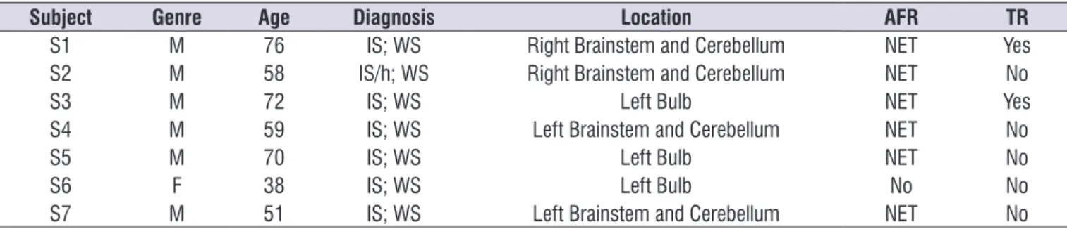
averaging 60.57 years (minimum of 38 and maximum of 76). In Table 1 the data of the subjects are reported.
From seven subjects, four (57%) presented systemic arterial hypertension (SAH), two (28.57%) had diabetes, one (14.28%) was obese, one (14, 28%) presented cardiomyopathy and one (14.24%) used to smoke. These were considered risk factors for Cerebral Vascular Accident (CVA). Only one patient (14.28%) had a diagnosis of previous stroke.
Table 1. Characterization of subjects with wallenberg syndrome
Subject Genre Age Diagnosis Location AFR TR
S1 M 76 IS; WS Right Brainstem and Cerebellum NET Yes
S2 M 58 IS/h; WS Right Brainstem and Cerebellum NET No
S3 M 72 IS; WS Left Bulb NET Yes
S4 M 59 IS; WS Left Brainstem and Cerebellum NET No
S5 M 70 IS; WS Left Bulb NET No
S6 F 38 IS; WS Left Bulb No No
S7 M 51 IS; WS Left Brainstem and Cerebellum NET No
Caption: Diagnosis: Medical diagnostic; Location: Location of the lesion from Neuroimaging; AFR: Alternative Feeding Route use; TR: Tracheostomy use ; M: male; F: female; IS: Ischemic stroke; WS: Wallenberg Syndrome; IS/h: Ischemic stroke with hemorrhagic transformation; NET: Nasoenteric tube.
At the time of phonological assessment, ive
(71.43%) subjects presented degree of severe dysphagia and two (29.57%) had moderate dysphagia. Six subjects (85.71%) needed AFR. From these ones,
ive (71.43%) used exclusively AFR (FOIS 1) and (14.28%) used AFR in pasty consistency (oral) (FOIS 2). One (14.28%) subject received exclusive oral feeding in paste consistency (FOIS 4).
Two (28.57%) subjects made use of tracheostomy and, one of them (50%) required prolonged mechanical ventilation.
Regarding speech therapy conduct, all patients received speech therapy indication and four (57.14%) needed further investigation by swallowing
video-luoroscopy (VFS), however, due to the routine of the
service, no subject carried out the objective evaluation of swallowing.
In this research, there was not oral intake evalu-ation (FOIS 1) or minimum (FOIS 2) for the six subjects, indicating the need for AFR initially. In the literature, other studies have described the inability of the patient to feed themselves orally only after the onset of OD signals8,9,12,14,23,24 . The AFR chosen for all patients in this
study was nasoenteric tube (NET), according to data from another reaearch25. However, due to the severity
of dysphagia and its slow recovery, gastrostomy indication would be the most appropriate to these subjects.
This work showed that adult subjects had lower levels of OD and greater oral intake levels at baseline, whereas subjects in middle-aged and elderly had severe OD degree at baseline and FOIS 1. Therefore, it is important to note that the physiological changes in the swallowing process, due to aging, associated with a vulnerability to chronic diseases makes them sensitive to swallowing disorders and greater negative impact
when aflicted with neurological diseases20,26.
In this study, all subjects had speech therapy indication. However, due to the discharge of two
subjects, only ive received speech therapy during hospitalization. Of these ive subjects, only two had
reduction in the degree of OD and evolution at the level
of oral intake. After hospital discharge, the ive reeval -uated subjects received referral to outpatient speech
DISCUSSION
OD can be deined as a secondary symptom of the
underlying disease, which prevents the correct food transportation. So, it is directly associated with the
interruption of food pleasure and it may cause deicits
in proper nutrition and hydration of patients affected by such symptom5,19,20.
Studies show that the WS affects the oral and oral preparatory phases, due to motor and sensory impair-ments, which associated with changes in the intrinsic and extrinsic muscles of the larynx results in signif-icant disturbances in the pharyngeal phase, which is considered the main phase of swallowing8,9, 12-14,21,22.
A study evaluated 20 patients with WS by electro-myography, verifying the occurrence of dysphagia in 95% of cases12. These indings are similar to the
present study, which found dysphagia in all the cases evaluated with WS. Still, in relation to the severity of OD, for the aforementioned research, 45% of the subjects were diagnosed with severe dysphagia, while in this study 71.42% received the same diagnosis. This difference between the studies may be attributed to the
use of different criteria for OD classiication, as in other
research, the degree of severe dysphagia was found by clinical evaluation, and a total of 11 patients with WS, was 63.63 % approaching more of this study data23.
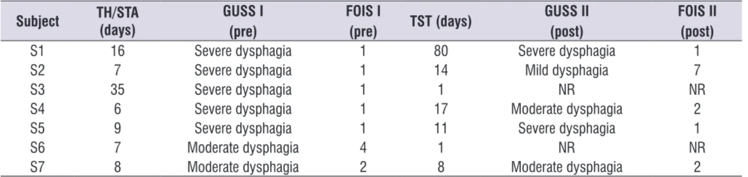
Table 2. Characterization of speech therapy evaluations of subjects with wallenberg syndrome
Subject TH/STA (days)
GUSS I (pre)
FOIS I
(pre) TST (days)
GUSS II (post)
FOIS II (post)
S1 16 Severe dysphagia 1 80 Severe dysphagia 1
S2 7 Severe dysphagia 1 14 Mild dysphagia 7
S3 35 Severe dysphagia 1 1 NR NR
S4 6 Severe dysphagia 1 17 Moderate dysphagia 2
S5 9 Severe dysphagia 1 11 Severe dysphagia 1
S6 7 Moderate dysphagia 4 1 NR NR
S7 8 Moderate dysphagia 2 8 Moderate dysphagia 2
Caption: TH/STA: time between hospitalization and speech therapy evaluation; GUSS I: initial degree of dysphagia; FOIS I: level of oral intake at the time of evaluation;
TST: time between speech therapy evaluation and discharge; GUSS II: dysphagia degree in discharge FOIS II: level of oral intake at the time of hospital discharge; NR:
not reevaluated.
other three subjects (60%) did not present improvement in the degree of dysphagia and level of oral intake. Two (28.57%) were discharged after clinical assessment, by appointment of the medical staff and therefore they did not receive speech therapy intervention while hospitalized. One (50%) of the subjects had oral
Clinical-magnetic resonance imaging correlations. Arch Neurol. 1993;50:609-14.
2. Rolak LA. Segredos em neurologia: respostas necessárias ao dia-a-dia: em rounds, na clínica, em exames orais e escritos. 2ª ed. Tradução: Francisco Tellechea Rotta. Porto Alegre: ArtMed; 2001.
3. Sanvito WL. Síndromes neurológicas. 3ª ed. São Paulo: Atheneu, 2008.
4. Norrving B, Cronqvist S. Lateral medullary infarction: prognosis in an unselected series. Neurology. 1991;41:244-8.
5. Santini CS. Disfagia neurogênica. In: Furkim AM, Santini CS. Disfagias orofaríngeas. 2ª ed. Carapicuíba: Pró-Fono, 2004. p. 19-34.
6. Gonçalves MIR, César SR. Disfagias neurogênicas: Avaliação. In: Ortiz KZ. Distúbios neurológicos adquiridos: fala e deglutição. 2ª ed. Barueri: Manole, 2010. p. 278-301.
7. Hickling KG, Howard R. A retrospective survey of treatment and mortality in aspiration pneumonia. Intensive Care Med. 1998;14:617-22.
8. Martino R, Terrault N, Ezerzer F, Mikulis D, Diamant NE. Dysphagia in a patient with lateral medullary syndrome: insight into the central control of swallowing. Gastroenterology. 2001;121:420-6. 9. Nicholson J Paralkar U, Lawton G, Sigston P.
Lateral medullary syndrome causing vocal cord palsy and stridor. JICS. 2009;10:218-9.
10. MacGowan DJ, Janal MN, Clark WC, Wharton RN, Lazar RM, Sacco RL et al. Central poststroke pain and Wallenberg’s lateral medullary infarction: frequency, character, and determinants in 63 patients. Neurology. 1997;49:120-5.
11. Nelles G, Contois KA, Valente SL, Higgins JL, Jacobs DH, Kaplan JD et al. Recovery following lateral medullary infarction. Neurology. 1998;50:1418-22.
12. Aydogdu I, Ertekin C, Tarlaci S, Turman B, Kiylioglu N, Secil Y. Dysphagia in Lateral Medullary Infarction (Wallenberg’s Syndrome): An Acute Disconnection Syndrome in Premotor Neurons Related to Swallowing Activity?. Stroke. 2001;32:2081-7.
13. Castillo AL, Barahona-Garrido J, Criales S, Chang-Menéndez S, Torre A. Wallenberg’s Syndrome: An Unusual Case of Dysphagia. Case Rep Gastroenterol. 2007;1:135-43.
14. El Mekkaoui A, Irhoudane H, Ibrahimi A, El Yousi M. Dysphagia caused by a lateral medullary infarction syndrome (Wallenberg’s syndrome). Pan Afr Med J. 2012;12:92.
therapy. The literature shows that the recovery of OD can be very slow and can take several months to years or even not present evolution8,12,27,28. Some studies
have compared dysphagia in WS with dysphagia in hemispheric stroke, showing that in WS the dysphagia tends to be more severe and its recovery slower12,29.
Still, in relation to speech therapy in patients who were referred for early evaluation, six and seven days showed the evolution in both the degree of OD, as the level of oral intake. It is known that early intervention in dysphagia minimizes the risk of complications, and
provide the beneits as speech therapy and nutritional
aspects16,30.
In a study of 208 with dysphagic patients from different etiologies it was found that after swallowing therapy, 30% of patients with WS still needed alternative feeding route29. In this study, except for one subject
who had complete evolution of swallowing passing from FOIS 1 to FOIS 7, the other four, even undergoing speech therapy, remained with some food restriction and needing also of AFR. In addition, all required speech therapy after hospital discharge, according to the opinion of the team that treated the cases.
Some studies suggest the beneit of different
therapies for dysphagia in WS such as repetitive transcranial magnetic stimulation; injection of botulinum toxin in the salivary glands; rehabilitation program based on techniques of oral tactile and thermal stimu-lation; postural maneuvers; pharyngeal maneuvers and facilitating strategies8,13,21,23,27.
CONCLUSION
This study concluded that OD in SW presented itself as a serious degree of disturbance to moderate,
requiring the use of AFR in most cases. It identiied the
importance of early assessment and speech therapy in order to prevent risks of lung and / or nutritional compli-cations, and rehabilitate the function of swallowing. The multidisciplinary effort can ensure comprehensive care and promote better quality of life to these subjects.
More investigations related to dysphagia in WS are required for different evaluation methods and especially
with larger groups of patients, as found in the scientiic
literature, most studies refers to the case of a single subject reports.
REFERENCES
28. Vigderman AM, Chavin JM, Korosky C, Tahmoush AJ. Aphagia due to pharyngeal constrictor paresis from acute lateral medullary infarction. J Neurol Sci. 1998;155:208-10.
29. Prosiegel M, Höling R, Heintze M, Wagner-Sonntag E, Wiseman K. Swallowing therapy: a prospective study on patients with neurogenic dysphagia due to unilateral paresis of the vagal nerve, Avellis’ syndrome, Wallenberg’s syndrome, posterior fossa tumours and cerebellar hemorrhage. Acta Neurochir; 2005;93:35-7.
30. Mendes FS, Tchakmakian LA. Qualidade de vida e interdisciplinaridade: a necessidade do programa de assistência domiciliar na prevenção das complicações em idosos com disfagia. O Mundo da Saúde. 2009;33:320-8.
15. Padovani AR, Moraes DP, Medeiros GC, Almeida TM, Andrade CRF. Intubação orotraqueal e disfagia: comparação entre pacientes com e sem dano cerebral. Einstein. 2008;6:343-9.
16. Cardoso MCAF, Fontoura EG. Valor da ausculta cervical em pacientes acometidos por disfagia neurogênica. Arq Int Otorrinolaringol. 2009;13:431-9.
17. Trapl M, Enderle P, Nowotny M, Teuschl Y, Matz K, Dachenhausen A et al. Dysphagia bedside screening for acute-stroke patients - the gugging swallowing screen. Stroke. 2007;38:2948-52.
18. Crary MA, Carnaby-Mann GD, Groher ME. Initial psychometric assessment of a functional oral intake scale for dysphagia in stroke patients. Arch Phys Med Rehabil. 2005;86:1516-20.
19. Groher ME. Dysphagia: diagnosis and management. 3ª ed. Boston: Butterworth, Heinemann, 1997.
20. Najas M (coord.). I Consenso brasileiro de nutrição e disfagia em idosos hospitalizados. Barueri: Manole, 2011.
21. Oshima F. Dysphagia with lateral medullary infarction (Wallenberg’s syndrome). Rinsho Shinkeigaku. 2011;51:1069-71.
22. Jotz GP, Dornelles S. Fisiologia da deglutição. In: Jotz GP, Angelis EC, Barros APB. Tratado da deglutição e disfagia: no adulto e na criança. 1ª ed. Rio de Janeiro: Revinter, 2010. p. 16-20.
23. Khedr EM, Abo-Elfetoh N. Therapeutic role of rTMS on recovery of dysphagia in patients with lateral medullary syndrome and brainstem infarction. J Neurol Neurosurg Psychiatry. 2010;81:495-9.
24. Saha R, Alam S, Hossain MA. Lateral medullary syndrome (Wallenberg’S syndrome): A case report. Faridpur Medical College Journal. 2010;5:35-6. 25. Nogueira SCJ, Carvalho APC, Melo CB, Morais
EPG, Chiari BM, Gonçalves MIR. Peril de
pacientes em uso de via alternativa de alimentação internados em um hospital geral. Rev CEFAC. 2013;15(1):94-104.
26. Chaimowicz F, Camargos MCS. Envelhecimento e saúde no Brasil. In: Freitas EV. Tratado de Geriatria e Gerontologia. 3ª ed. Rio de Janeiro: Guanabara-Koogan, 2011. p. 99-106.