Educational Material of Dental Anatomy Applied to
Study the Morphology of Permanent Teeth
Selma SIÉSSERE1,2
Mathias VITTI1
Luiz Gustavo de SOUSA1
Marisa SEMPRINI1
Simone Cecílio Hallak REGALO1
1Department of Morphology, Stomatology and Physiology, Faculty of Dentistry of Ribeirão Preto,
University of São Paulo, Ribeirão Preto, SP, Brazil
2Department of Morphological Sciences, Faculty of Dentistry of Uberaba, University of Uberaba, Uberaba, MG, Brazil
The purpose of this report is to present educational material that would allow the dental student to learn to easily identify the morphologic characteristics of permanent teeth, and how they fit together (occlusion). In order to do this, macro models of permanent teeth with no attrition were carved in wax and later molded with alginate. These molds were filled with plaster, dental stone and/or cold-cured acrylic resin. The large individual dental stone tooth models were mounted on a wax base, thus obtaining maxillary and mandibular arches which were occluded. These dental arches were molded with plaster or dental stone. The authors suggest that these types of macro models allow an excellent visualization of the morphologic characteristics of permanent teeth and occlusion. Dental students are able to carve the permanent dentition in wax with great facility when they can observe macro models.
Key Words: macro models, dental morphology, permanent teeth.
Correspondence: Dr. Mathias Vitti, Faculdade de Odontologia de Ribeirão Preto, USP, Avenida do Café s/n, Monte Alegre, 14040-904 Ribeirão Preto, SP, Brasil. e-mail: mvitti@forp.usp.br
INTRODUCTION
Dental anatomy, as a branch of biology, com-prises the study and organization of the tooth as an isolated entity and as an integrant of both the dental and the masticatory systems. Although anatomy, in general, seems to be a descriptive and static science, dental anatomy escapes from this rule, because it needs to explain the reason for the existence of dynamic func-tions of the teeth (1). Teeth, dental arches, and peri-odontal tissues constitute the major part of most dental practices (1).
Teeth are mainly mineralized structures, situ-ated in the initial section of the digestive system. Their origin is the oral mucosa epithelium. Human teeth are distributed in the maxilla and mandible. Among their many functions are skull-facial growth, chewing, de-glutition, phonation, aesthetics, and protection to soft tissues. Human teeth have a rich variety of anatomic
characteristics and thus deserve detailed study (2). The professional (surgeon/dentist) who is com-mitted to the preservation of human teeth should have a clear understanding of the characteristics and funda-mentals of dental morphology and must develop enough manual dexterity to reproduce any part of the dental system, maintaining perfect correlation with the whole. Of great importance is a knowledge of function and anatomic dental elements, a knowledge that is inti-mately related to most dental areas. The use of com-puter-graphics to aid in teaching three-dimensional dental anatomy (3) and the development of a computer-assisted learning program designed to teach the anatomy of adult dentition (4) are current realities in teaching. Nonetheless, drawing and dental carving are consid-ered to be very practical and objective methods for teaching and motivating dental students to obtain this knowledge (5).
educational material that was used for teaching and learning dental morphology in the discipline of Dental Anatomy of the Faculty of Dentistry of Ribeirão Preto (FORP-USP).
MATERIAL AND METHODS
In agreement with the morphology of natural teeth without attrition, macro models were carved out using Horus dental carving wax (Petrópolis, RJ, Brazil) associated with Horus utility wax. A digital caliper rule (Mitutoyo Absolut Digimatic, Japan) was used to ob-tain the measurements of the natural permanent teeth of both the maxillary and mandibular arches of the same individual. The following measures were made: length of crown, width of crown in mesiodistal (MD) and buccolingual (BL) directions, width of tooth necks in MD and BL directions, and cervical curves mesial (M) and distal (D). The length, width and thickness of the
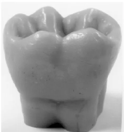
roots were also measured. Areas of greater width between the points used as a reference to obtain tooth measurements were also considered for both the crown and the root. The obtained measurements were then magnified 6X to establish the proportion between the macro models and the natural teeth (6:1) and the wax carving was then made. In this phase, only the crown was carved (Figure 1). After carving, the wax was smoothed using a wax carver and silk stockings. These wax macro models were then molded with alginate (Jeltrate, Dentsply, Petrópolis, RJ, Brazil), thus obtain-ing the mold (Figure 2). Next, the mold was filled with Wilson plaster stone type III (São Paulo, SP, Brazil), which was leaked with the aid of a plaster vibrator, to avoid the inclusion of air bubbles. Hence, macro mod-els of permanent teeth crowns were obtained (Figure 3). After all macro models of the crowns of all permanent teeth were finished, they were mounted one by one in a base of wax #7, thus obtaining the maxillary and mandibular arches (Figure 4). These macro models were mounted in occlusion.
These wax mounted arches were molded again using Jeltrate Dentsply alginate and the mold was then immediately filled with plaster stone type III (Wilson), obtaining the maxillary and mandibular arches totally in plaster (Figure 5).
Complete macro models (crowns and roots) were
Figure 1. Crown macro model in wax carving of a first mandibular left molar. Lingual view.
Figure 2. Crown alginate mold.
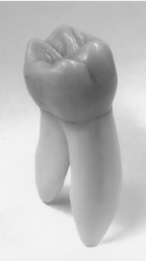
also made from auto-polymerized acrylic. In order to obtain the macro models in auto-polymerized acrylic, macro models were initially carved using dental carv-ing wax. The crown and the root were carved in agree-ment with the morphology and initially obtained mea-surements of the natural permanent teeth, which were later magnified 6X (Figure 6). In the same way, the wax was smoothed and then the mold was obtained with the Jeltrate alginate (Figure 7). Colorless auto polymerized acrylic resin (Jet, São Paulo, SP, Brazil) was used to immediately fill the mold, with the aid of a plaster
Figure 7. The alginate mold of the macro model in wax. Figure 6. Macro model (crown and root) in wax carving of a first maxillary right molar. Distopalatine view.
Figure 5. Plaster arches in occlusion. a) Macro models. b) Models with normal size.
Figure 4. Macro models mounted in wax base. Top: maxillary arch. Bottom: mandibular arch.
vibrator.
stone and water were also used together with a felt wheel, coupled to a buffing wheel, providing better smoothening and burnishing of the macro model (Fig-ure 8).
studies, dental carving techniques and methods are extremely important. Research and activities in dental anatomy carving can also be directly related to the practice of Restorative Dentistry. The importance of the recognition of morphologic and anatomy-functional characteristics of teeth, seeking adaptation to indi-vidual conditions, has been acknowledged (6).
The development of alternative methods, such as computer-graphics, to aid in teaching three-dimen-sional dental anatomy (3) and Tooth Morphology, which is a computer-assisted learning program designed to teach the anatomy of the adult dentition (4) are impor-tant for motivating and teaching students. The Tooth Morphology program, in combination with interactive class meetings, has replaced traditional dental anatomy lectures (4); however, it does not replace the practice of dental sculpting.
The purpose of carving is the reintegration, by means of total or partial reconstruction, of one or more dental elements in its form and function inside the arch, re-establishing the lost balance in the physiology of mastication. Secondarily, it is a precious auxiliary method in the assimilation or re-memorization of the anatomical knowledge and a process of manual ability, which is useful and necessary in professional activities (5).
Professionals require the knowledge of dental element morphology on a daily basis as a direct conse-quence of the need to constantly restructure the dental organ and reinstate its function. Dental form is ex-tremely varied and difficult to reproduce. The normal anatomical form of teeth assures the efficiency of mas-tication (5).
The external anatomy of teeth should be very well known. Theoretical studies are not enough. The student should study the detailed description of the tooth with copies of them in their hands. Besides the study of extracted natural teeth, macro models made of plaster or resins and dental arch models help to under-stand the aspects that must be taught. The drawing and carving of teeth in wax are also valuable means of learning dental anatomy, besides developing psycho-motor ability (7).
Macro models facilitate the assimilation or re-memorization of anatomy because they show all ana-tomical details that need to be reproduced. In this way, the student who practices dental carving exercises is able to develop normal anatomical form of teeth, rees-Figure 8. Macro model in autopolymerized acrylic resin of a first
mandibular left molar. Distolingual view.
RESULTS
This work resulted in the production of plaster crown models of the maxillary and mandibular arches, mounted with all dental crowns in occlusion, as well as macro models in autopolymerized acrylic with the crown and dental root.
This educational material is reproduced yearly in plaster models for each student of the Dental Anatomy Discipline of FORP-USP and used during the practical classes of this discipline for teaching and learning to aid the student during individual dental carving, as well as for the interpretation of dental occlusion.
DISCUSSION
tablishing the function of the dental element. Macro models allow a better visualization of the morphologic characteristics of permanent teeth and students of den-tal anatomy of the Dentistry course of FORP-USP are able to easily carve permanent teeth into wax guided by the macro models. Macro models of the dental arches allow a better visualization and an easier understanding of the occlusion of permanent teeth.
RESUMO
O objetivo deste trabalho é de apresentar um material didático que permita ao aluno de odontologia, identificar com maior facilidade, as características morfológicas dos dentes permanentes assim como entender mais facilmente a oclusão dos dentes. Para isto, foram confeccionados macromodelos de dentes permanentes sem desgaste, que foram obtidos da seguinte maneira: foram esculpidos macromodelos em cera; posteriormente estes foram moldados com alginato e os moldes foram preenchidos com gesso pedra e resina acrílica. Os macromodelos de gesso pedra foram montados em oclusão em uma base de cera #7 sendo obtidas desta maneira as arcadas superior e inferior. Estas arcadas montadas em cera foram moldadas novamente e o molde foi preenchido com gesso pedra, obtendo então as duas arcadas totalmente em gesso. Os autores concluíram que os macromodelos permitem uma melhor visualização das características morfológicas dos dentes permanentes bem como da oclusão; os alunos conseguem esculpir dentes permanentes em cera, com maior facilidade, quando observam macromodelos.
ACKNOWLEDGEMENTS
We thank Luisa Caliri Juzzo for translating this paper to the English Language.
REFERENCES
1. Figún ME, Garino RR. Anatomia Funcional Odontológica e Aplicada. 3rd ed. Rio de Janeiro: Editora Guanabara Koogan, 1994.
2. Cantisano W, Palhares WR, Santos HJ. Anatomia Dental e Escultura. 3rd ed. Rio de Janeiro: Editora Guanabara Koogan, 1987.
3. Pao YC, Reinhardt RA, Krejci RF, Taylor DT. Computer-graph-ics aided instruction of three-dimensional dental anatomy. J Dent Educ 1984;48:315-317.
4. Bogacki RE, Best A, Abbey LM. Equivalence study of dental anatomy computer-assisted learning program. J Dent Educ 2004;68:867-871.
5. Santos Jr J, Fichman DM. Escultura Dental na Clínica e no Laboratório. 4th ed. São Paulo: Artes Médicas, 1982.
6. Eugênio OS. Anatomia e Escultura Dental: Teoria e Prática de Ensino. São Paulo: Editora Santos, 1995.
7. Madeira MC. Anatomia do Dente. 2nd ed. São Paulo: Editora Sarvier, 2000.

