Association between age and survival in a
cohort of Brazilian patients with operable
breast cancer
Associação entre idade e sobrevida em uma
coorte de pacientes brasileiras com câncer
de mama operável
Asociación entre edad y supervivencia en una
cohorte de pacientes brasileñas con cáncer
de mama operable
1 Faculdade de Medicina, Universidade Federal de Minas Gerais, Belo Horizonte, Brasil.
2 Centro de Desenvolvimento e Planejamento Regional, Universidade Federal de Minas Gerais, Belo Horizonte, Brasil.
Correspondence D. Balabram
Faculdade de Medicina, Universidade Federal de Minas Gerais.
Av. Professor Alfredo Balena 190, Belo Horizonte, MG 30130-100, Brasil. [email protected]
Débora Balabram 1 Cassio M. Turra 2 Helenice Gobbi 1
Abstract
Whether age is an independent prognostic fac-tor in breast cancer is a matter of debate. This is a retrospective cohort study of 767 breast cancer patients, stages I-III, treated at the Hospital das Clínicas, Minas Gerais Federal University, Belo Horizonte, Minas Gerais State, Brazil, from 2001 to 2008, aiming to study the relationship be-tween age and survival. We included variables related to patients, tumors, and types of treat-ment. Different sets of Cox models were used for survival analysis. Hazard ratios (HR) and 95%CI were calculated. The relationship between age and breast cancer survival did not change sub-stantially in any of them. In the model that ac-counted for all variables, women aged 70 and older (HR = 1.51, 95%CI: 1.04-2.18), and 35 or younger (HR = 1.78, 95%CI: 1.05-3.01) had short-er cancshort-er specific survival than patients aged be-tween 36 and 69. In addition, older age, having at least one comorbidity, and being white were associated with a higher risk of dying from other causes. In conclusion, shorter breast cancer sur-vival is expected among the youngest and oldest patients.
Neoplasm Staging; Ethnicity and Health; Age Factors; Breast Neoplasms
Resumo
É discutível se idade é um fator prognóstico in-dependente para câncer de mama. Conduzimos uma coorte retrospectiva de 767 pacientes com câncer de mama, estádios I-III, tratadas no Hos-pital das Clínicas, Universidade Federal de Mi-nas Gerais, Belo Horizonte, MiMi-nas Gerais, Brasil, de 2001 a 2008, para estudar a relação entre ida-de e sobrevida. Incluímos variáveis relacionadas às pacientes, aos tumores e ao tratamento. Dife-rentes conjuntos de modelos de Cox foram cons-truídos. As razões de risco (RR) e IC95% foram calculados. A relação entre idade e sobrevida por câncer de mama não foi alterada substancial-mente entre os modelos de Cox. No modelo com todas as variáveis explicativas, as mulheres de 70 anos ou mais (RR = 1,51; IC95%: 1,04-2,18) e até 35 anos (RR = 1,78; IC95%: 1,05-3,01) tive-ram sobrevida causa-específica mais curta que as de 36-69 anos. Idades a partir de 70 anos, ter ao menos uma comorbidade e ser branca foram associadas a risco maior de óbito por outras cau-sas. Em conclusão, as pacientes mais jovens e as mais idosas parecem ter sobrevida mais curta por câncer de mama.
Introduction
Breast cancer is the most common malignant neoplasm among women in Brazil, with an ex-pected incidence of 57,120 new cases for the year 2014 1. Age is the strongest risk factor for the dis-ease 2 and thus, breast cancer incidence is in-creasing with population aging in Brazil 3,4.
Many studies have reported that older women (≥ 70 years of age) have less aggressive breast cancer, including a higher frequency of lower grade tumors and positivity for hormone receptors 5,6,7. However, they may receive less than standard treatment, due to the presence of comorbidities or to a belief in a less aggres-sive disease in this age subgroup 6,8,9. Compared to the elderly, young women (≤ 35 years of age) have more frequently higher grade breast cancer and negativity for hormone receptors 7,10. Yet, it remains unclear whether age is an independent prognostic factor for the lower survival among younger and older patients 10,11 or if the increase in mortality risk is associated with different tu-mor features in these groups.
In a previous study 12, we found that women 70 years of age and older have a higher risk of dying from breast cancer, independent of tumor related factors, in comparison with patients 36 to 69 years of age. Other studies have also dem-onstrated a higher chance of dying from breast cancer for both older and younger age groups 2,8,10,13,14. Furthermore, older age is related to a higher prevalence of comorbidities, which may reduce overall and disease-specific survival 15,16. Here, we examine in more detail the relationship between age and mortality from breast cancer and other causes of death, by looking at the role played by several intervening variables, includ-ing skin color, comorbidities, tumor factors and use of systemic treatments. We use data from patients treated at a public Brazilian hospital be-tween the years 2001 and 2008.
Methods
Study design and population
We obtained data from a retrospective cohort study of patients with breast cancer, stages I-III, who underwent surgery for breast cancer treat-ment at the Hospital das Clínicas of the Minas Gerais Federal University, Belo Horizonte, Minas Gerais State, Brazil (HC-UFMG), from the years 2001 to 2008. The UFMG Ethics Research Com-mittee approved the study’s protocol on March 7, 2012 (project CAAE number 0660.0.203.000-11).
Among the 1,004 patients who underwent surgery for treatment of invasive breast cancer at the HC-UFMG between 2001 and 2008, we excluded 75 women for whom treatment was not paid for by the public health system. Of the remaining 929 individuals, we excluded cases without medical records (n = 76), patients who underwent surgery only for palliative purposes (stage IV disease, n = 7), individuals with recur-rent breast cancer (n = 14), patients who had in-complete information on tumor stage (n = 30), and 35 cases with missing data on the indepen-dent variables. After excluding these cases, our cohort analysis contained 767 patients.
Variables
We examined four sets of determinants of mortal-ity: patient demographic characteristics, health status, tumor characteristics, and systemic treat-ments (hormone and chemotherapy).
Besides age, measured in three categories (up to 35, 36 to 69, and 70 years and older), we includ-ed tumor size and lymph node status according to the American Joint Committee on Cancer Staging Manual 17,18, as well as tumor type and grade 18. From the patients’ medical records we obtained information on skin color and comorbidities. Skin color was assigned by the attending physi-cian as white, black and brown skin. We dichot-omized the patients into white and non-white (black or brown skin). As for the comorbidities, we used the Charlson comorbidity index, which combines mortality risk levels associated with different chronic conditions, such as diabetes mellitus, chronic obstructive pulmonary disease, congestive heart failure, and dementia 19. Each condition receives a different score (1, 2, 3 or 6), depending on the risk of death associated with it. The scores are then added up to provide a total score for each individual 19. In our study cohort, all patients had at least a score of 2 because of the presence of tumor without metastasis. Most of them (625 patients, 81.5%) had no other major comorbidities and thus, maintained a score of 2. 114 patients (14.9%) had a comorbidity that resulted in one extra point (final score of 3). The remaining patients (28, 3.7%) had scores of 4, 5 and 6. In order to reduce the small variability of counts in the index, we constructed a dichoto-mous variable, which is equal to one for patients who had at least a score of 3, and zero for those with a score that is equal to 2.
therapy (41 patients, 5.5%). We used information on estrogen receptor status to impute the miss-ing values of hormone therapy. We assumed that patients with estrogen receptor (ER) positive tu-mors (21 cases) received treatment, whereas pa-tients with ER negative tumors (20 cases) did not. One may notice that we chose not to add ER as an independent variable in our models, since there were too many missing values for this measure (55 cases, 7.2%). For imputing missing data for chemotherapy we applied a relatively more com-plex algorithm, based on four variables: disease stage, age, use of hormone therapy, and presence of comorbidities. These variables are frequently used to predict the benefit of prescribing chemo-therapy for patients with breast cancer 20,21. We assumed that patients with stage I disease did not undergo chemotherapy. Also, we considered that stage II patients received chemotherapy, only if they were younger than 70 years of age, had no major comorbidities, and had not received en-docrine therapy. We further assumed that stage II patients who were older than 70 years of age received chemotherapy if they did not receive hormone therapy and had no comorbidities. We considered all stage III patients who were younger than 70 years of age to have received chemotherapy. Stage III patients older than 70 years only received chemotherapy if they did not receive hormone therapy and had no major co-morbidities. After the imputation procedure, we determined that 20 patients received chemother-apy, while the other 16 did not. Since the number of missing cases is somewhat small, alternative imputation procedures for both the hormone and chemotherapies proved to have only minor effects on our results.
We retrieved data on cause and date of death as checked in the Mortality Information System (SIM) of the Brazilian Ministry of Health 3, from 2001 to 2011. When the cause of death was un-known or the patient died without assistance (7 cases, 2.8% of total of deaths), breast cancer was considered to be the cause 22,23. We used a proba-bilistic record linkage strategy to identify patients in our database who had died up to December 31st, 2011 24. The program used was the RecLink version 3.0 24. Survival time was counted from the first day of treatment (surgery or chemotherapy) until the date of death or the end of the study pe-riod. The patient’s first and last name, their moth-er’s name and their date of birth were retrieved from both databases (the study’s and the SIM database), and different linkage strategies were used to find patients who had died 12. Patients not found in the MIS database were considered to be alive at the end of the observation period. We classified the causes of death according to
the International Classification of Diseases, 10th revision (ICD-10) 25. The linkage procedure was described in detail in an earlier study 12.
Statistical analysis
We used the exact two-sided linear-by-linear as-sociation statistic to compare the distribution of patient and tumor characteristics across age groups. The significance level was defined as 0.05. Mean and median survival times were cal-culated. We use Cox proportional hazards model for survival analysis. The hazard ratios (HR) and 95% confidence intervals (95%CI) for each inde-pendent variable were calculated.
We tested interaction terms between age and each one of the covariates in our final model, but none of them was significant. Further, add-ing interaction terms did not improve our mod-el as indicated by the log-likmod-elihood statistic. We confirmed the proportional hazard assumption for all variables in each of the models by verify-ing Schoenfeld residuals against survival time. All statistical analyses were performed with the IBM SPSS software, version 21.0 (IBM Corp., Ar-monk, USA).
In the first set of regression models, we ex-amined cause-specific survival, by censoring pa-tients who died from causes not related to breast cancer. We specified four Cox models to explore the effects of age on mortality. Model 1 includes only age. Model 2 adds other patient’s character-istics (skin color and the Charlson comorbidity index). The tumor characteristics were added in Model 3, and in Model 4 the use of chemo and hormone therapy were included as well. In the second set of regression models, we followed the same sequence of Cox proportional hazard mod-els as in the first set, but considered survival only from causes not related to the disease.
Results
of deaths). The prevalence of comorbidities in-creased with age (39.4% of patients 70 years and older had at least one comorbidity).
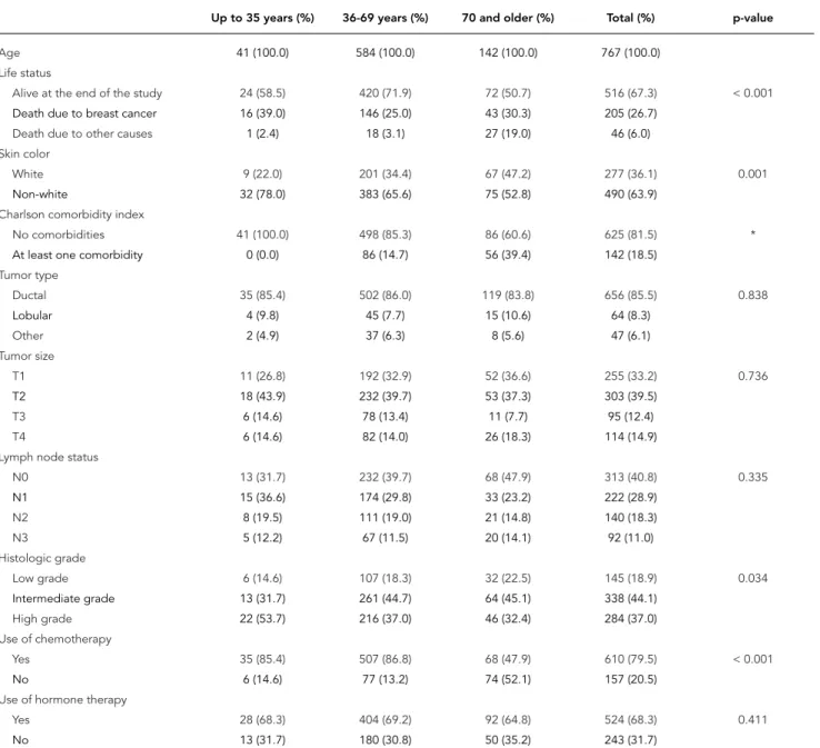
Table 1 shows the distribution of the explana-tory variables by age. Younger patients were more frequently non-white (78% non-white ver-sus 22% white) than the oldest age group (52.8% non-white versus 47.2% white). The distributions of tumor type, tumor size, and lymph node status were not different across age groups. However, when lymph node status was dichotomized in
Table 1
Age and covariates of Brazilian breast cancer patients treated from 2001 to 2008 (n = 767).
Up to 35 years (%) 36-69 years (%) 70 and older (%) Total (%) p-value
Age 41 (100.0) 584 (100.0) 142 (100.0) 767 (100.0)
Life status
Alive at the end of the study 24 (58.5) 420 (71.9) 72 (50.7) 516 (67.3) < 0.001
Death due to breast cancer 16 (39.0) 146 (25.0) 43 (30.3) 205 (26.7)
Death due to other causes 1 (2.4) 18 (3.1) 27 (19.0) 46 (6.0)
Skin color
White 9 (22.0) 201 (34.4) 67 (47.2) 277 (36.1) 0.001
Non-white 32 (78.0) 383 (65.6) 75 (52.8) 490 (63.9)
Charlson comorbidity index
No comorbidities 41 (100.0) 498 (85.3) 86 (60.6) 625 (81.5) *
At least one comorbidity 0 (0.0) 86 (14.7) 56 (39.4) 142 (18.5)
Tumor type
Ductal 35 (85.4) 502 (86.0) 119 (83.8) 656 (85.5) 0.838
Lobular 4 (9.8) 45 (7.7) 15 (10.6) 64 (8.3)
Other 2 (4.9) 37 (6.3) 8 (5.6) 47 (6.1)
Tumor size
T1 11 (26.8) 192 (32.9) 52 (36.6) 255 (33.2) 0.736
T2 18 (43.9) 232 (39.7) 53 (37.3) 303 (39.5)
T3 6 (14.6) 78 (13.4) 11 (7.7) 95 (12.4)
T4 6 (14.6) 82 (14.0) 26 (18.3) 114 (14.9)
Lymph node status
N0 13 (31.7) 232 (39.7) 68 (47.9) 313 (40.8) 0.335
N1 15 (36.6) 174 (29.8) 33 (23.2) 222 (28.9)
N2 8 (19.5) 111 (19.0) 21 (14.8) 140 (18.3)
N3 5 (12.2) 67 (11.5) 20 (14.1) 92 (11.0)
Histologic grade
Low grade 6 (14.6) 107 (18.3) 32 (22.5) 145 (18.9) 0.034
Intermediate grade 13 (31.7) 261 (44.7) 64 (45.1) 338 (44.1)
High grade 22 (53.7) 216 (37.0) 46 (32.4) 284 (37.0)
Use of chemotherapy
Yes 35 (85.4) 507 (86.8) 68 (47.9) 610 (79.5) < 0.001
No 6 (14.6) 77 (13.2) 74 (52.1) 157 (20.5)
Use of hormone therapy
Yes 28 (68.3) 404 (69.2) 92 (64.8) 524 (68.3) 0.411
No 13 (31.7) 180 (30.8) 50 (35.2) 243 (31.7)
* Fisher’s exact text was not performed because one of the values is equal to 0.
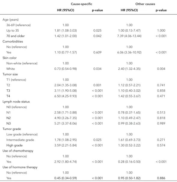
oldest cohort). The use of hormone therapy did not change across age categories. Table 2 shows crude hazard ratios for each of the covariates, in models of disease-specific and mortality associ-ated with other causes.
Table 3 shows the results for cause-specific Cox regression models. According to model 1, both patients aged 35 years old and younger (HR = 1.81; 95%CI: 1.08-3.03), and 70 years old and older (HR = 1.42; 95%CI: 1.01-2.00), had low-er disease-specific survival than women 36 to 69 years old. The coefficients for age changed little in Model 2, indicating that most of the effect of age is not captured by the presence of at least
one comorbidity and patients’ skin color. Model 2 also shows that non-white women have a high-er risk of dying than white women (HR = 0.71; 95%CI: 0.53-0.96). Having at least one comorbid-ity was not associated with lower disease-specif-ic survival (HR = 1.06; 95%CI: 0.73-1.54).
Model 3 (Table 3) shows that multiple tu-mor characteristics are significantly associated with the risk of dying during the observation period. Higher tumor grade, larger tumor size, and higher number of positive nodes were all as-sociated with higher mortality risks. The effect of the youngest age group, however, became no longer statistically significant (HR = 1.64; 95%CI:
Table 2
Factors related to breast cancer-specific survival (n = 767) and death due to other causes (n = 726) in Brazilian breast cancer patients stages I-III, treated from 2001 to 2008, univariate analysis.
Cause-specific Other causes
HR (95%CI) p-value HR (95%CI) p-value
Age (years)
36-69 (reference) 1.00 1.00
Up to 35 1.81 (1.08-3.03) 0.025 1.00 (0.13-7.47) 1.000
70 and older 1.42 (1.01-2.00) 0.042 7.39 (4.06-13.44) < 0.001
Comorbidities
No (reference) 1.00 1.00
Yes 1.10 (0.77-1.57) 0.609 6.06 (3.36-10.92) < 0.001
Skin color
Non-white (reference) 1.00 1.00
White 0.73 (0.54-0.98) 0.034 2.40 (1.32-4.35) 0.004
Tumor size
T1 (reference) 1.00 1.00
T2 2.04 (1.35-3.08) 0.001 1.12 (0.57-2.21) 0.741
T3 3.11 (1.90-5.08) < 0.001 1.10 (0.40-3.02) 0.858
T4 6.50 (4.25-9.93) < 0.001 1.42 (0.55-3.67) 0.471
Lymph node status
N0 (reference) 1.00 1.00
N1 2.58 (1.71-3.88) < 0.001 0.78 (0.37-1.65) 0.513
N2 4.90 (3.26-7.35) < 0.001 1.10 (0.49-2.47) 0.818
N3 5.21 (3.37-8.06) < 0.001 0.99 (0.38-2.63) 0.989
Tumor grade
Low grade (reference) 1.00 1.00
Intermediate grade 1.78 (1.08-2.95) 0.025 1.61 (0.69-3.73) 0.271
High grade 3.59 (2.21-5.84) < 0.001 1.30 (0.52-3.22) 0.574
Use of chemotherapy
No (reference) 1.00 1.00
Yes 2.92 (1.80-4.74) < 0.001 0.28 (0.16-0.50) < 0.001
Use of hormone therapy
No (reference) 1.00 1.00
Yes 0.45 (0.34-0.59) < 0.001 0.95 (0.50-1.82) 0.886
0.97-2.76). On the other hand, the coefficient for the oldest age group remained virtually un-changed and statistically significant in Model 3 (HR = 1.44; 95%CI: 1.01-2.06). The patient’s skin color (HR = 0.79; 95%CI: 0.58-1.07), and the presence of at least one comorbidity (HR = 1.19; 95%CI: 0.82-1.72), were not statistically associ-ated with death due to breast cancer.
When adding chemo and hormone thera-pies (Model 4, Table 3), the youngest age group became again associated with a higher risk of dying due to breast cancer (HR = 1.78; 95%CI: 1.05-3.01), while the effect for the oldest age group became slightly greater (HR = 1.51; 95%CI: 1.04-2.18). Tumor related factors remained
sig-Table 3
Cox regression models of factors related to breast cancer-specific survival in Brazilian breast cancer patients stages I-III, treated from 2001 to 2008 (n = 767).
Model 1 Model 2 Model 3 Model 4
HR (95%CI) p-value HR (95%CI) p-value HR (95%CI) p-value HR (95%CI) p-value
Age (years)
36-69 (reference) 1.00 1.00 1.00 1.00
Up to 35 1.81 (1.08-3.03) 0.025 1.77 (1.05-2.97) 0.032 1.64 (0.97-2.76) 0.065 1.78 (1.05-3.01) 0.031 70 and older 1.42 (1.01-2.00) 0.042 1.46 (1.03-2.08) 0.034 1.44 (1.01-2.06) 0.043 1.51 (1.04-2.18) 0.028 Comorbidities
No (reference) 1.00 1.00 1.00
Yes 1.06 (0.73-1.54) 0.753 1.19 (0.82-1.72) 0.37 1.28 (0.87-1.86) 0.209 Skin color
Non-white (reference) 1.00 1.00 1.00
White 0.71 (0.53-0.96) 0.027 0.79 (0.58-1.07) 0.127 0.83 (0.61-1.13) 0.242 Tumor size
T1 (reference) 1.00 1.00
T2 1.19 (0.77-1.83) 0.437 1.15 (0.74-1.77) 0.542
T3 1.75 (1.05-2.93) 0.033 1.60 (0.95-2.69) 0.076
T4 3.69 (2.34-5.82) < 0.001 3.31 (2.08-5.26) < 0.001 Lymph node status
N0 (reference) 1.00 1.00
N1 1.94 (1.26-2.97) 0.005 1.87 (1.21-2.89) 0.003
N2 2.93 (1.90-4.52) < 0.001 2.90 (1.87-4.50) < 0.001 N3 3.92 (2.48-6.20) < 0.001 4.02 (2.52-6.43) < 0.001 Tumor grade
Low grade (reference) 1.00 1.00
Intermediate grade 1.46 (0.88-2.42) 0.145 1.40 (0.84-2.33) 0.191 High grade 2.71 (1.65-4.44) < 0.001 2.12 (1.27-3.54) 0.004 Use of chemotherapy
No (reference) 1.00
Yes 1.31 (0.74-2.29) 0.354
Use of hormone therapy
No (reference) 1.00
Yes 0.59 (0.44-0.80) 0.001
95%CI: 95% confidence interval; HR: hazard ratio.
nificantly associated with survival. Chemother-apy was not statistically associated with longer survival among the patients, although the use of hormone therapy seemed to have a statistically significant protective effect (HR = 0.59; 95%CI: 0.44-0.80).
Table 4
Cox regression models of factors related to death due to causes other than breast cancer in Brazilian patients with breast cancer stages I-III, treated from 2001 to 2008 (n = 726).
Model 1 Model 2 Model 3 Model 4
HR (95%CI) p-value HR (95%CI) p-value HR (95%CI) p-value HR (95%CI) p-value
Age (years)
36-69 (refence) 1.00 1.00 1.00 1.00
70 and older 7.42 (4.08-13.49) < 0.001 4.73 (2.52-8.89) < 0.001 5.23 (2.74-9.97) < 0.001 4.24 (2.06-8.73) < 0.001
Comorbidities
No (reference) 1.00 1.00 1.00
Yes 3.90 (2.10-7.23) < 0.001 4.09 (2.16-7.75) < 0.001 3.72 (1.92-7.24) 0.001
Skin color
Non-white (reference) 1.00 1.00 1.00
White 2.05 (1.12-3.75) 0.02 2.22 (1.19-4.14) 0.012 2.08 (1.11-3.89) 0.023
Tumor size
T1 (reference) 1.00 1.00
T2 0.91 (0.43-1.94) 0.815 0.99 (0.46-2.13) 0.975
T3 1.44 (0.50-4.19) 0.501 1.77 (0.57-5.52) 0.324
T4 0.82 (0.29-2.33) 0.706 1.03 (0.33-3.16) 0.964
Lymph node status
N0 (reference) 1.00 1.00
N1 0.98 (0.45-2.16) 0.967 1.10 (0.49-2.48) 0.819
N2 1.12 (0.45-2.79) 0.803 1.22 (0.48-3.11) 0.683
N3 1.59 (0.57-4.42) 0.371 1.98 (0.67-5.88) 0.217
Tumor grade
Low grade (reference) 1.00 1.00
Intermediate grade 2.00 (0.83-4.86) 0.124 2.05 (0.84-5.04) 0.117
High grade 1.97 (0.74-5.25) 0.175 2.08 (0.73-5.94) 0.172
Use of chemotherapy
No (reference) 1.00
Yes 0.57 (0.25-1.33) 0.195
Use of hormone therapy
No (reference) 1.00
Yes 0.88 (0.43-1.82) 0.738
95%CI: 95% confidence interval; HR: hazard ratio.
risk from other causes of death was significantly increased for white women and for those who had at least one comorbidity. The variables as-sociated with tumor characteristics and systemic treatments were not predictors of mortality from other causes of death in any of the models.
Discussion
In the current study, we showed that the relation-ship between age and breast cancer survival re-mained statistically significant after including all the control variables available for our analysis. When age was considered alone, we found that
the oldest age group (70 years old and older, HR = 1.42; 95%CI: 1.01-2.00) and the youngest one (up to 35 years old, HR = 1.81; 95%CI: 1.08-3.03) had higher risk of dying due to breast cancer than pa-tients 36 to 69 years old. When tumor and patient characteristics, as well as use of systemic thera-pies were added to the model, the survival disad-vantage of the youngest (up to 35 years old, HR = 1.78; 95%CI: 1.05-3.01) and the oldest groups (70 years old and older, HR = 1.51; 95%CI: 1.04-2.18) remained statistically significant.
that tumor characteristics explain to some ex-tent the survival disadvantage among the young-est patients. These results are in accordance with previous research that has shown that younger patients have more aggressive tumors, whereas older patients have lower grade diseases, but a higher frequency of comorbidities, and more advanced stages at diagnosis 2,7,10,13,14,26,27. In our study, the presence of comorbidities was not associated with a higher hazard of dying from breast cancer, as shown in the study by Berglund et al. 15.
Some of the crude hazard ratios for tumor re-lated factors (tumor size and grade, and lymph node status) in the univariate analysis (Table 2) were substantially larger than the adjusted ef-fects in the multivariate models shown in Table 3. One possible explanation for this pattern is the existence of high correlation among the predic-tor variables. Yet, in tests that we performed us-ing the variance inflation factor, we found mul-ticollinearity to be within the limits of tolerance. Keeping the predictor variables together in our final model is also in accordance with the litera-ture that has shown they are independent prog-nostic factors in cancer survival.
The appropriate treatment for elderly wom-en with breast cancer remains a matter of debate. Since older women are usually not included in treatment trials, the benefits of therapy for them are more difficult to evaluate 2,26. Also, the in-cidence of toxicity after adjuvant treatments 2 and the presence of comorbidities 15,16 is high-er among the eldhigh-erly, thus reducing the use of these types of therapies. On the other hand, some studies have shown that less than standard treat-ment can be harmful for older patients 6,8,9, and thus, the individualization of treatment strate-gies is recommended. Chemotherapy was less frequently used among older patients (p-value < 0.001) in our cohort and it did not seem to offer mortality protection among patients of any age group. The benefits of chemotherapy are known to be more important among hormone recep-tor-negative patients 20,21, and since around 70% of our study comprises patients with hormone receptor-positive tumors, the cohort size may be too small to show the protective effects of this type of treatment. Other treatment modalities, like type of surgical treatment and use of radia-tion therapy, were omitted from our analysis, since they have a weaker association with overall survival than the other measures 20,21,26.
A similar debate exists surrounding the ideal age to interrupt breast cancer screening. In Bra-zil, the recommended age span for screening by the Public Health System is from 50 to 69 years of age 28. According to the International Society of
Geriatric Oncology (SIOG), the decision to main-tain screening over the age of 70 should be “based on risks and benefits, patient preference, physi-ological age, and life expectancy” 26 (p. e152). Un-fortunately we cannot test directly for delayed di-agnosis, although in our sample the distribution of patients by tumor size and lymph node status, compared to countries where screening is avail-able, suggests that there are more advanced cases in Brazil, particularly among the elderly 29,30.
One of the limitations of our study is that we drew our data from pathology records, which means we have selected a sample of patients which were at least fit enough to undergo sur-gical treatment. Patients with a lower health status who could not have undergone surgery were excluded from our cohort study from the start, which precludes us from generalizing our conclusions to all breast cancer patients. In ad-dition, we obtained data on comorbidities and adjuvant therapies from medical records. There-fore, we were not able to identify which chemo and hormone therapy regimens were applied to each patient, although we recognize that they vary depending on disease characteristics and comorbidities.
con-clude, based on the lower instantaneous hazard from other causes of deaths among non-whites, that this group truly experiences lower incidence of this types of deaths.
Our finding that tumor related characteristics were not significantly associated with mortality due to other causes of death is not surprising and indicates the quality of our data, particu-larly the accuracy of the classification of causes of death. One should note that the risk of dying from diseases other than breast cancer increases with time since diagnosis 38,39, especially after ten years. Since the median follow-up time in our study was much shorter, we already expect-ed a larger proportion of deaths (82% of total of deaths) due to breast cancer compared to other studies. However, in at least one study of Ameri-can women, which followed patients for a period of time (2000 to 2007) shorter than the observa-tion period in our study, the proporobserva-tion of breast cancer deaths was relatively lower (only 56%) 40.
Multiple factors are involved in mortality from breast cancer and there is still much to be learned about the biological, medical and socioeconomic mechanisms responsible for improving survival. The current study has extended previous research for Brazil in showing that age is an independent predictor of cause-specific mortality, at least in the presence of the numerous control variables available for our analysis. Of course, survival dif-ferentials by age may depend on a variety of other key factors not included in our models such as the socioeconomic status of patients 30, access to health care 31,32, and more detailed data on types of treatment employed 12,17,41,42, which reinforces the need for further analysis. Understanding the pathways linking age to mortality due to breast cancer should help doctors, epidemiologists and policy makers to propose specific measures to improve the chances of survival for women of dif-ferent age groups, particularly in a context of pro-found changes in the population age structure.
Resumen
Es discutible si la edad es un factor pronóstico indepen-diente para el cáncer de mama. Se realizó sobre una co-horte retrospectiva de 767 pacientes con cáncer de ma-ma, etapas I-III, atendidas en el Hospital de Clínicas, Universidad Federal de Minas Gerais, Belo Horizonte, Minas Gerais, Brasil, entre 2001 y 2008, para estudiar la relación entre edad y supervivencia. Incluimos va-riables relacionadas con las pacientes, los tumores y el tratamiento. Se construyeron diferentes conjuntos de modelos de Cox. Se calcularon los cocientes de riesgo (CR) e IC95%. La relación entre edad y supervivencia del cáncer de mama no ha cambiado substancialmente en los modelos. En el modelo con todas las variables, las mujeres de 70 años o más (CR = 1,51; IC95%: 1,04-2,18) y 35 años o menos (CR = 1,78; IC95%: 1,05-3,01) tuvie-ron menor supervivencia por cáncer de mama que las de 36 a 69 años. Tener edad avanzada, al menos una comorbilidad, y ser de piel blanca se asociaron a un mayor riesgo de morir por otras causas. En conclusión, las mujeres más jóvenes y las mayores parecen tener menor supervivencia de cáncer de mama.
Estadificacíon de Neoplasias; Origem Étnico y Salud; Factores de Edad; Neoplasias de la Mama
Contributors
D. Balabram was responsible for the study design, data collection and analysis, and for writing the paper. C. M. Turra was responsible for the study design, analysis of data and aided in paper writing. H. Gobbi aided in the study design, reviewed specimen slides and aided in paper writing. All authors have read and approved the submitted version of the manuscript.
Acknowledgments
References
1. Instituto Nacional de Câncer José Alencar Gomes da Silva. INCA e Ministério da Saúde apresentam estimativas de câncer para 2014. http://www2. inca.gov.br/wps/wcm/connect/agencianoticias/ site/home/noticias/2013/inca_ministerio_saude_ apresentam_estimativas_cancer_2014 (accessed on 20/Jul/2014).
2. Turner N, Zafarana E, Becheri D, Mottino G, Bigan-zoli L. Breast cancer in the elderly: which lessons have we learned? Future Oncol 2013; 9:1871-81. 3. Instituto Nacional de Câncer José Alencar Gomes da
Silva. Atlas de mortalidade por câncer. http://mor talidade.inca.gov.br/Mortalidade/ (accessed on 24/Jun/2012).
4. Instituto Brasileiro de Geografia e Estatística. Atlas do Censo Demográfico 2010. http://www.ibge.gov. br/home/geociencias/geografia/atlas.shtm?c=5 (accessed on 15/Sep/2014).
5. Gennari R, Curigliano G, Rotmensz N, Robertson C, Colleoni M, Zurrida S, et al. Breast carcinoma in elderly women: features of disease presentation, choice of local and systemic treatments compared with younger postmenopasual patients. Cancer 2004; 101:1302-10.
6. Schonberg MA, Marcantonio ER, Ngo L, Li D, Sil-liman RA, McCarthy EP. Causes of death and rl.ive survival of older women after a breast cancer diag-nosis. J Clin Oncol 2011; 29:1570-7.
7. Dutra MC, Rezende MA, de Andrade VP, Soares FA, Ribeiro M V, de Paula EC, et al. Imunofenótipo e evolução de câncer de mama: comparação entre mulheres muito jovens e mulheres na pós-meno-pausa. Rev Bras Ginecol Obstet 2009; 31:54-60. 8. Schonberg MA, Marcantonio ER, Li D, Silliman RA,
Ngo L, McCarthy EP. Breast cancer among the old-est old: tumor characteristics, treatment choices, and survival. J Clin Oncol 2010; 28:2038-45. 9. Yood MU, Owusu C, Buist DSM, Geiger AM, Field
TS, Thwin SS, et al. Mortality impact of less-than-standard therapy in older breast cancer patients. J Am Coll Surg 2008; 206:66-75.
10. Thomas GA, Leonard RC. How age affects the bi-ology of breast cancer. Clin Oncol (R Coll Radiol) 2009; 21:81-5.
11. Colzani E, Liljegren A, Johansson ALV, Adolfsson J, Hellborg H, Hall PFL, et al. Prognosis of patients with breast cancer: causes of death and effects of time since diagnosis, age, and tumor characteris-tics. J Clin Oncol 2011; 29:4014-21.
12. Balabram D, Turra CM, Gobbi H. Survival of pa-tients with operable breast cancer (Stages I-III) at a Brazilian public hospital – a closer look into cause-specific mortality. BMC Cancer 2013; 13:434. 13. Beadle BM, Woodward WA, Buchholz TA. The
im-pact of age on outcome in early-stage breast can-cer. Semin Radiat Oncol 2011; 21:26-34.
14. Jatoi I, Anderson WF, Rosenberg PS. Qualitative age-interactions in breast cancer: a tale of two dis-eases? Am J Clin Oncol 2008; 31:504-6.
15. Berglund A, Wigertz A, Adolfsson J, Ahlgren J, For-nander T, Wärnberg F, et al. Impact of comorbid-ity on management and mortalcomorbid-ity in women diag-nosed with breast cancer. Breast Cancer Res Treat 2012; 135:281-9.
16. Jørgensen TL, Hallas J, Friis S, Herrstedt J. Comor-bidity in elderly cancer patients in relation to over-all and cancer-specific mortality. Br J Cancer 2012; 106:1353-60.
17. Edge SB, Byrd DR, Compton CC, Fritz AG, Greene FL, Trotti A. AJCC cancer staging manual. 7th Ed. New York: Springer; 2009.
18. Lakhani SR, Ellis IO, Schnitt SJ, Tan PH, van de Vijver MJ. WHO classification of tumours of the breast. 4th Ed. Lyon: IARC Press; 2012.
19. Charlson ME, Pompei P, Ales KL, MacKenzie CR. A new method of classifying prognostic comorbidity in longitudinal studies: development and valida-tion. J Chronic Dis 1987; 40:373-83.
20. Peto R, Davies C, Godwin J, Gray R, Pan HC, Clarke M, et al. Comparisons between different polyche-motherapy regimens for early breast cancer: me-ta-analyses of long-term outcome among 100,000 women in 123 randomised trials. Lancet 2012; 379:432-44.
21. Darby S, McGale P, Correa C, Taylor C, Arriagada R, Clarke M, et al. Effect of radiotherapy after breast-conserving surgery on 10-year recurrence and 15-year breast cancer death: meta-analysis of in-dividual patient data for 10,801 women in 17 ran-domised trials. Lancet 2011; 378:1707-16. 22. Dignam JJ, Huang L, Ries L, Reichman M, Mariotto
A, Feuer E. Estimating breast cancer-specific and other-cause mortality in clinical trial and popula-tion-based cancer registry cohorts. Cancer 2009; 115:5272-83.
23. Howlader N, Ries LAG, Mariotto AB, Reichman ME, Ruhl J, Cronin KA. Improved estimates of can-cer-specific survival rates from population-based data. J Natl Cancer Inst 2010; 102:1584-98. 24. Camargo Jr. KR, Coeli CM. Reclink: aplicativo para
o relacionamento de bases de dados, implemen-tando o método probabilistic record linkage. Cad Saúde Pública 2000; 16:439-47.
25. World Health Organization. International Clas-sification of Diseases (ICD). http://www.who.int/ classifications/icd/en/ (accessed on 03/Sep/2011). 26. Biganzoli L, Wildiers H, Oakman C, Marotti L,
Loibl S, Kunkler I, et al. Management of elderly patients with breast cancer: updated recommen-dations of the International Society of Geriatric Oncology (SIOG) and European Society of Breast Cancer Specialists (EUSOMA). Lancet Oncol 2012; 13:e148-60.
27. Ring A, Reed M, Leonard R, Kunkler I, Muss H, Wildiers H, et al. The treatment of early breast can-cer in women over the age of 70. Br J Cancan-cer 2011; 105:189-93.
28. Instituto Nacional de Câncer José Alencar Gomes da Silva. SISMAMA – informação para o avanço das ações de controle do câncer de mama no Bra-sil. http://bvsms.saude.gov.br/bvs/publicacoes/ inca/Sismama.pdf (accessed on 31/Mar/2013). 29. National Cancer Institute. SEER Stat Fact Sheets:
30. Liedke PER, Finkelstein DM, Szymonifka J, Barrios CH, Chavarri-Guerra Y, Bines J, et al. Outcomes of breast cancer in Brazil related to health care cover-age: a retrospective cohort study. Cancer Epide-miol Biomarkers Prev 2014; 23:126-33.
31. Alves C, Silva MSD, Pinto LM, Toralles MBP, Tava-res-Neto J. Definition and use of the variable “race” by medical students in Salvador, Brazil. São Paulo Med J 2010; 128:206-10.
32. Pena SDJ, Di Pietro G, Fuchshuber-Moraes M, Genro JP, Hutz MH, Kehdy FSG, et al. The genomic ancestry of individuals from different geographical regions of Brazil is more uniform than expected. PLoS One 2011; 6:e17063.
33. Pena SDJ. Razões para banir o conceito de raça da medicina brasileira. Hist Ciênc Saúde-Mangui-nhos 2005; 12:321-46.
34. Smith EC, Ziogas A, Anton-Culver H. Delay in sur-gical treatment and survival after breast cancer di-agnosis in young women by race/ethnicity. JAMA Surg 2013; 148:516-23.
35. Chor D. Health inequalities in Brazil: race matters. Cad Saúde Pública 2013; 29:1272-5.
36. Chor D, Lima CRA. Aspectos epidemiológicos das desigualdades raciais em saúde no Brasil. Cad Saú-de Pública 2005; 21:1586-94.
37. Haller B, Schmidt G, Ulm K. Applying competing risks regression models: an overview. Lifetime Da-ta Anal 2013; 19:33-58.
38. Riihimäki M, Thomsen H, Brandt A, Sundquist J, Hemminki K. Death causes in breast cancer pa-tients. Ann Oncol 2012; 23:604-10.
39. Du XL, Fox EE, Lai D. Competing causes of death for women with breast cancer and change over time from 1975 to 2003. Am J Clin Oncol 2008; 31:105-16.
40. Cho H, Mariotto AB, Mann BS, Klabunde CN, Feuer EJ. Assessing non-cancer-related health status of US cancer patients: other-cause survival and comorbidity prevalence. Am J Epidemiol 2013; 178:339-49.
41. Oliveira EXG, Melo ECP, Pinheiro RS, Noronha CP, Carvalho MS. Acesso à assistência oncológi-ca: mapeamento dos fluxos origem-destino das internações e dos atendimentos ambulatoriais. O caso do câncer de mama. Cad Saúde Pública 2011; 27:317-26.
42. Silber JH, Rosenbaum PR, Clark AS, Giantonio BJ, Ross RN, Teng Y, et al. Characteristics associated with differences in survival among black and white women with breast cancer. JAMA 2013; 310:389-97.
Submitted on 28/Jul/2014