ABSTRACT: The reduction in caries prevalence has not occurred uniformly for all dental surfaces. As the occlusal surfaces are still the most likely sites for the development of lesions, new methods of diagnosis are still being evalu-ated. This study compared a laser luorescence (LF) system (DIAGNOdent) with the Ekstrand’s visual system for in vitro detection of occlusal caries. A total of 57 extracted molars with macroscopically intact occlusal surfaces were selected. Two-examiners assessed 110 sites by visual inspection (VI) and LF. After ten days from the irst measure -ment, all teeth were re-evaluated through the same methods by each examiner. Caries extension was histologically assessed (X 40). The methods were compared by means of sensitivity, speciicity, intra- and inter-examiner repro -ducibility and area under the ROC curve. The kappa’s test showed good intra- and inter-examiner repro-ducibility for both methods. VI and LF showed similar sensitivities for both examiners, however, VI showed higher speciici -ties than LF. The overall analysis, as demonstrated by the area under the ROC curve, showed that VI had a better performance than the LF device. It was concluded that the Ekstrand’s visual system is more reliable than the LF device. LF should be considered only as an adjuvant for occlusal caries diagnosis.
DESCRIPTORS: Lasers; Fluorescence; Dental caries; Molar; Diagnostic techniques and procedures.
RESUMO: A redução da prevalência de cáries não ocorreu uniformemente para todas as superfícies dentárias. Como as superfícies oclusais ainda são as mais susceptíveis ao desenvolvimento de lesões, novos métodos de diagnóstico ainda estão sendo avaliados. Este estudo comparou um sistema de luorescência a laser (DIAGNO -dent – DD) com o método visual de Ekstrand na detecção de cárie oclusal. Um total de 57 terceiros molares com superfícies oclusais macroscopicamente intactas foram selecionados. Dois examinadores examinaram 110 sítios por inspecção visual (IV) e DD. Após dez dias da primeira mensuração, todos os dentes foram novamente avaliados pelos mesmos métodos. A extensão de cárie foi validada por exame histológico (40 X). Os dados foram analisados quanto a sensibilidade, especiicidade, reprodutibilidade intra e interexaminador e área sob a curva ROC. O teste kappa demonstrou boa reprodutibilidade intra e interexaminadores para ambos os métodos. A IV e o DD apresen-taram sensibilidade semelhante para ambos os examinadores, entretanto, a IV apresentou maior especiicidade que o DD. A análise geral, através da área sob a curva ROC, mostrou que a IV teve um melhor desempenho que o DD. Concluiu-se que o critério visual proposto por Ekstrand é mais coniável para o diagnóstico de cáries oclusais. O DD deve ser considerado apenas como um coadjuvante no exame de cárie em superfícies oclusais.
DESCRITORES: Lasers; Fluorescência; Cárie dentária; Molar; Técnicas de diagnóstico e procedimentos.
INTRODUCTION
Despite the fact that the prevalence of den-tal caries has declined considerably, it is still a problem of great importance, mainly in Brazil. The reduction in caries prevalence has not occurred uniformly for all dental surfaces and the occlusal surfaces are still the most likely sites for the de-velopment of lesions.
Several methods of dental caries diagnosis have been used for more than half a century. Al-though there are known drawbacks, visual inspec-tion (VI) alone has been claimed to be the best diagnostic method in populations with low caries prevalence, but it is unable to correctly detect car-ies lesions because of its low sensitivity12. On the
* Dental clinicians, Joaçaba, Santa Catarina, Brazil.
** Associate Professor, Department of Dental Materials, University of São Paulo.
*** Assistant Professors, Department of Dental Materials and Restorative Dentistry, Oeste de Santa Catarina University.
Occlusal caries diagnosis in permanent teeth: an
in vitro
study
Diagnóstico de cárie oclusal em dentes permanentes: estudo
in
vitro
Gisele Angnes* Vivian Angnes*
Rosa Helena Miranda Grande** Márcio Battistella***
other hand, the use of a sharp probe along with the visual method does not appear to improve the diagnostic accuracy16,21. It may contaminate other
sound sites14, damage the fissure3 as well as
facili-tate the lesion’s progression29.
The main drawbacks of the conventional
meth-ods are that they still rely on the dentist’s subjec -tive interpretation, that it is difficult to evaluate a lesion’s progression and that some clinicians decide on an unwarranted invasive intervention. In the search for more accurate diagnostic ap-proaches, investigators have used alternative non-invasive and instrument-based techniques for detecting and quantifying demineralization lesions5,11,18. These techniques include electrical
conductance measurements, light scattering and quantitative laser/light induced fluorescence (LF). A new device is the KaVo DIAGNOdent® (KaVo,
Bib-erach, Germany), which generates a laser light that is absorbed by both inorganic and organic tooth substances and by metabolites from oral bacte-ria11. In the presence of caries, light with a higher
wavelength is re-emitted, and the changes are reg-istered in a digital number scale. Promising results have been published with this LF device2,18,19,24,27.
However, its accuracy still lacks further studies that should be conducted in order to verify how far this method works in other samples.
The LF device can be considered a valuable
tool as an adjunct to visual inspection mainly for
long-term monitoring of caries and for assessing the outcomes of preventive interventions, as the caries progress can be quantitatively measured; however its performance still needs validation.
Therefore the aim of this study was to validate histologically the use of DIAGNOdent (LF) for the detection and quantification of caries on intact oc-clusal surfaces; to compare the use of this device with VI and to evaluate the inter- and intra-exam-iner reproducibility of both diagnostic methods.
MATERIALS AND METHODS
A total of 57 molars presenting macroscopi-cally intact occlusal surfaces were selected and cleaned to remove any debris. Inclusion criteria for teeth in this study were the apparent absence of occlusal restorations and fissure sealants, absence of hypoplastic pits and frank occlusal cavitation. The teeth were stored in a physiological saline so-lution before the beginning of the study. The Ethics Committee, School of Dentistry, University of São Paulo, approved the study.
All teeth were properly identified. The occlusal surfaces were cleaned with pumice slurry and co-piously washed with water. Then, a drawing was done and all sites identified. Data were collected at 110 suspected sites in the fissures. Two examiners watched the same instructive session on diagnos-tic procedures using two representative teeth for each VI scoring system4 (Table 1). They were also
trained on how to use the LF device, according to the manufacturer’s directions.
Visual examination (VI)
After removing each tooth one-by-one from distilled water, the sites were examined under a
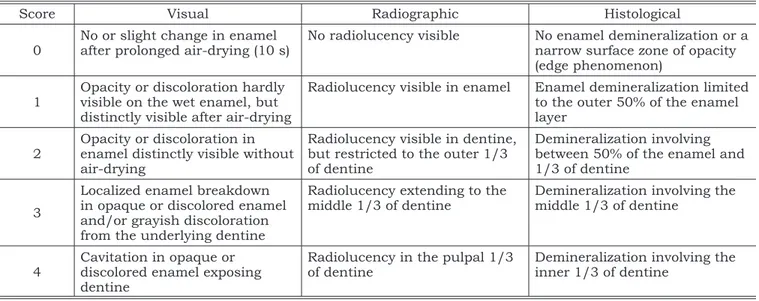
TABLE 1 - Criteria used for visual, radiographic and histological examination4.
Score Visual Radiographic Histological
0
No or slight change in enamel after prolonged air-drying (10 s)
No radiolucency visible No enamel demineralization or a narrow surface zone of opacity (edge phenomenon)
1
Opacity or discoloration hardly visible on the wet enamel, but distinctly visible after air-drying
Radiolucency visible in enamel Enamel demineralization limited to the outer 50% of the enamel layer
2
Opacity or discoloration in enamel distinctly visible without air-drying
Radiolucency visible in dentine, but restricted to the outer 1/3 of dentine
Demineralization involving between 50% of the enamel and 1/3 of dentine
3
Localized enamel breakdown in opaque or discolored enamel and/or grayish discoloration from the underlying dentine
Radiolucency extending to the middle 1/3 of dentine
Demineralization involving the middle 1/3 of dentine
4
Cavitation in opaque or discolored enamel exposing dentine
Radiolucency in the pulpal 1/3 of dentine
standard dental operating light at an eye-tooth distance of 20 cm. If no visible signs were seen on the wet occlusal surface, the examiners were al-lowed to dry the teeth with compressed air.
DIAGNOdent readings (LF)
The measurements with the LF device were made after calibration of the device with the ce-ramic standard. The assessment of the teeth with the LF fiber-tip was performed according to the distributor’s instructions. The laser tip (A tip) was positioned on a sound enamel region to provide a baseline measurement. After that, the laser tip was positioned on the target site and rotated around its long axis; the highest value was then recorded.
To verify intra-examiner reproducibility the examiners re-performed all examinations after a period of 7 to 10 days.
Validation
The sites were sectioned in a buccal to lin-gual direction using a 0.3 mm thick diamond saw mounted in a microtome (Labcut 1010, Extec Co., CT, USA). An experienced examiner evaluated the two sections of each site under a stereomicroscope (40 X) and reflected light (SZPT Olympus, Tokyo, Japan), and the side with more extensive altera-tions was classified according the Ekstrand’s cri-teria4.
Statistical analysis
Reproducibility of the VI system was assessed using unweighted kappa statistics. This was per-formed for repeated readings carried out by each examiner (intra-examiner reproducibility) and for
the second series of scores made by pairs of ex-aminers (inter-examiner reproducibility). Kappa values from 0.4 to 0.75 denote good reproduc-ibility6. The same procedure was performed for LF
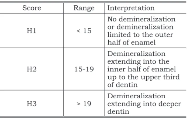
values only after categorization of the measure-ments (Table 2).
The scale that correlates the lesion extension and the range of the LF values was obtained by performing three ROC analyses. This analysis was performed after dichotomization of the histological scores into three cuts-offs: H1 (no demineraliza-tion or demineralizademineraliza-tion limited to the outer half of enamel), H2 (demineralization extending to the inner half of enamel up to the upper third of den-tin) and H3 (demineralization extending into ½ of dentin). The best cut-offs for each dichotomiza-tion were obtained, allowing the composidichotomiza-tion of Table 2.
Sensitivity and specificity were calculated us-ing the threshold between 2 and 3 for VI (Table 1). For the categorized LF data, threshold was set be-tween H2 and H3 (Table 2). The McNemar’s Change test was applied to compare the performance of the diagnostic methods for each examiner. ROC analy-sis was also conducted to compare the diagnostic performance of the three methods for occlusal car-ies diagnosis. In addition, a non-parametric statis-tical test was applied to estimate the significance of areas under ROC curves8.
RESULTS
Histological examination showed that 20 sites were classified as score 0; 24 as score 1; 50 as score 2; 14 as score 3; and 2 as score 4. Hence, 16 out of 110 sites were classified as “carious”, which represents approximately 14.5% of the sample.
Table 3 gives unweighted kappa values for in-ter-examiner reproducibility for each ranked scale. Kappa statistics showed good reproducibility for all methods.
TABLE 2 - Ranked scale used in the DIAGNOdent ex-amination.
Score Range Interpretation
H1 < 15
No demineralization or demineralization limited to the outer half of enamel
H2 15-19
Demineralization extending into the inner half of enamel up to the upper third of dentin
H3 > 19
Demineralization extending into deeper dentin
TABLE 3 - Unweighted kappa values for intra- and in-ter-examiner reproducibility for ranked scoring sys-tems for each of the diagnostic methods.
Diagnostic methods
Intra-examiner reproducibility
Inter-examiner reproducibility examiner
1
examiner 2
examiners 1-2
Visual 0.75 0.74 0.52
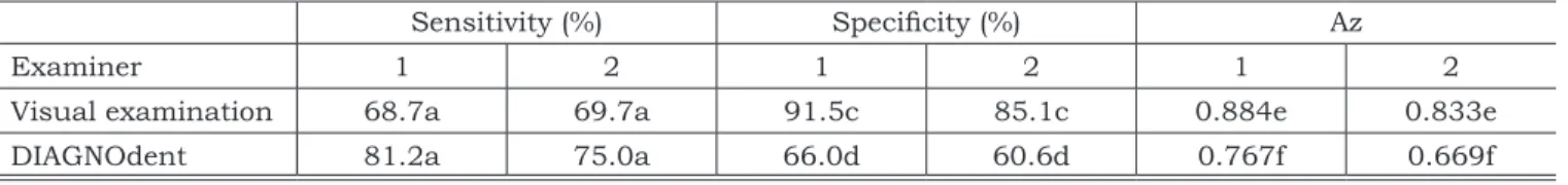
Sensitivities, specificities and areas under ROC curve (Az) are shown in Table 4. VI and the LF showed similar sensitivities (p > 0.05) for both examiners, however, VI showed higher specificities than LF (p < 0.05). The overall analysis, seen by the area under the ROC curve, shows that VI had a better performance than the LF device.
DISCUSSION
The greater sensitivity of VI compared with LF for the detection of carious lesions was not found in some previous studies1,17,18. This is likely to be
due to the selected VI system employed in these studies, which attempted to predict the severity of a lesion (i.e, no lesion, enamel lesion and dentine lesion) instead of using systems that attempted to characterize some features on the occlusal sur-face4.
As shown in Table 4, the sensitivity (68.7%; 69.7%) and specificity (85.1%; 91.5%) values for the VI are within the range of the values provided by the literature. When nine studies of VI for the diagnosis of occlusal caries were reviewed, the sen-sitivity ranged from 0.12 to 0.80, and the speci-ficity, from 0.67 to 0.9713. The great variability of
sensitivity and specificity values for VI found in the literature may reflect other variables such as the scoring systems used, the conditions of the samples, cut-off points, and validation methods. Disease prevalence in the selected sample can be another variable. An increase of the number of teeth with visually apparent cavitation can in-crease the sensitivities values15.
The VI has been considered an invaluable tool in the last decades. Due to its high specificity, this diagnostic method avoids overtreatment, mainly in low caries prevalence populations. However, only recently, the VI has also demonstrated high sensitivity values2,24, which is important to avoid
undertreatment. This has been due to the employ-ment of the VI proposed by Ekstrand et al.4 (1997).
These authors demonstrated, in a laboratory set-ting, a good correlation between occlusal signs and
the histological depth of the lesion, which implies that the poor sensitivity values of other workers’ VI could be attributed to a failure in selecting ap-propriate VI criteria.
However, the application of this ranked scor-ing system requires extensive trainscor-ing of represen-tative macroscopic occlusal signs of each score be-forehand. This previous calibration procedure is of paramount importance in order to obtain positive results. In fact, one may consider that the bad per-formance of the Ekstrand’s visual system4 in other
recent published papers10,22,23 could conceivably be
attributed to the lack of experience in using this VI system. The LF device can be considered an
ad-juvant tool for occlusal caries diagnosis. However,
this system cannot be used as a substitute for VI, since its sole use could lead to overtreatment. Ac-cording to the Ekstrand’s visual system4, the
pres-ence of discoloration, visible without air-drying, is an indicator of demineralization involving between 50% of the enamel and the outer 1/3 of dentine. Thus, all sites presenting this visual pattern are considered sound by the visual inspection, while for the LF diagnostic method there is a high ten-dency of being considered caries-affected, which results in a high rate of false-positive answers. Re-cent evidence has shown that the LF device tends to overscore discolored sites7,9,25.
Although some researchers have detected a good to excellent performance of the LF de-vice17,18,19,24,27, others have not observed this
trend22,23. Several factors may account for this
difference, such as the recommended cut-offs for interpretation of the LF measurements9 and the
storage medium of the sample before the beginning of the study20,26,28.
If we carefully examine the best cut-off points used for LF readings in clinical studies, one should observe that values higher than 19-20 indicate dentinal involvement and are currently used as a threshold between “sound” and “carious” sites1,19,30,
which is in agreement with the present investiga-tion. Therefore, when LF readings were higher than 20, it is likely that the lesion has extended into
TABLE 4 - Performance of the diagnostic methods in diagnosing occlusal carious lesions using threshold between
2 and 3 in each scoring system: sensitivity, speciicity and area under ROC curve (Az) (*).
Sensitivity (%) Speciicity (%) Az
Examiner 1 2 1 2 1 2
Visual examination 68.7a 69.7a 91.5c 85.1c 0.884e 0.833e
DIAGNOdent 81.2a 75.0a 66.0d 60.6d 0.767f 0.669f
dentine. One should bear in mind, however, that the use of this criterion could lead to overtreatment in discolored sites.
CONCLUSION
A meticulous VI enables the dentist to score earliest signs of caries changes or to differentiate
between fissures with or without discoloration, staining or opacities. The LF showed a good perfor-mance on occlusal caries diagnosis. However, due to its relative high cost and performance inferior to that obtained through the Ekstrand’s visual crite-ria, it should be considered only as an alternative method for occlusal caries diagnosis.
REFERENCES
1. Alwas-Danowska HA, Plasschaert AJM, Suliborski S, Ver-donschot EH. Reliability and validity issues of laser fluores-cence measurements in occlusal caries diagnosis. J Dent 2002;30:129-34.
2. Attrill DC, Ashley PF. Occlusal caries detection in primary teeth: a comparison of DIAGNOdent with conventional methods. Brit Dent J 2001;190:440-3.
3. Ekstrand K, Qvist V, Thylstrup A. Light microscope study of the effect of probing in occlusal surfaces. Caries Res 1987;21:368-74.
4. Ekstrand KR, Ricketts DNJ, Kidd EAM. Reproducibility and accuracy of three methods for assessment of demineraliza-tion depth on the occlusal surface: an in vitro examination. Caries Res 1997;31:224-31.
5. Ferreira-Zandoná AG, Anloui M, Beiswanger BB, Isaacs RL, Kafrawy AH, Eckert GJ, et al. An in vitro comparison between laser fluorescence and visual examination for de-tection of demineralization in occlusal pits and fissures. Caries Res 1998;32:210-8.
6. Fleiss IL. Statistical Methods for rates and proportions. 2nd ed. New York: Wiley; 1981.
7. Francescut P, Lussi A. Correlation between fissure discol-oration, DIAGNOdent measurements and caries depth: an in vitro study. Pediatr Dent 2003;25:559-64.
8. Hanley JA, McNeil BJ. A method of comparing the areas under Receiver Operating Characteristic curves derived from the same cases. Radiology 1983;148:839-43. 9. Heinrich-Weltzien R, Kühnisch J, Oehme T, Ziehe A,
Stös-ser L, García-Godoy F. Comparison of different DIAGNO-dent cut-off limits for in vivo detection of occlusal caries. Oper Dent 2003;28:672-80.
10. Heinrich-Weltzien R, Weerheijm KL, Kühnisch J, Oeh -me T, Stösser L. Clinical evaluation of visual, radiographic and laser fluorescence methods for detection of occlusal caries. J Dent Child 2002;69:127-32.
11. Hibst R, Paulus R, Lussi A. Detection of occlusal car-ies by laser fluorescence: basic and clinical investigations. Med Laser Appl 2001;16:205-13.
12. Huysmans MC, Longbottom C, Pitts N. Electrical methods in occlusal caries diagnosis: an in vitro compari-son with visual inspection and bite-wing radiography. Car-ies Res 1998;32:324-9.
13. Ie YL, Verdonschot EH. Performance of diagnostic systems in occlusal caries detection compared. Community Dent Oral Epidemiol 1994;22:187-91.
14. Loesche WJ, Svanberg ML, Pape HR. Intraoral trans-mission of Streptococcus mutans by a dental explorer. J Dent Res 1979;58:1765-70.
15. Lussi A. Impact of including and excluding cavitated lesions when evaluating methods for the diagnosis of oc-clusal caries. Caries Res 1996;30:389-93.
16. Lussi A. Validity of diagnostic and treatment decisions of fissure caries. Caries Res 1991;25:296-303.
17. Lussi A, Francescut P. Performance of conventional and new methods for the detection of occlusal caries in deciduous teeth. Caries Res 2003;37:2-7.
18. Lussi A, Imwinkelried S, Pitts NB, Longbottom C, Reich E. Performance and reproducibility of a laser fluo-rescence system for detection of occlusal caries in vitro. Caries Res 1999;33:261-6.
19. Lussi A, Megert B, Longbottom C, Reich E, Fran-cescut P. Clinical performance of a laser fluorescence de-vice for detection of occlusal caries lesions. Eur J Oral Sci 2001;109:14-9
20. Mendes FM, Pinheiro SL, Bengtson AL. Effect of altera-tion in organic material of the occlusal caries on DIAGNO-dent readings. Braz Oral Res 2004;18:141-4.
21. Penning C, van Amerongen JP, Seef RE, ten Cate JM. Validity of probing for fissure caries diagnosis. Caries Res 1992;26:445-9.
22. Pereira AC, Verdonschot EH, Huysmans MCDNJM. Caries Detection methods: can they aid decision making for invasive sealant treatment? Caries Res 2001;35:83-9. 23. Reis A, Zach VL Jr, de Lima AC, de Lima Navarro MF,
Grande RHM. Occlusal caries detection: a comparison of DIAGNOdent and two conventional diagnostic methods. J Clin Dent 2004;15:76-82.
24. Rocha RO, Ardenghi TM, Oliveira LB, Rodrigues CRMD, Ciamponi AL. In vivo effectiveness of laser-fluo-rescence compared to visual inspection and radiography for detection of occlusal caries in primary teeth. Caries Res 2003;18:32-5.
25. Sheehy EC, Brailsford SR, Kidd EAM, Beighton D, Zoitopoulos L. Comparison between visual examination and a laser fluorescence system for in vivo diagnosis of occlusal caries. Caries Res 2001;35:421-6.
26. Shi XQ, Tranæus S, Angmar-Månsson B. Compari-son of QLF and DIAGNOdent for quantification of smooth surface caries. Caries Res 2001;35:21-6.
27. Shi XQ, Welander U, Angmar-Månsson B. Occlusal caries detection with Kavo DIAGNOdent and radiography: an in vitro comparison. Caries Res 2000;34:151-8. 28. Takamori K, Hokari N, Okumura Y, Watanabe S.
29. van Dorp CS, Exterkate RA, ten Cate JM. The effect of dental probing on subsequent enamel demineralization. ASDC J Dent Child 1988;55:343-7.
30. Verdonschot EH, Abdo H, Frankenmolen FWA. The in vivo performance of a laser fluorescence device compared to visual inspection in occlusal caries diagnosis [abstract]. Caries Res 1999;33:283.